. 2020 Feb 4;10(1):1778.
doi: 10.1038/s41598-020-58374-z.

Porphyromonas gingivalis triggers the shedding of inflammatory endothelial microvesicles that act as autocrine effectors of endothelial dysfunction

Affiliations

Porphyromonas gingivalis triggers the shedding of inflammatory endothelial microvesicles that act as autocrine effectors of endothelial dysfunction

Isaac Maximiliano Bugueno et al. Sci Rep. .

Abstract

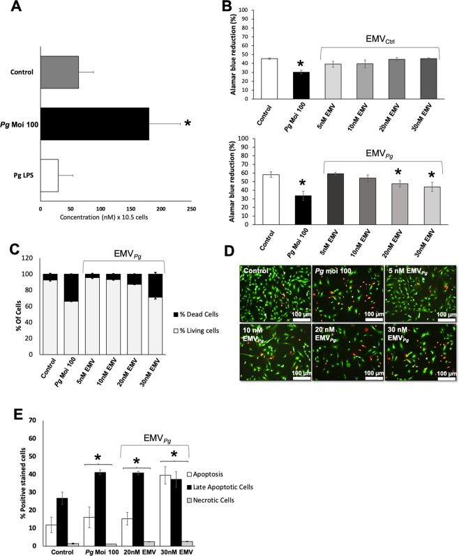
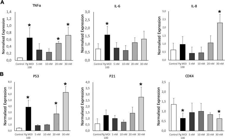
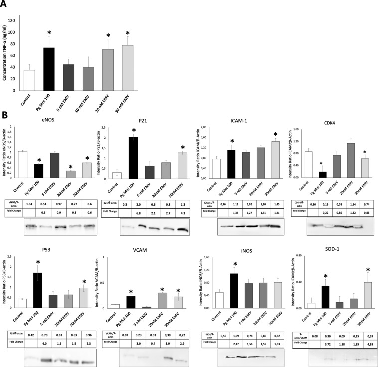
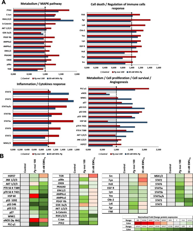
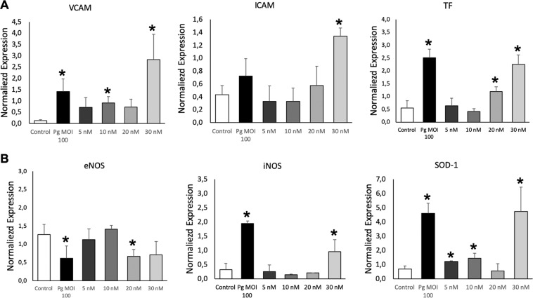
A link between periodontitis and atherothrombosis has been highlighted. The aim of this study was to determine the influence of Porphyromonas gingivalis on endothelial microvesicles (EMVPg) shedding and their contribution to endothelial inflammation. Endothelial cells (EC) were infected with P. gingivalis (MOI = 100) for 24 h. EMVPg were isolated and their concentration was evaluated by prothrombinase assay. EMVPg were significantly increased in comparison with EMVCtrl shedded by unstimulated cells. While EMVCtrl from untreated EC had no effect, whereas, the proportion of apoptotic EC was increased by 30 nM EMVPg and viability was decreased down to 25%, a value elicited by P. gingivalis alone. Moreover, high concentration of EMVPg (30 nM) induced a pro-inflammatory and pro-oxidative cell response including up-regulation of TNF-α, IL-6 and IL-8 as well as an altered expression of iNOS and eNOS at both mRNA and protein level. An increase of VCAM-1 and ICAM-1 mRNA expression (4.5 folds and 3 folds respectively (p < 0.05 vs untreated) was also observed after EMVPg (30 nM) stimulation whereas P. gingivalis infection was less effective, suggesting a specific triggering by EMVPg. Kinasome analysis demonstrated the specific effect induced by EMVPg on main pro-inflammatory pathways including JNK/AKT and STAT. EMVPg are effective pro-inflammatory effectors that may have detrimental effect on vascular homeostasis and should be considered as potential autocrine and paracrine effectors involved in the link between periodontitis and atherothrombosis.


Conflict of interest statement

The authors declare no competing interests.

Figures

Figure 1
Figure 1
P. gingivalis promotes EMV shedding and alters endothelial cell viability. (A) The generation of EMV from naïve EC (control) and following 24 h of infection with P. gingivalis (Pg) (MOI = 100) or stimulation with Pg-LPS (1μg/ml) was measured in the supernatant by prothrombinase assay. Concentrations are represented by mean +/− SD from 3 independent experiments; *p < 0.05 vs control (unstimulated EC). (B) Metabolic activity of EC infected with Pg (MOI = 100) or exposed to EMVCtrl (upper panel) and EMVPg (lower panel) (5, 10, 20 and 30 nM) for 24 h measured by AlamarBlue assay. Results are expressed as mean +/− SD from 3 independent experiments; *p < 0.05 vs control (unstimulated EC). (C) Live-Dead assay to evaluate the ratio of live EC versus dead EC in cells exposed to P. gingivalis (Pg) (MOI:100) and EMVPg (5, 10, 20 and 30 nM) for 24 h. Results are expressed as percentage of live and dead EC (mean +/−SD). (D) Immunofluorescence imaging of live-dead staining (green: live EC, red: dead EC) for each condition after 24 hours of exposure. (E) Evaluation of type of cell death by flow cytometry after P. gingivalis infection and EMVPg (20 and 30 nM) exposure for 24 h. EC were labelled with Annexin-V FITC and propidium iodide (IPPE). All data were expressed as mean ± SD. *(p < 0.05 vs untreated cells).
Figure 2
Figure 2
EMVPg trigger inflammatory endothelial response. (A) The mRNA expression of inflammatory markers TNF-α, IL-6 and IL-8 in EC exposed to P. gingivalis (Pg) (MOI:100) and EMVPg (5, 10, 20 and 30 nM) for 24 h was measured by qRT-PCR. (B) The gene expression of cell cyle related markers p53, p21 and CDK4 EC in EC exposed to P. gingivalis (Pg) (MOI:100) and EMVPg (5, 10, 20 and 30 nM) for 24 h. All data were expressed as mean ± SD from 3 independent experiments. *p < 0.05 between cells infected or stimulated against control (unstimulated cells).
Figure 3
Figure 3
EMVPg induce pro-inflammatory and pro-oxidative protein expression similar to P. gingivalis infection. (A) TNF-α secretion in supernatant by EC in response to P. gingivalis (Pg) (MOI:100) and EMVPg (5, 20 and 30 nM) for 24 h was measured by ELISA. (B) Intra-cellular protein expression of eNOS, P21, ICAM-1, CDK4, P53, VCAM, iNOS and SOD-1 in response to P. gingivalis (Pg) (MOI:100) and EMVPg (5, 20 and 30 nM) for 24 h was evaluated by Western Blot. All data were expressed as mean ± SD from 3 independent experiments and normalized against internal control β-actin. *p < 0.05 between cells infected or stimulated against control (unstimulated cells).
Figure 4
Figure 4
Exposure to EMVPg modulates significantly inflammatory pathways related to kinases activation. Analysis of kinases activation induced by P. gingivalis infection (Pg) (MOI:100) and EMVPg (30 nM) for 24 h evaluated by phospho-kinase array. The density of spots was measured by MyImageTM Analysis Softwate 2.0 (Thermofisher) for each molecule and each condition. (A) Graphical representation of the kinases expression in untreated EC, in EC following P. gingivalis infection (Pg) (MOI:100) and EMVPg (30 nM) stimulation for 24 h. (B) Heat-map representation of the kinases expression normalized against untreated control (untreated EC).
Figure 5
Figure 5
EMVPg induce expression of atherothrombosis and oxidative stress markers expression. (A) The mRNA expression of endothelial markers VCAM, ICAM and Tissular factor (TF) in EC exposed to P. gingivalis (Pg) (MOI:100) and EMVPg (5, 10, 20 and 30 nM) for 24 h was measured by qRT-PCR. (B) The gene expression of oxidative stress markers eNOS, iNOS and SOD-1 in EC exposed to P. gingivalis (Pg) (MOI:100) and EMVPg (5, 10, 20 and 30 nM) for 24 h. All data were expressed as mean ± SD from 3 independent experiments. *p < 0.05 between cells infected or stimulated against control (unstimulated cells).

References

    1. Hajishengallis G, Lamont RJ. Breaking bad: Manipulation of the host response by Porphyromonas gingivalis. Eur. J. Immunol. 2014;44:328–338. doi: 10.1002/eji.201344202. - DOI - PMC - PubMed
    1. Kassebaum NJ, et al. Global burden of severe periodontitis in 1990–2010: a systematic review and meta-regression. J. Dent. Res. 2014;93:1045–1053. doi: 10.1177/0022034514552491. - DOI - PMC - PubMed
    1. Hajishengallis G. Periodontitis: from microbial immune subversion to systemic inflammation. Nat. Rev. Immunol. 2015;15:30–44. doi: 10.1038/nri3785. - DOI - PMC - PubMed
    1. Linden GJ, Lyons A, Scannapieco FA. Periodontal systemic associations: review of the evidence. J. Clin. Periodontol. 2014;40:8–19. doi: 10.1111/jcpe.12064. - DOI - PubMed
    1. Huck O, Elkaim R, Davideau JL, Tenenbaum H. Evaluating periodontal risk for patients at risk of or suffering from atherosclerosis: recent biological hypotheses and therapeutic consequences. Arch. Cardiovasc. Dis. 2011;104:352–358. doi: 10.1016/j.acvd.2011.02.002. - DOI - PubMed

Publication types

MeSH terms

Substances

LinkOut - more resources