. 2020 Feb 4;10(1):1807.
doi: 10.1038/s41598-020-58368-x.

A New Function for Perivascular Adipose Tissue (PVAT): Assistance of Arterial Stress Relaxation

Affiliations

A New Function for Perivascular Adipose Tissue (PVAT): Assistance of Arterial Stress Relaxation

Stephanie W Watts et al. Sci Rep. .

Abstract

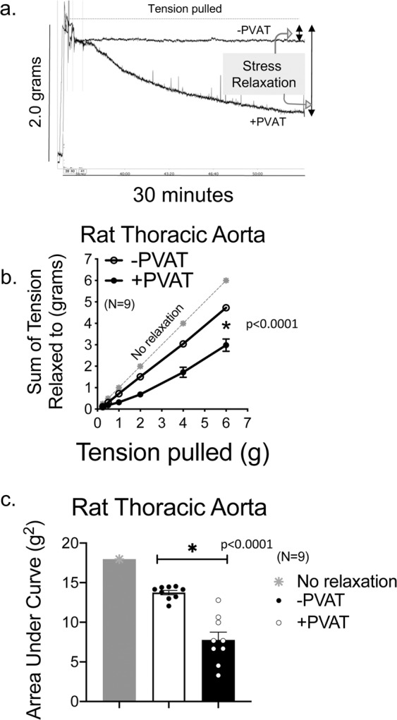
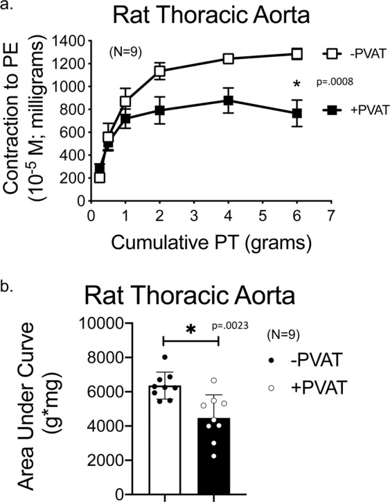
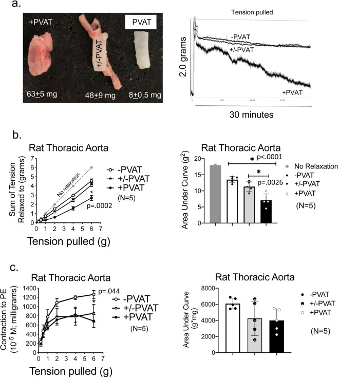
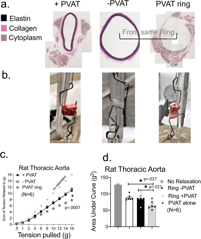
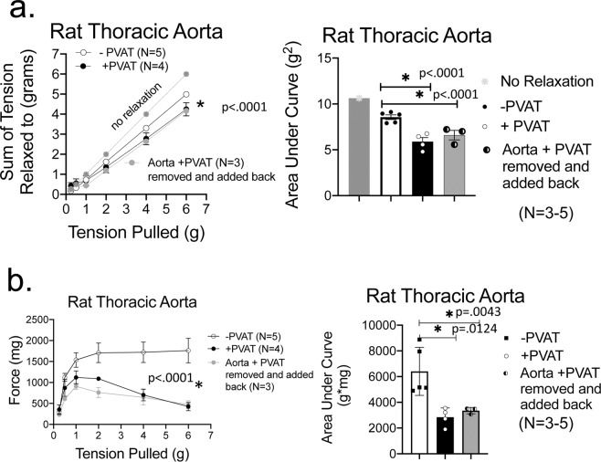
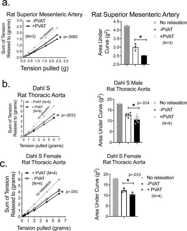
In health, PVAT secretes anti-contractile factors that relax the underlying artery. PVAT's contributions to vascular function include more than production of vasoactive substances. We hypothesized that PVAT benefits the artery by assisting the function of stress (-induced) relaxation. Thoracic aorta rings from Sprague Dawley rats were mounted in isolated tissue baths with (+) and without (-) PVAT. A cumulative length tension (0-6 grams) was generated. The tension to which the tissue stress relaxed over 30 minutes was recorded; the tension lost was stress relaxation. The presence of PVAT increased the amount of stress relaxation (final tension in mgs; aortic ring -PVAT = 4578 ± 190; aortic ring + PVAT = 2730 ± 274, p < 0.05). PVAT left attached but not encompassing the aorta provided no benefit in cumulative stress relaxation (aortic ring +/- PVAT = 4122 ± 176; p > 0.05 vs -PVAT). A PVAT ring separated from the aorta demonstrated more profound stress relaxation than did the aortic ring itself. Finally, PVAT-assisted stress relaxation was observed in an artery with white fat (superior mesenteric artery) and in aorta from both male and female of another rat strain, the Dahl S rat. Knowledge of this new PVAT function supports PVAT as an essential player in vascular health.


Conflict of interest statement

The authors declare no competing interests.

Figures

Figure 1
Figure 1
PVAT assists stress relaxation in thoracic aorta. (a) A representative tracing of isometric tension of aortic ring + and −PVAT over a 30-minute stretch step in a protocol using PE challenges between each stretch step. (b) Quantitation of the cumulative tension achieved with each stretch step vs no relaxation (gray line). (c) Quantitation of the area under the curve for aortic rings +PVAT and aortic rings −PVAT vs that of no relaxation (gray). Final mass: aortic rings +PVAT = 61.7 +/- 3.7 mgs; aortic rings −PVAT = 7.9 +/- 0.5 mg. Points/bars represent means +/- SEM for the number of animals in parentheses. *Significant differences as determined by a two-tail unpaired Students t-test (p value reported in figure) for maximum values.
Figure 2
Figure 2
PVAT anti-contractile effect is present through a range of passive tensions. (a) Quantitation of the magnitude of PE-induced contraction with each stretch step. (b) Quantitation of the area under the curve for aortic rings +PVAT and aortic rings −PVAT. Tissue masses are stated in legend for Fig. 1. Points/bars represent means± SEM for the number of animals in parentheses. *Significant differences as determined by a two-tail unpaired Students t-test (p value reported in figure) for maximum values.
Figure 3
Figure 3
Detachment of PVAT from absolute length of ring abolishes its assistance of arterial stretch relaxation. (a) Image of aortic rings from the same animal +PVAT, +/−PVAT (intact only on ends), and −PVAT. Numbers beneath each vessel image is the vessel mass presented as means ± SEM for the N = 5 vessels experimented on to generate data in this figure. Quantitation of cumulative stress relaxation (b) and active contraction to PE (c) reporting absolute values (left) and area under the curve (right). Points/bars represent means ± SEM for the number of animals in parentheses. *Significant differences (p value reported in figure) as determined by a one-way ANOVA with Tukey’s post hoc determinations comparing averages as indicated by the tied bar.
Figure 4
Figure 4
PVAT independent of vessel demonstrates profound stress relaxation. (a) Histology of PVAT separated from proper vessel to validate the adventitia is still present in the vessel. Representative of six separate rats. (b) Image of an aortic ring +PVAT, aortic ring −PVAT and PVAT ring alone when placed between two L-shaped hooks and with no tension applied. (c) Quantitation of the cumulative tension achieved with each stretch step in aortic ring +/− its PVAT or PVAT ring alone. (d) Quantitation of the area under the curve for aortic ring +PVAT, aortic ring −PVAT and PVAT ring. Final mass: aortic rings +PVAT = 51.6 ± 3.6 mg; aortic rings −PVAT = 8.1 ± 0.7 mg; PVAT ring = 38.3 ± 10.7 mg. Points/bars represent means ± SEM for the number of animals in parentheses. *Significant differences (p value reported in figure) as determined by a one-way ANOVA with Tukey’s post hoc determinations comparing averages as indicated by the tied bar.
Figure 5
Figure 5
PVAT assistance of stress relaxation is independent of direct connections to artery. (a) Quantitation of the cumulative tension achieved with each stretch step in aortic ring +PVAT, aortic ring −PVAT and a ring in which PVAT was physically detached from artery, removed, and then placed back on artery (aorta ring +PVAT ring removed and added back) as a line graph (left) and area under the curve (right). (b) Response of vessels to a maximum concentration of PE after each stretch step as a line graph (left) and area under the curve (right). Final masses: aortic ring +PVAT = 35.6 ± 7.1, aortic ring −PVAT = 4.8 ± 0.9, aortic ring +PVAT ring removed and added back = 28.2 ± 2.2 mg. *Significant differences (p value reported in figure) as determined by a one-way ANOVA with Tukey’s post hoc determinations comparing averages as indicated by the tied bar.
Figure 6
Figure 6
PVAT assisted stress relaxation occurs in a different artery and different rat strain. Quantitation (line graph, left; area under the curve right) of cumulative stress relaxation in the isolated superior mesenteric artery (a) and thoracic aorta of the Dahl S male rat (b) and female rat (c). +PVAT indicates PVAT was present, −PVAT indicates PVAT was removed. Final masses: Superior mesenteric artery +PVAT = 162 ± 17 mg; mesenteric artery −PVAT: 2.1 ± 0.45 mg; Dahl S male aortic ring +PVAT = 68 ± 4.5 mgs Dahl S male aortic ring −PVAT = 12.0 ± 1.1 mgs; Dahl S female aortic ring +PVAT = 50.7 ± 4.8 mg, Dahl S female aortic ring −PVAT = 12.3 ± 2.1 mg. Points/bars represent means ± SEM for the number of animals in parentheses. *Significant differences as determined by a two-tail unpaired Students t-test (p value reported in figure).

Similar articles

Cited by

References

    1. Cheng CK, Bakar HA, Gollasch M, Huang Y. Perivascular adipose tissue: the sixth man of the cardiovascular system. Cardiovasc. Drugs Ther. 2018;32:481–502. doi: 10.1007/s10557-018-6820-z. - DOI - PMC - PubMed
    1. Pond CM. Accuracy and artistry in anatomical illustration of perivascular adipose tissue. Front. Physiol. 2017;8:990. doi: 10.3389/fphys.2017.00990. - DOI - PMC - PubMed
    1. Soltis EE, Cassis LA. Influence of perivascular adipose tissue on rat aortic smooth muscle responsiveness. Clin. Exp. Hypertens. A. 1991;13:277–296. - PubMed
    1. Lohn M, et al. Periadventital fat releases a vascular relaxing factor. FASEB J. 2002;16:1057–1063. doi: 10.1096/fj.02-0024com. - DOI - PubMed
    1. Agabati-Rosei C, et al. Modulation of vascular reactivity by perivascular adipose tissue (PVAT) Curr. Hypertens. Rep. 2018;20:44. doi: 10.1007/s11906-018-0835-5. - DOI - PubMed

Publication types