

Development of Magnetic Probe for Sentinel Lymph Node Detection in Laparoscopic Navigation for Gastric Cancer Patients
- PMID: 32019961
- PMCID: PMC7000689
- DOI: 10.1038/s41598-020-58530-5
Development of Magnetic Probe for Sentinel Lymph Node Detection in Laparoscopic Navigation for Gastric Cancer Patients
Abstract
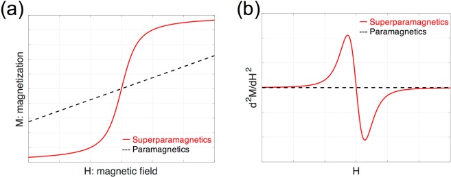
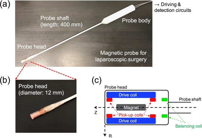
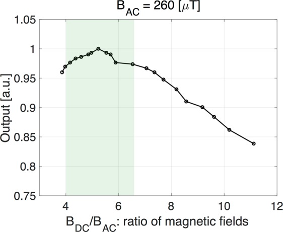
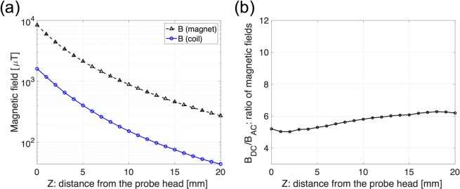
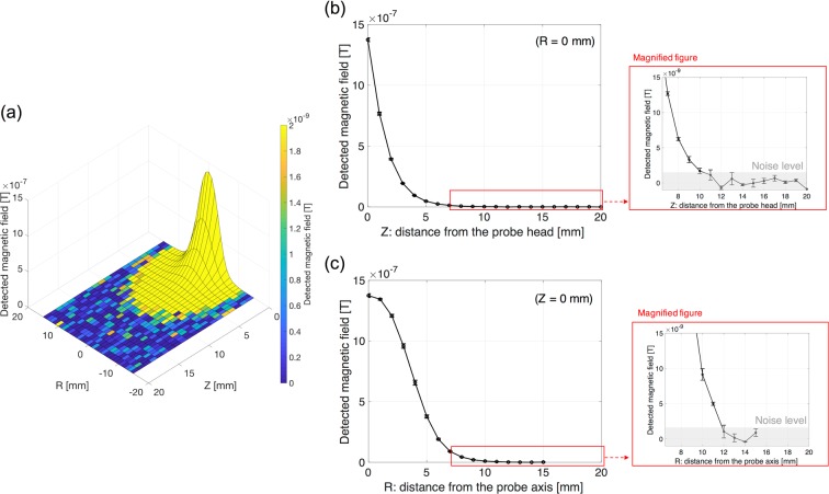
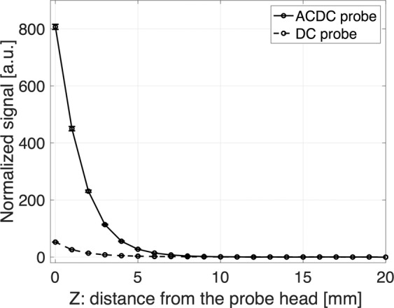
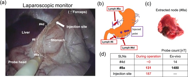
New laparoscopic sentinel lymph node navigation using a dedicated magnetic probe and magnetic nanoparticle tracer for gastric cancer patients allows minimally invasive surgeries. By identifying the sentinel lymph nodes containing magnetic nanoparticles, patients can avoid excessive lymph node extraction without nuclear facilities and radiation exposure. This paper describes the development of the laparoscopic magnetic probe, ACDC-probe, for laparoscopic sentinel lymph node identification utilizing the nonlinear response of the magnetic nanoparticles magnetized by an alternating magnetic field with a static magnetic field. For highly sensitive detection, the ratio of static to alternating magnetic fields was optimized to approximately 5. The longitudinal detection length was approximately 10 mm for 140 μg of iron, and the detectable amount of iron was approximately 280 ng at a distance of 1 mm. To demonstrate the feasibility of laparoscopic detection using the ACDC-probe and magnetic tracers, an experiment was performed on a wild swine. The gastric sentinel lymph node was clearly identified during laparoscopic navigation. These results suggest that the newly developed ACDC-probe is useful for laparoscopic sentinel lymph node detection and this magnetic technique appears to be a promising method for future sentinel lymph node navigation of gastric cancer patients.
Conflict of interest statement
The authors declare no competing interests.
Figures


Similar articles
-
Sentinel lymph node navigation surgery for gastric cancer: Does it really benefit the patient?World J Gastroenterol. 2016 Mar 14;22(10):2894-9. doi: 10.3748/wjg.v22.i10.2894. World J Gastroenterol. 2016. PMID: 26973385 Free PMC article. Review.
-
Sentinel Node Navigation in Gastric Cancer: Where Do We Stand?J Gastrointest Cancer. 2019 Jun;50(2):201-206. doi: 10.1007/s12029-019-00217-w. J Gastrointest Cancer. 2019. PMID: 30815770 Review.
-
Laparoscopic sentinel node navigation surgery versus laparoscopic gastrectomy with lymph node dissection for early gastric cancer: short-term outcomes of a multicentre randomized controlled trial (SENORITA).Br J Surg. 2020 Oct;107(11):1429-1439. doi: 10.1002/bjs.11655. Epub 2020 Jun 3. Br J Surg. 2020. PMID: 32492186 Clinical Trial.
-
Pilot-study on the feasibility of sentinel node navigation surgery in combination with thoracolaparoscopic lymphadenectomy without esophagectomy in early esophageal adenocarcinoma patients.Dis Esophagus. 2017 Nov 1;30(11):1-8. doi: 10.1093/dote/dox097. Dis Esophagus. 2017. PMID: 28881907
-
Feasibility of Regional Lymphadenectomy for Stomach-Preserving Surgery in Early Gastric Cancer Omitting Sentinel Node Navigation: A Post Hoc Analysis of the SENORITA Trial.Ann Surg Oncol. 2024 Oct;31(10):6939-6946. doi: 10.1245/s10434-024-15950-1. Epub 2024 Jul 31. Ann Surg Oncol. 2024. PMID: 39085549 Free PMC article. Clinical Trial.
Cited by
-
Preclinical feasibility of robot-assisted sentinel lymph node biopsy using multi-modality magnetic and fluorescence guidance in the head and neck.Head Neck. 2022 Dec;44(12):2696-2707. doi: 10.1002/hed.27177. Epub 2022 Sep 8. Head Neck. 2022. PMID: 36082404 Free PMC article.
-
Functional roles of magnetic nanoparticles for the identification of metastatic lymph nodes in cancer patients.J Nanobiotechnology. 2023 Sep 21;21(1):337. doi: 10.1186/s12951-023-02100-0. J Nanobiotechnology. 2023. PMID: 37735449 Free PMC article. Review.
-
A Fluorescent and Magnetic Hybrid Tracer for Improved Sentinel Lymphadenectomy in Prostate Cancer Patients.Biomedicines. 2023 Oct 13;11(10):2779. doi: 10.3390/biomedicines11102779. Biomedicines. 2023. PMID: 37893150 Free PMC article.
-
Preclinical evaluation of sentinel node localization in the stomach via mannose-labelled magnetic nanoparticles and indocyanine green.Surg Endosc. 2023 Aug;37(8):6185-6196. doi: 10.1007/s00464-023-10099-6. Epub 2023 May 10. Surg Endosc. 2023. PMID: 37165173 Free PMC article.
-
A new bimodal approach for sentinel lymph node imaging in prostate cancer using a magnetic and fluorescent hybrid tracer.Eur J Nucl Med Mol Imaging. 2024 Aug;51(10):2922-2928. doi: 10.1007/s00259-023-06522-8. Epub 2023 Nov 24. Eur J Nucl Med Mol Imaging. 2024. PMID: 37999812 Free PMC article.
References
Publication types
MeSH terms
LinkOut - more resources
Full Text Sources
Medical
Miscellaneous
