. 2020 Mar;43(3):930-942.
doi: 10.3892/or.2020.7480. Epub 2020 Jan 27.

Linc00261 inhibits metastasis and the WNT signaling pathway of pancreatic cancer by regulating a miR‑552‑5p/FOXO3 axis

Affiliations

Linc00261 inhibits metastasis and the WNT signaling pathway of pancreatic cancer by regulating a miR‑552‑5p/FOXO3 axis

Tengxiang Chen et al. Oncol Rep. 2020 Mar.

Abstract

The biological function of long non‑coding RNA00261 (Linc00261) has been widely investigated in various types of cancer. The aim of the present study was to explore the role of Linc00261 in pancreatic cancer (PC). The expression of Linc00261 in patients with PC and PC cell lines was assessed using reverse transcription‑quantitative PCR and the association of Linc00261 expression with survival was analyzed in the online database, GEPIA. The effects of Linc00261 on PC cell metastasis in vitro and in vivo were determined using a wound healing assay, Transwell invasion assays and a nude mouse model of liver metastasis. The relationship between Linc00261, the miR‑552‑5p/forkhead box O3 (FOXO3) axis and the Wnt signaling pathway were determined using bioinformatics analysis, dual luciferase assay and western blotting. Linc00261 expression was significantly decreased in PC tissues and cell lines, and reduced expression was associated with less favorable outcomes in patients with PC. Linc00261 overexpression inhibited migration and invasion of PC cells in vitro, whereas knockdown of Linc00261 increased migration and invasion. Linc00261 overexpression also decreased metastasis of PC cells in vivo. Linc00261 was revealed to directly bind to microRNA (miR)‑552‑5p and to decrease the expression of miR‑552‑5p. In addition, Linc00261 overexpression increased the expression of FOXO3, a target gene of miR‑552‑5p, as well as inhibited the Wnt signaling pathway. Overexpression of miR‑552‑5p in Linc00261‑overexpressing PC cells increased migration and invasion, as well as decreased the expression of FOXO3 and members of the Wnt signaling pathway. Collectively, the present study demonstrated that Linc00261 inhibited metastasis and the Wnt signaling pathway of PC by regulating the miR‑552‑5p/FOXO3 axis. Linc00261 may suppress the development of PC, and serve as a potential biomarker and effective target for the diagnosis and treatment of PC.

Keywords: pancreatic cancer; metastasis; Linc00261; microRNA-552-5p; forkhead box O3.


Figures

Figure 1.
Figure 1.
Linc00261 is significantly downregulated in PC. (A) The expression of Linc00261 in PC tissues and adjacent healthy tissues was analyzed using the online database, GEPIA. (B) Association between the prognosis and the expression of Linc00261 was analyzed using the online database, GEPIA. (C) Expression of Linc00261 was measured in 54 PC tissues and 54 adjacent non-tumor tissues using reverse transcription-quantitative PCR. (D) The expression of Linc00261 in the PC cell lines, CFPAC-1, AsPC-1, MIA-PaCa2, Capan-2, BXPC-3 and PANC-1 was significantly decreased compared with the HPDE cells. *P<0.05, **P<0.01. PC, pancreatic cancer; HPDE, human pancreatic epithelium cells.
Figure 2.
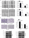
Figure 2.
Linc00261 overexpression inhibits the migration and invasion of PC cells in vitro. PANC-1 and MIA-PaCa2 were transfected with empty vector or Linc00261 overexpression lentivirus. (A) A wound healing assay was used to assess the effect of Linc00261 overexpression on the migration of PC cells. (B) A Transwell invasion assay was used to detect the effect of Linc00261 overexpression on the invasion of PC cells. (C) Western blotting was used to detect the expression of three epithelial-mesenchymal transition markers, E-cadherin, N-cadherin and vimentin, in Linc00261-overexpressing cells. *P<0.05, **P<0.01. PC, pancreatic cancer; LV-Linc00261, Linc00261-overexpressing cells; NC, negative control.
Figure 3.
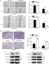
Figure 3.
Linc00261 knockdown increases the migration and invasion of PC cells in vitro. PANC-1 and MIA-PaCa2 cells were transfected with scrambled shRNA and targeted Linc00261 shRNA, and used as the NC group and Linc00261-knockdown group, respectively. (A) A wound healing assay was used to assess the effects of Linc00261 knockdown on the migration of PC cells. (B) A Transwell invasion assay was used to assess the effects of Linc00261 knockdown on the invasion of PC cells. (C) Western blotting was used to detect the expression of three epithelial-mesenchymal transition markers, E-cadherin, N-cadherin and vimentin in Linc00261-knockdown cells. **P<0.01. PC, pancreatic cancer; sh, short hairpin; NC, negative control represented by sh-Control; sh-Linc00261, Linc00261 knockdown.
Figure 4.
Figure 4.
Linc00261 overexpression reduces metastasis of PC cells in vivo. (A) Images of the liver metastasis model. (B) Quantitative analysis of the average number of visible liver metastases. n=6 per group. (C) Kaplan-Meier survival curves for the mice in both groups in the mouse model of metastasis. (D) Changes in body weight in the mice in the mouse model of metastasis. *P<0.05. PC, pancreatic cancer.
Figure 5.
Figure 5.
miR-552-5p is a target of Linc00261 in PC. (A) Linc00261 predicted binding site in the 3′UTR of miR-552-5p mRNA. (B) miR-552-5p mimics reduced luciferase activity in PC cells, this was not observed with the NC mimics. (C) Expression of Linc00261 was inversely associated with expression of miR-552-5p in PC tissues. (D) miR-552-5p inhibition increased the mRNA expression levels of Linc00261 in PC cells. (E) Overexpression of Linc00261 decreases the expression of miR-552-5p. (F) The mRNA expression levels of miR-552-5p were detected in PC tissues and adjacent non-tumor tissues using reverse transcription-quantitative PCR. (G) mRNA expression levels of miR-552-5p in the CFPAC-1, AsPC-1, MIA-PaCa2, Capan-2, BXPC-3 and PANC-1 PC cells were significantly higher compared with HPDE cells. *P<0.05, **P<0.01. PC, pancreatic cancer; UTR, untranslated region; NC, negative control; miR, microRNA; HPDE, human pancreatic epithelium cells.
Figure 6.
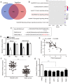
Figure 6.
FOXO3 is a target of miR-552-5p in PC. (A) Using TargetScan and miRwalk online databases, the target genes of miR-552-5p were predicted and 332 genes were considered relevant based on the results from both databases. (B) Bubble chart showing the pathways in which miR-552-5p targeted genes are enriched in. (C) miR-552-5p was predicted to bind to the 3′UTR of FOXO3 mRNA. The mutated site is highlighted. (D) miR-552-5p mimics reduced the luciferase intensity in PC cells, whereas mutation of the associated element in 3′-UTR of FOXO3 mRNA abolished the effect of miR-552-5p mimic on luciferase activity. (E) The expression of miR-552-5p was inversely associated with expression of FOXO3 in PC tissues. (F) The mRNA expression levels of FOXO3 in PC tissues and adjacent non-tumor tissues. (G) The mRNA expression levels of FOXO3 in CFPAC-1, AsPC-1, MIA-Paca2, Capan-2, BXPC-3 and PANC-1 PC cells were significantly decreased compared with HDPE cells. *P<0.05, **P<0.01. PC, pancreatic cancer; miR, microRNA; UTR, untranslated region; HPDE, human pancreatic epithelium cells; FOXO3, forkhead box O3.
Figure 7.
Figure 7.
Linc00261 regulates the β-catenin/TCF4 signaling pathway via miR-552-5p/FOXO3. Cells were divided into four groups: NC lentivirus; LV-Linc00261; miR-552-5p mimics; and LV-Linc00261+miR-552-5p mimics. (A) The mRNA expression levels of FOXO3 in each group were detected using reverse transcription-quantitative PCR. (B) The protein expression levels of FOXO3, β-catenin and TCF4 in each group were detected using western blotting. (C) The mRNA expression levels of E-cadherin, N-cadherin and vimentin in each group. (D) The protein expression levels of E-cadherin, N-cadherin and vimentin in each group. *P<0.05, **P<0.01. miR, microRNA; FOXO3, forkhead box O3; NC, negative control; LV-Linc00261, Linc00261 overexpression lentivirus; miR, microRNA.
Figure 8.
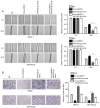
Figure 8.
miR-552-5p overexpression reverses the inhibitory effects of Linc00261. Cells were divided into four groups: NC lentivirus; LV-Linc00261; miR-552-5p mimics; and LV-Linc00261+miR-552-5p mimics. (A) Wound healing assays were used to assess the migratory ability of each group. (B) Transwell invasion assays were used to assess the invasive ability of each group. *P<0.05. miR, microRNA; NC, negative control; LV-Linc00261, Linc00261 overexpression lentivirus.

References

    1. Lee SH, Chang PH, Chen PT, Lu CH, Hung YS, Tsang NM, Hung CY, Chen JS, Hsu HC, Chen YY, Chou WC. Association of time interval between cancer diagnosis and initiation of palliative chemotherapy with overall survival in patients with unresectable pancreatic cancer. Cancer Med. 2019;8:3471–3478. doi: 10.1002/cam4.2254. - DOI - PMC - PubMed
    1. Skau Rasmussen L, Vittrup B, Ladekarl M, Pfeiffer P, Karen Yilmaz M, Østergaard Poulsen L, Østerlind K, Palnæs Hansen C, Bau Mortensen M, Viborg Mortensen F, et al. The effect of postoperative gemcitabine on overall survival in patients with resected pancreatic cancer: A nationwide population-based Danish register study. Acta Oncol. 2019;58:864–871. doi: 10.1080/0284186X.2019.1581374. - DOI - PubMed
    1. Elaileh A, Saharia A, Potter L, Baio F, Ghafel A, Abdelrahim M, Heyne K. Promising new treatments for pancreatic cancer in the era of targeted and immune therapies. Am J Cancer Res. 2019;9:1871–1888. - PMC - PubMed
    1. Mitschke J, Burk UC, Reinheckel T. The role of proteases in epithelial-to-mesenchymal cell transitions in cancer. Cancer Metastasis Rev. 2019;38:431–444. doi: 10.1007/s10555-019-09808-2. - DOI - PubMed
    1. Hu X, Zhai Y, Kong P, Cui H, Yan T, Yang J, Qian Y, Ma Y, Wang F, Li H, et al. FAT1 prevents epithelial mesenchymal transition (EMT) via MAPK/ERK signaling pathway in esophageal squamous cell cancer. Cancer Lett. 2017;397:83–93. doi: 10.1016/j.canlet.2017.03.033. - DOI - PubMed