

New insight into podocyte slit diaphragm, a therapeutic target of proteinuria
- PMID: 32020343
- PMCID: PMC7040068
- DOI: 10.1007/s10157-020-01854-3
New insight into podocyte slit diaphragm, a therapeutic target of proteinuria
Abstract
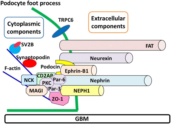
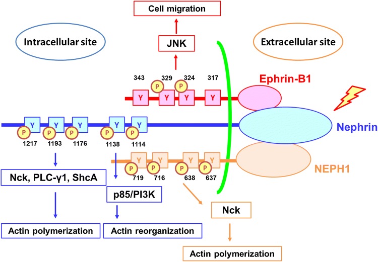
Dysfunction of slit diaphragm, a cell-cell junction of glomerular podocytes, is involved in the development of proteinuria in several glomerular diseases. Slit diaphragm should be a target of a novel therapy for proteinuria. Nephrin, NEPH1, P-cadherin, FAT, and ephrin-B1 were reported to be extracellular components forming a molecular sieve of the slit diaphragm. Several cytoplasmic proteins such as ZO-1, podocin, CD2AP, MAGI proteins and Par-complex molecules were identified as scaffold proteins linking the slit diaphragm to the cytoskeleton. In this article, new insights into these molecules and the pathogenic roles of the dysfunction of these molecules were introduced. The slit diaphragm functions not only as a barrier but also as a signaling platform transfer the signal to the inside of the cell. For maintaining the slit diaphragm function properly, the phosphorylation level of nephrin is strictly regulated. The recent studies on the signaling pathway from nephrin, NEPH1, and ephrin-B1 were reviewed. Although the mechanism regulating the function of the slit diaphragm had remained unclear, recent studies revealed TRPC6 and angiotensin II-regulating mechanisms play a critical role in regulating the barrier function of the slit diaphragm. In this review, recent investigations on the regulation of the slit diaphragm function were reviewed, and a strategy for the establishment of a novel therapy for proteinuria was proposed.
Keywords: Nephrin; Podocyte; Proteinuria; Slit diaphragm; Thrapeutic target.
Conflict of interest statement
The authors have declared no competing interest.
Figures
References
-
- Madison JR, Spies C, Schatz IJ, Masaki K, Chen R, Yano K, et al. Proteinuria and risk for stroke and coronary heart disease during 27 years of follow-up: the Honolulu Heart Program. Arch Intern Med. 2006;166(8):884–889. - PubMed
-
- Tryggvason K, Pettersson E. Causes and consequences of proteinuria: the kidney filtration barrier and progressive renal failure. J Intern Med. 2003;254(3):216–224. - PubMed
-
- Kawachi H, Shimizu F. Molecular composition and function of the slit diaphragm: nephrin, the molecule responsible for proteinuria. Clin Exp Nephrol. 2000;4(3):161–172.
-
- Kawachi H, Suzuki K, Miyauchi N, Hashimoto T, Otaki Y, Shimizu F. Slit diaphragm dysfunction in proteinuric states: identification of novel therapeutic targets for nephrotic syndrome. Clin Exp Nephrol. 2009;13(4):275–280. - PubMed
Publication types
MeSH terms
LinkOut - more resources
Full Text Sources
Miscellaneous
