. 2020 Jul;48(7):2053-2063.
doi: 10.1007/s10439-020-02460-w. Epub 2020 Feb 4.

Phototunable Viscoelasticity in Hydrogels Through Thioester Exchange

Affiliations

Phototunable Viscoelasticity in Hydrogels Through Thioester Exchange

Benjamin J Carberry et al. Ann Biomed Eng. 2020 Jul.

Abstract

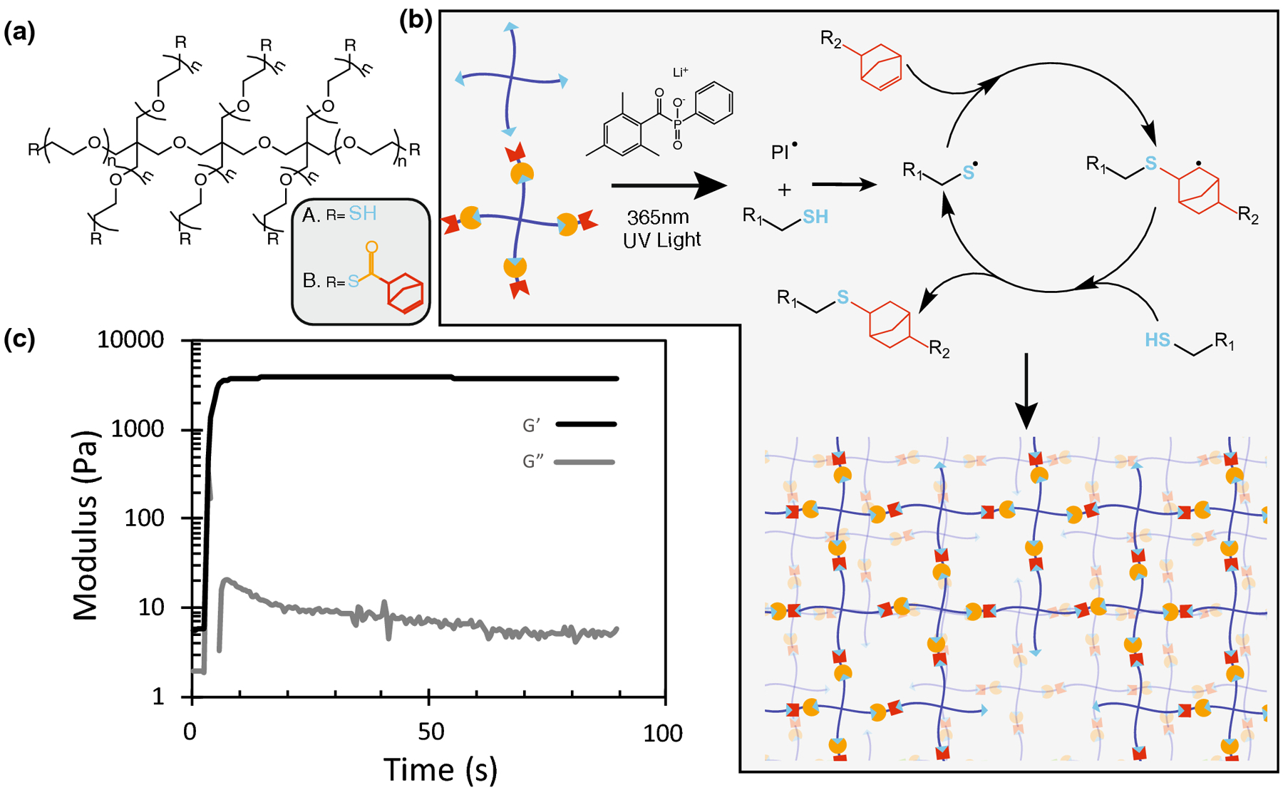
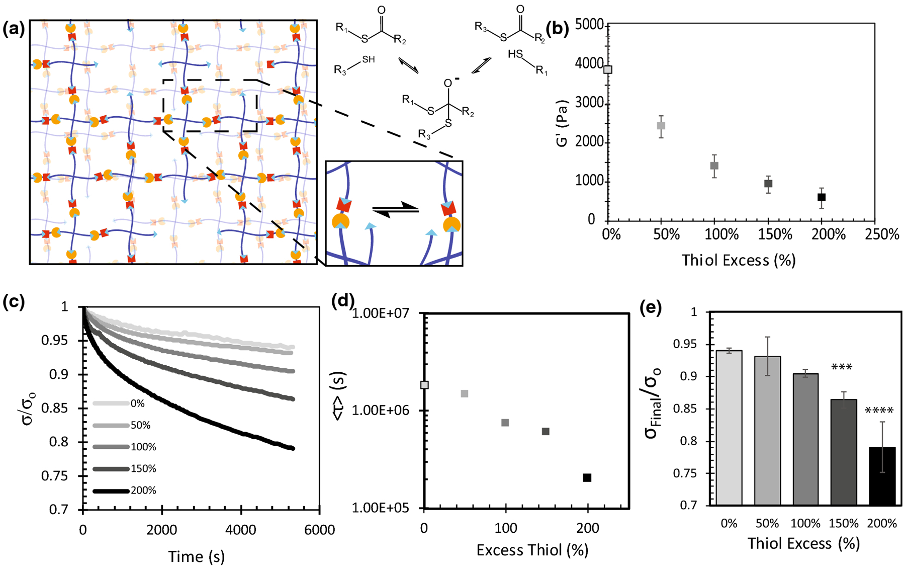
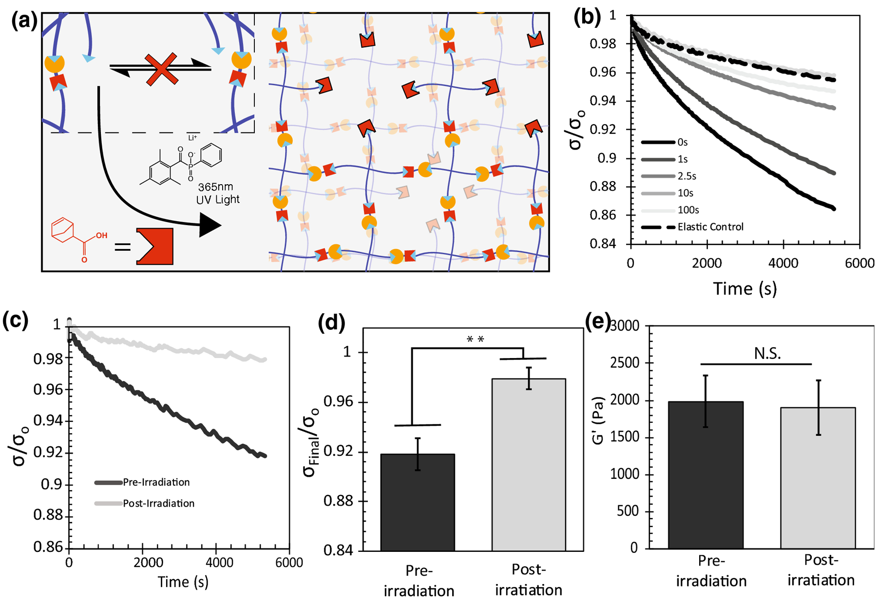
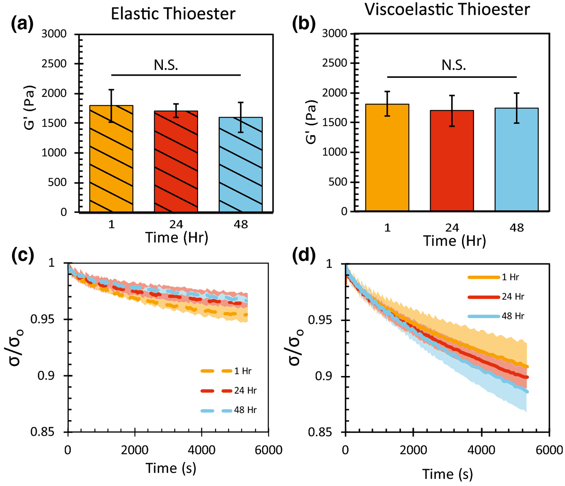
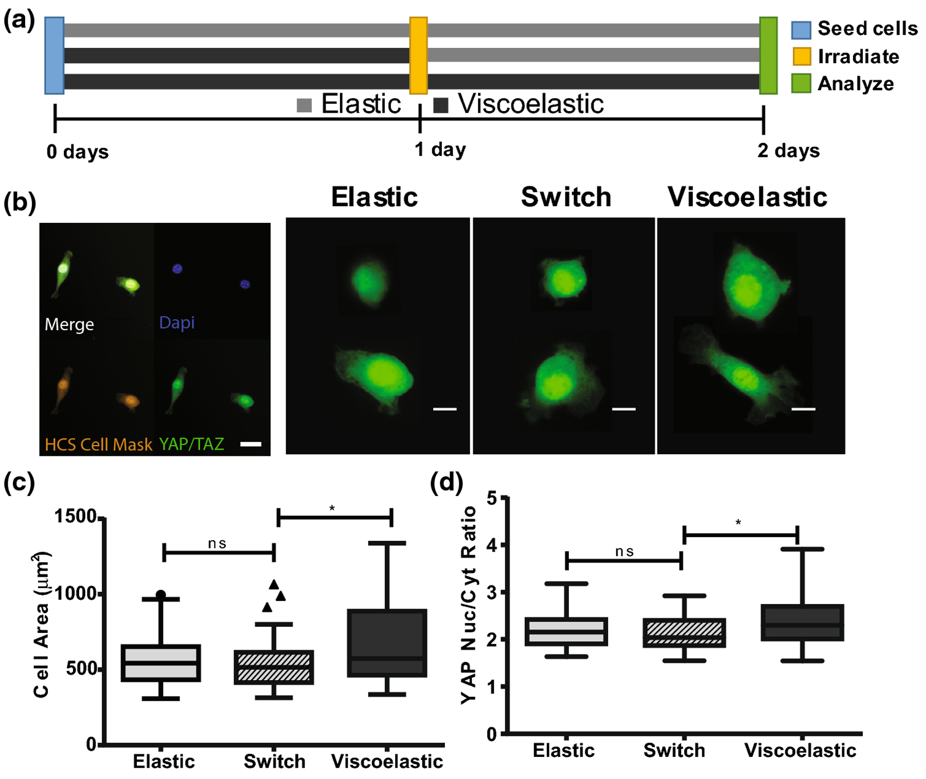
Mechanical cues are delivered to resident cells by the extracellular matrix and play an important role in directing cell processes, ranging from embryonic development and cancer metastasis to stem cell differentiation. Recently, cellular responses to viscoelastic and elastic mechanical cues have been studied; however, questions remain as to how cells identify and transduce these cues differently. We present a synthetic cell culture substrate with viscoelastic properties based on thioester exchange chemistry that can be modulated in situ with the photoinitiated thiol-ene 'click' reaction. With this method, stress relaxation in thioester hydrogels with an average relaxation time of 740,000 s can be switched off in the presence of cells without change to the elastic modulus. NIH 3T3 fibroblasts, cultured for 48 h on viscoelastic compared to elastic thioester substrates, displayed increased cell area (660-560 μm2) and increased nuclear to cytoplasmic YAP/TAZ ratios (2.4 to 2.2) when cultured on elastic compared to viscoelastic hydrogels, respectively. Next, when the viscoelasticity was switched off after 24 h, the fibroblasts responded to this change and exhibited an average cell area of 540 μm2, and nuclear to cytoplasmic YAP/TAZ ratio of 2.1, approaching that of the control elastic gels. Phototunable viscoelastic thioester hydrogels provide a tunable materials system to investigate time-dependent cellular responses to viscoelasticity and should prove useful for understanding the dynamics of mechanoresponsive cellular pathways.

Keywords: Covalent adaptable network; Mechanotransduction; NIH 3T3 fibroblasts; Photoresponsive biomaterial; Tunable viscoelasticity.


Figures

FIGURE 1
FIGURE 1
Multifunctional PEG thiol and thioester norbornene macromers rapidly form hydrogels via the photoinitiated thiol-ene polymerization. (a) Molecular structure of 8 arm 20 kDa PEG Thiol and 8 arm 20 kDa PEG Thioester norbornene. (b) Macromers participate in the radical mediated thiol-ene ‘click’ reaction catalyzed by the photoinitiator LAP to give rise to a fully formed hydrogel network. (c) A 3 wt% stoichiometric mixture of the two PEG macromers containing 1mM LAP rapidly polymerizes to a final modulus of 3500 Pa after 60s of irradiation with 10[mW][cm]−2 365nm UV light.
FIGURE 2
FIGURE 2
Viscoelastic properties of thioester crosslinked hydrogels depend on the concentration of unreacted thiols in the network. (a) Gel stoichiometry was tuned to ensure unreacted thiol species remained after network formation. These thiols then facilitated the exchange of crosslinks via the thioester exchange reaction. (b) In-situ shear elastic modulus of thioester hydrogels with a stoichiometric thiol excess ranging from 0–200%. (c) In-situ stress relaxation properties of thioester gels with a stoichiometric thiol excess ranging from 0–200%. Each profile is an average of n=3 replicates. (d) Average stress relaxation profiles (2c) were fitted to a stretched exponential equation (1) <τ> was calculated using equation (2) and the fitted parameters τk and β. (e) Final fraction of stress remaining after 5400s of relaxation. Reported error bars are the standard deviation, (***) indicates p < 0.001, (****) indicates p<0.0001, n=3.
FIGURE 3
FIGURE 3
The photoinitiated thiol-ene reaction can be used to consume thiol function groups in the thioester network to tune stress relaxation with light. (a) By swelling LAP and norbornene COOH, the photoinitiated thiol-ene reaction can consume all network thiol species, interrupting the thioester exchange and bringing viscoelastic behavior to a halt. (b) Swollen stress relaxation properties of hydrogels swollen with 1mM LAP and 10mM Norbornene but irradiated with 10[mW][cm]−2 365nm UV light for different amounts of time. (n=1) (c) Swollen stress relaxation properties of swollen hydrogels before and after 100s of irradiation with 10[mW][cm]−2 365nm UV light. (n=3) (d) Final fraction of stress remaining after 5400s of relaxation. (n=3) (e) Swollen shear elastic modulus of swollen hydrogels before and after 100s of irradiation with 10[mW][cm]−2 365nm UV light. (n=3) Reported error bars are the standard deviation, (**) indicates p < 0.01, (N.S.) indicates no significance.
FIGURE 4
FIGURE 4
Swollen thioester hydrogels containing a 100% excess of network thiols were either treated with norbornene, LAP and light to be made purely elastic (striped/dashed), or left untreated as viscoelastic hydrogels (solid). (a,b) Shear elastic modulus of thioester hydrogels swollen in phosphate buffered saline. (c,d) Stress relaxation profile of swollen thioester hydrogels measured over 5400s. Final fraction of stress remaining after 5400s of relaxation. Each curve represents an n of 3 and the reported shaded region is the standard deviation, (N.S.) indicates no significance.
FIGURE 5
FIGURE 5
(a) NIH 3T3 fibroblasts were seeded onto swollen 2D thioester hydrogels and cultured for 48 hours. Cells were seeded onto an elastic thioester condition, a viscoelastic thioester condition and a switch condition where viscoelastic hydrogels were made elastic after 24 hours of culture. (b) Cells were fixed at 48 hours and stained for HCS Cell Mask (orange), DAPI (blue) and YAP/TAZ (green); pictured scalebar is 30 microns. YAP/TAZ staining for select cells is shown; the red circle indicates the outline of the nucleus as identified by DAPI staining. Scale bar for single cell images is 10 microns. (c) Spread cell area is visualized by a Tukey boxplot. (d) Nuclear to cytoplasmic ratio of YAP. Reported statistics were calculated using a one tailed student’s t-test, and a population of n=50–60 cells for each condition. Greater than 40 cells were quantified per hydrogel, then 10 cells were selected at random and added to the final population sampling for that condition. (*p<0.05, **p<0.01, ns nonsignificant)

References

    1. Engler AJ, Sen S, Sweeney HL & Discher DE Matrix Elasticity Directs Stem Cell Lineage Specification. Cell 126, 677–689 (2006). - PubMed
    1. Guilak F et al. Control of Stem Cell Fate by Physical Interactions with the Extracellular Matrix. Cell Stem Cell 5, 17–26 (2009). - PMC - PubMed
    1. Ingber DE Mechanical control of tissue morphogenesis during embryological development. Int. J. Dev. Biol 50, 255–266 (2006). - PubMed
    1. Liu F et al. Mechanosignaling through YAP and TAZ drives fibroblast activation and fibrosis. Am. J. Physiol. Cell. Mol. Physiol 308, L344–L357 (2015). - PMC - PubMed
    1. Zanconato F, Cordenonsi M & Piccolo S YAP/TAZ at the Roots of Cancer. Cancer Cell 29, 783–803 (2016). - PMC - PubMed

LinkOut - more resources