Review
. 2020 Feb 4;11(1):9.
doi: 10.1186/s13244-019-0819-2.

CEUS LI-RADS: a pictorial review

Affiliations
Review

CEUS LI-RADS: a pictorial review

Tommaso Vincenzo Bartolotta et al. Insights Imaging. .

Abstract

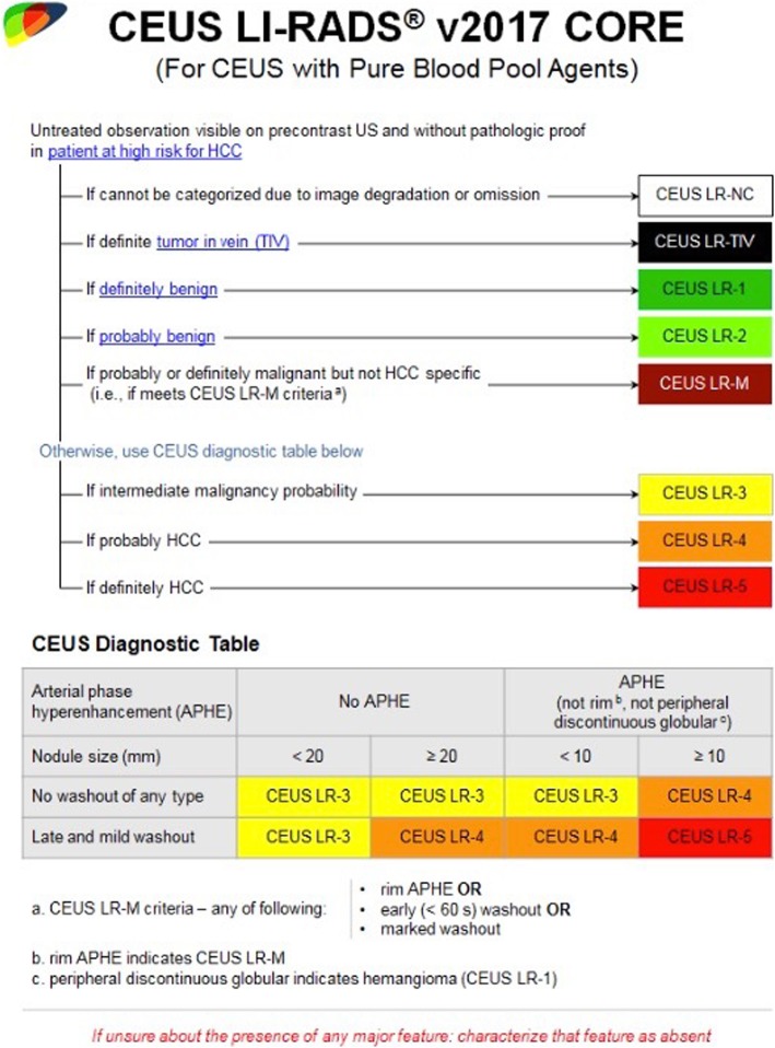
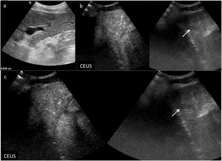
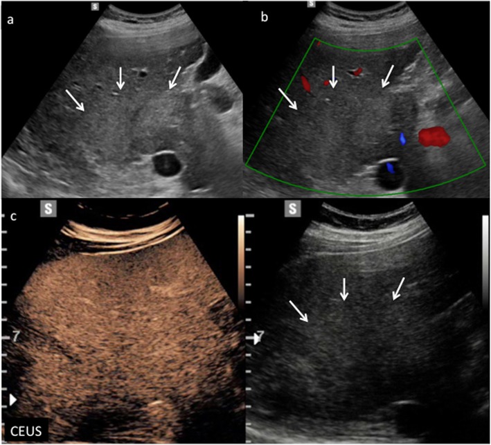
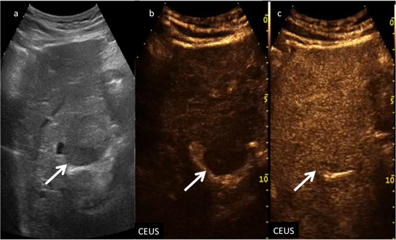
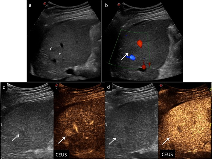
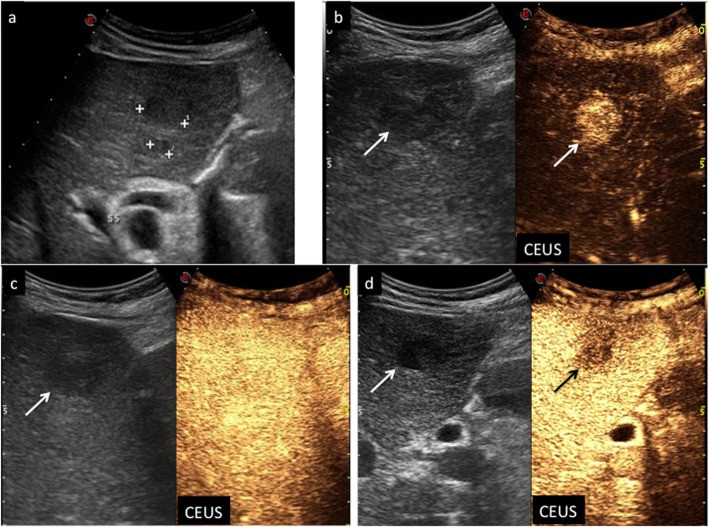
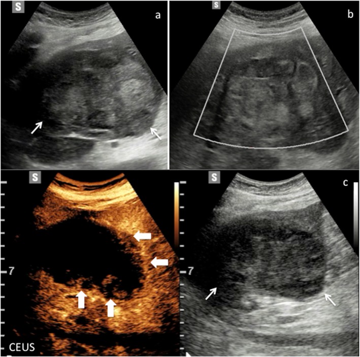
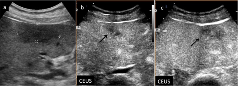
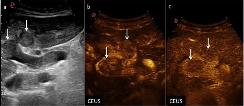
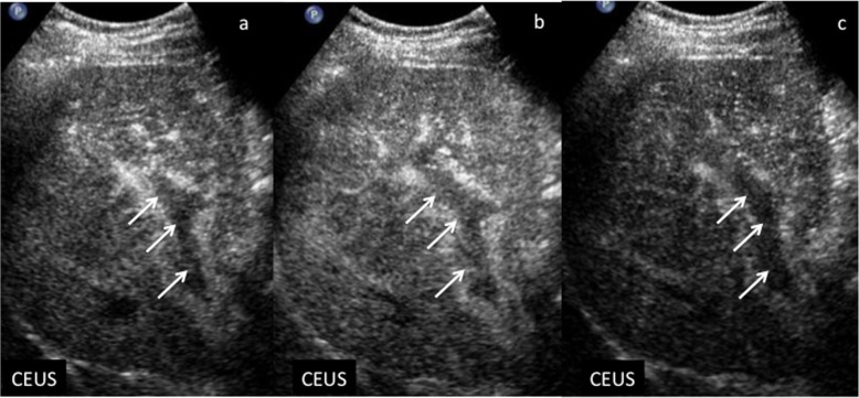
Contrast-enhanced ultrasound (CEUS) greatly improved the diagnostic accuracy of US in the detection and characterization of focal liver lesions (FLLs), and it is suggested and often included in many international guidelines as an important diagnostic tool in the imaging work-up of cirrhotic patients at risk for developing hepatocellular carcinoma (HCC). In particular, CEUS Liver Imaging Reporting and Data System (LI-RADS) provides standardized terminology, interpretation, and reporting for the diagnosis of HCC. The aim of this pictorial essay is to illustrate CEUS features of nodules discovered at US in cirrhotic liver according to LI-RADS categorization.

Keywords: Cholangiocarcinoma; Cirrhosis; Contrast-enhanced ultrasonography; Hepatocellular carcinoma; Liver tumor characterization.


Conflict of interest statement

One author (TVB) is a scientific advisor for Samsung.

Figures

Fig. 1
Fig. 1
ACR CEUS LI-RADS categorization and diagnostic algorithm
Fig. 2
Fig. 2
CEUS LI-RADS 1 (definitely benign). Complex cyst in a 60-year-old woman with chronic hepatitis B viral infection. Baseline US image (a) shows a heterogeneous and moderately hypoechoic lesion sized 6.2 cm in the segment VI–VII (arrows). At CEUS (b), the lesion shows a complete lack of enhancement throughout the vascular phases (arrows)
Fig. 3
Fig. 3
CEUS LI-RADS 1 (definitely benign). Hemangioma in a 49-year-old woman with chronic hepatitis B viral infection. Baseline US image (a) shows a hypoechoic lesion surrounded by a tiny peripheral hyperechoic rim sized 1.3 cm in the segment VII (calipers). CEUS in the early arterial phase (b) depicts peripheral globular enhancement (arrows) followed by a complete centripetal fill-in in the late arterial phase (c) (arrows). The lesion shows sustained contrast-enhancement in the extended portal-venous phase (d) (arrows)
Fig. 4
Fig. 4
CEUS LI-RADS 2 (probably benign). Atypically located area of hypersteatosis in a 47-year-old woman with chronic hepatitis B viral infection. Baseline US image (a) shows a hyperechoic oval-shaped area sized 2.8 cm in the segment IV in a fatty liver (arrow). At CEUS in the extended portal venous phase (b), the area is constantly isoechoic with respect to the surrounding liver parenchyma, as well as throughout the vascular study (arrow)
Fig. 5
Fig. 5
CEUS LI-RADS 2 (probably benign). Regenerative nodule in a 68-year-old woman with virus B-related cirrhosis. Baseline US image (a) shows a small hypoechoic lesion sized 0.9 cm in the segment III (calipers). At CEUS, the nodule is constantly isoechoic with respect to the surrounding liver parenchyma during the arterial (a) and extended portal venous phase (b, c)
Fig. 6
Fig. 6
CEUS LI-RADS 2 (probably benign). Non mass-like area in a 57-year-old man with virus C-related cirrhosis. Baseline US image (a) shows a slightly hyperechoic area with indistinct margins sized 7 cm in the segment V–VI (arrows). The area does not show any vascular signal at color-Doppler (b) (arrows). At CEUS (c), it appears constantly isoechoic with respect to the surrounding liver parenchyma throughout the vascular study (arrows)
Fig. 7
Fig. 7
CEUS LI-RADS 3 (intermediate malignancy probability). Nodule in a 74-year-old man with virus B-related cirrhosis. Baseline US image (a) shows a slightly hypoechoic lesion sized 3 cm in the segment IV (arrow). At CEUS, the nodule appears moderately hypoechoic during the arterial phase (b) and isoechoic to the adjacent liver parenchyma in the extended portal venous (c) phase (arrows)
Fig. 8
Fig. 8
CEUS LI-RADS 4 (probably HCC). Nodule in a 52-year-old woman with virus C-related cirrhosis. Baseline US image (a) shows a slightly hypoechoic lesion sized 1.5 cm in the segment VIII (calipers). No vascular signal is detectable at color-Doppler (b) (arrow). At CEUS, the nodule is highly hypervascular during the arterial phase (c) (arrow) and isoechoic in the extended portal venous phase (d) (arrow)
Fig. 9
Fig. 9
CEUS LI-RADS 5 (definitely HCC). Nodule in a 72-year-old man with virus B-related cirrhosis. Baseline US image (a) shows two moderately hypoechoic lesions sized, respectively, 2.2 cm and 0.7 cm in the segment V (calipers). At CEUS, the largest one is markedly hypervascular during the arterial phase (b) (arrows), becoming isoechoic 45 s after contrast medium injection (c) (arrow) and showing moderate wash-out 120 s after contrast medium injection (d) (arrows)
Fig. 10
Fig. 10
CEUS LI-RADS M (probably or definitely malignant but not HCC specific) nodule. Cholangiocarcinoma in a 87-year-old man with virus B-related cirrhosis. Baseline US image shows a markedly heterogeneous lesion sized 8.5 cm in the segment VI (a) (arrows). No vascular signal is detectable at color-Doppler (b). At CEUS (c), the nodule shows rim enhancement surrounding a constantly avascular area (arrows)
Fig. 11
Fig. 11
CEUS LI-RADS M (probably or definitely malignant but not HCC specific) nodule. Metastasis in a 32-year-old woman with virus B-related cirrhosis. Baseline US image (a) shows a hypoechoic lesion with not well-defined margins sized 5 cm in the segment IV (calipers). At CEUS, the lesion is heterogeneously vascularized during the arterial phase (b) (arrow) showing early wash-out: 34 s after contrast medium injection (c) (arrow)
Fig. 12
Fig. 12
CEUS LI-RADS TIV (tumor in vein). Neoplastic thrombosis in a 84-year-old man with virus B-related cirrhosis. Baseline US image shows (a) multiple thrombi in the lumen of portal vein (arrows). At CEUS, marked enhancement is evident in their context during the early arterial phase (b) (arrows), followed by a clear-cut wash-out in the extended portal venous phase (c) (arrows)
Fig. 13
Fig. 13
Non-neoplastic thrombosis in a 62-year-old man with virus B-related cirrhosis. At CEUS, no enhancement is appreciable in the context of the right branch of portal vein during the arterial (a), portal venous (b), and late (c) phases, respectively (arrows)

References

    1. World Health organization, International Agency for Research on Cancer, Global Cancer Observatory Cancer Today – IARC, Lyon, France, 2018 available via http://gco.iarc.fr/today/data/factsheets/cancers/11-Liver-fact-sheet.pdf. Last accessed 8 Jan 2019
    1. European Association for the Study of the Liver (2018) EASL clinical practice guidelines: management of hepatocellular carcinoma. European Association for the Study of the Liver. J Hepatol 69(1):182–236 - PubMed
    1. Heimbach Julie K., Kulik Laura M., Finn Richard S., Sirlin Claude B., Abecassis Michael M., Roberts Lewis R., Zhu Andrew X., Murad M. Hassan, Marrero Jorge A. AASLD guidelines for the treatment of hepatocellular carcinoma. Hepatology. 2017;67(1):358–380. doi: 10.1002/hep.29086. - DOI - PubMed
    1. Omata Masao, Cheng Ann-Lii, Kokudo Norihiro, Kudo Masatoshi, Lee Jeong Min, Jia Jidong, Tateishi Ryosuke, Han Kwang-Hyub, Chawla Yoghesh K., Shiina Shuichiro, Jafri Wasim, Payawal Diana Alcantara, Ohki Takamasa, Ogasawara Sadahisa, Chen Pei-Jer, Lesmana Cosmas Rinaldi A., Lesmana Laurentius A., Gani Rino A., Obi Shuntaro, Dokmeci A. Kadir, Sarin Shiv Kumar. Asia–Pacific clinical practice guidelines on the management of hepatocellular carcinoma: a 2017 update. Hepatology International. 2017;11(4):317–370. doi: 10.1007/s12072-017-9799-9. - DOI - PMC - PubMed
    1. Thomsen Henrik S. Nephrogenic Systemic Fibrosis: History and Epidemiology. Radiologic Clinics of North America. 2009;47(5):827–831. doi: 10.1016/j.rcl.2009.05.003. - DOI - PubMed

LinkOut - more resources