Bilateral lung lobe torsions in a cat
- PMID: 32020935
- PMCID: PMC6973216
Bilateral lung lobe torsions in a cat
Abstract
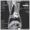
A 13-year-old spayed female domestic longhair cat was presented for tachypnea and was identified to have reduced lung sounds over the left hemithorax. Thoracic ultrasound examination and computed tomography identified changes consistent with bilateral lung lobe torsion. A median sternotomy confirmed torsion of both the cranial portion of the left cranial lung lobe and the right middle lung lobe. The affected lobes were resected. Pleural fluid analysis was indicative of a modified transudate and histopathology was consistent with a subacute to chronic torsion with no evidence of neoplasia or infection. The patient recovered without complication. Lung lobe torsion is an uncommon presentation across all species and is especially rare in cats. To the authors' knowledge, bilateral lung lobe torsion has not been previously reported in small animals.
Torsions bilatérales de lobes pulmonaires chez un chat. Une chatte domestique à poils longs âgées de 13 ans fut présentée pour tachypnée et on identifia une diminution des bruits respiratoires du côté de l’hémithorax gauche. Une échographie thoracique et un examen par tomodensitométrie (CT) identifièrent des changements compatibles avec une torsion bilatérale de lobes pulmonaires. Une sternotomie médiane confirma la torsion des portions crâniales du lobe pulmonaire crânial gauche et du lobe pulmonaire moyen droit. Les lobes affectés furent excisés. L’analyse du liquide pleural était indicatrice d’un transsudat modifié et l’histopathologie était compatible avec une torsion subaigüe à chronique sans évidence de néoplasie ou d’infection. La chatte récupéra sans complication. La torsion des lobes pulmonaires est une présentation peu commune chez toutes les espèces et est spécialement rare chez les chats. Selon les auteurs, une torsion bilatérale des lobes pulmonaires n’a pas encore été rapportée chez les petits animaux.(Traduit par Dr Serge Messier).
Copyright and/or publishing rights held by the Canadian Veterinary Medical Association.
Figures


References
-
- Tobias KM, Johnston SA. Veterinary Surgery Small Animal. St. Louis, Missouri: Saunders; 2012. pp. 1762–1766.
-
- Brenner OJ, Ettinger SN, Stefanacci JD. What is your diagnosis? Chronic fibrosing pleuritis, pleural effusion, and lobar consolidation. J Am Vet Med Assoc. 2000;216:1555–1556. - PubMed
-
- Schultz RM, Peters J, Zwingenberger A. Radiography, computed tomography and virtual bronchoscopy in four dogs and two cats with lung lobe torsion. J Small Anim Pract. 2009;50:360–363. - PubMed
-
- d’Anjou MA, Tidwell AS, Hecht S. Radiographic diagnosis of lung lobe torsion. Vet Radiol Ultrasound. 2005;46:478–484. - PubMed
Publication types
MeSH terms
LinkOut - more resources
Full Text Sources
Medical
Miscellaneous
