. 2020 Jan 7:14:51-60.
doi: 10.2147/DDDT.S228751. eCollection 2020.

Protective Effects of Chlorogenic Acid on Cerebral Ischemia/Reperfusion Injury Rats by Regulating Oxidative Stress-Related Nrf2 Pathway

Affiliations

Protective Effects of Chlorogenic Acid on Cerebral Ischemia/Reperfusion Injury Rats by Regulating Oxidative Stress-Related Nrf2 Pathway

Dequan Liu et al. Drug Des Devel Ther. .

Retraction in

Abstract

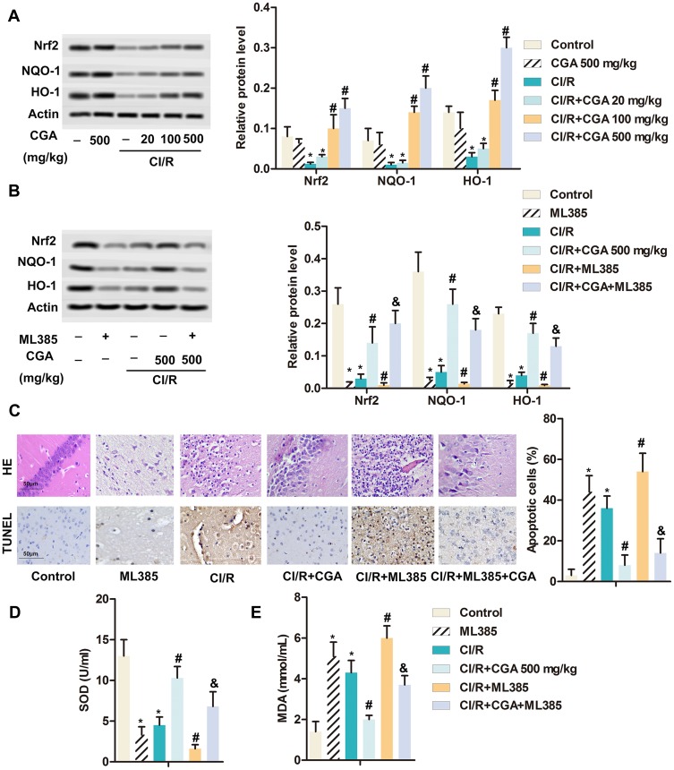
Introduction: Cerebral ischemia-reperfusion (CI/R) injury is caused by blood flow recovery after ischemic stroke. Chlorogenic acid (CGA, 5-O-caffeoylquinic acid) is a major polyphenol component of Coffea canephora, Coffea arabica L. and Mate (Ilex paraguariensis A. StHil.). Previous studies have shown that CGA has a significant neuroprotective effect and can improve global CI/R injury. However, the underlying molecular mechanism of CGA in CI/R injury has not been fully revealed.

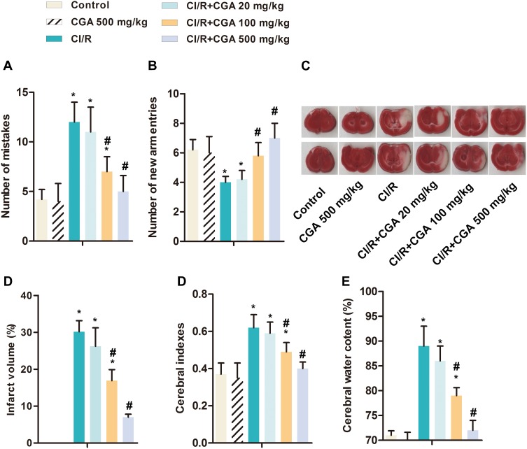
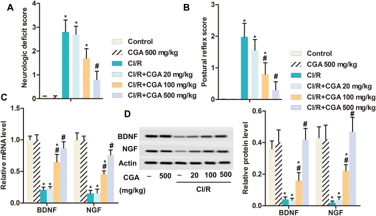
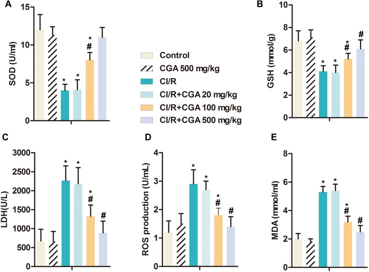
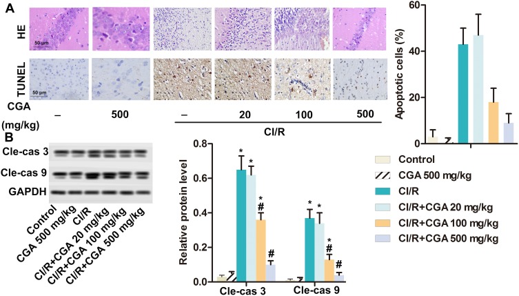
Materials: In this study, CI/R rat model was constructed. The rats were randomly divided into nine groups with ten in each group: Control, CGA (500 mg·kg-1), CI/R, CI/R + CGA (20 mg·kg-1), CI/R + CGA (100 mg·kg-1), CI/R + CGA (500 mg·kg-1), ML385 (30 mg·kg-1), CI/R + ML385 (30 mg·kg-1), CI/R + CGA + ML385. Cerebral infarction volume was detected by TTC staining. Brain pathological damage was detected by H&E staining. Apoptosis of cortical cells was detected by TUNEL staining. The expression of related proteins was detected by RT-qPCR and Western blotting.

Results: Step-down test and Y maze test showed that CGA dose-dependently mitigated CI/R-induced brain damage and enhanced learning and spatial memory. Besides, CGA promoted the expression of BDNF and NGF in a dose-dependent manner and alleviated CI/R-induced nerve injury. Moreover, CGA increased the activity of SOD and the level of GSH, as well as decreased production of ROS and LDH and the accumulation of MDA. Notably, CGA attenuated oxidative stress-induced brain injury and apoptosis and inhibited the expression of apoptosis-related proteins (cleaved caspase 3 and caspase 9). Additionally, CGA reversed CI/R induced inactivation of Nrf2 pathway and promoted Nrf2, NQO-1 and HO-1 expression. Nrf2 pathway inhibitor ML385 destroyed this promotion.

Discussion: All the data indicated that CGA had a neuroprotective effect on the CI/R rats by regulating oxidative stress-related Nrf2 pathway.

Keywords: NF-E2-related factor 2 pathway; cerebral ischemia/reperfusion injury; chlorogenic acid; neuroprotection; oxidative stress.


Conflict of interest statement

The authors report no conflicts of interest in this work.

Figures

Figure 1
Figure 1
CGA mitigates brain damage in rats with CI/R. Rats were randomly divided into six groups with ten in each group: Control group; CGA group; CI/R group; CI/R + CGA (20 mg·kg−1) group; CI/R + CGA (100 mg·kg-1) group; CI/R + CGA (500 mg·kg-1) group. (A) The number of mistakes in jumping platform was detected by Step-down test. (B) The number of new arm entries was detected by Y maze test. (C) Cerebral infarction volume. (D) Cerebral indexes. (E) Cerebral water content. *p<0.05 vs control group; #p<0.05 vs CI/R group.
Figure 2
Figure 2
CGA alleviates nerve injury in rats with CI/R. Rats were randomly divided into six groups with ten in each group: Control group, CGA group; CI/R group; CI/R + CGA (20 mg·kg−1) group; CI/R + CGA (100 mg·kg-1) group; CI/R + CGA (500 mg·kg−1) group. (A) Neurologic deficit score. (B) Postural reflex score. (C) The mRNA level of BDNF and NGF was measured by RT-qPCR. (D) The protein level of BDNF and NGF was measured by Western blotting. Actin was used as internal reference. *p<0.05 vs control group; #p<0.05 vs CI/R group.
Figure 3
Figure 3
CGA alleviates oxidative stress induced by CI/R in rats. Rats were randomly divided into six groups with ten in each group: Control group, CGA group; CI/R group; CI/R + CGA (20 mg·kg−1) group; CI/R + CGA (100 mg·kg-1) group; CI/R + CGA (500 mg·kg-1) group. (A) SOD. (B) GSH. (C) LDH. (D) ROS production. (E) MDA. *p<0.05 vs control group; #p<0.05 vs CI/R group.
Figure 4
Figure 4
CGA improves the pathological damage of hippocampal neurons and reduces apoptosis in rats with CI/R. Rats were randomly divided into six groups with ten in each group: Control group, CGA group; CI/R group; CI/R + CGA (20 mg·kg−1) group; CI/R + CGA (100 mg·kg−1) group; CI/R + CGA (500 mg·kg-1) group. (A) The pathological damage of neurons in rat hippocampus was measured by H&E staining and the apoptosis of cerebral cortex cells was detected by TUNEL staining. (B) The protein level of BDNF and NGF was measured by Western blotting. Actin was used as internal reference. *p<0.05 vs control group; #p<0.05 vs CI/R group.
Figure 5
Figure 5
Nrf2/NQO-1/HO-1 pathway is involved in the role of CGA in CI/R rats. (A) Rats were randomly divided into six groups with ten in each group: Control group; CGA group; CI/R group; CI/R + CGA (20 mg·kg−1) group; CI/R + CGA (100 mg·kg-1) group; CI/R + CGA (500 mg·kg-1) group. The protein level of Nrf2, NQO-1 and HO-1 was measured by Western blotting. Actin was used as internal reference. (*p<0.05 vs control group; #p<0.05 vs CI/R group). (B) Rats were randomly divided into five groups with ten in each group: Control group; ML385 group; CI/R group, CI/R + CGA (500 mg·kg−1) group; CI/R + ML385 (30 mg·kg−1) group. The protein level of Nrf2, NQO-1 and HO-1 was measured by Western blotting. Actin was used as internal reference. (*p<0.05 vs control group; #p<0.05 vs CI/R group; &p<0.05 vs CI/R+ML385 group). (C) The pathological damage of neurons in rat hippocampus was measured by H&E staining and the apoptosis of cerebral cortex cells was detected by TUNEL staining. (D) SOD. (E) MDA. *p<0.05 vs control group; #p<0.05 vs CI/R group; &p<0.05 vs CI/R+ML385 group.

References

    1. Khoshnam SE, Winlow W, Farbood Y, Moghaddam HF, Farzaneh M. Emerging roles of microRNAs in ischemic stroke: as possible therapeutic agents. J Stroke. 2017;19:166–187. doi: 10.5853/jos.2016.01368 - DOI - PMC - PubMed
    1. Jiang D, Sun X, Wang S, Man H. Upregulation of miR-874-3p decreases cerebral ischemia/reperfusion injury by directly targeting BMF and BCL2L13. Biomed Pharmacother. 2019;117:108941. doi: 10.1016/j.biopha.2019.108941 - DOI - PubMed
    1. Wang J, Chen T, Shan G. miR-148b regulates proliferation and differentiation of neural stem cells via Wnt/β-catenin signaling in rat ischemic stroke model. Front Cell Neurosci. 2017;11:329. doi: 10.3389/fncel.2017.00329 - DOI - PMC - PubMed
    1. Eryildiz ES, Ozdemir AO. Factors associated with early recovery after intravenous thrombolytic therapy in acute ischemic stroke. Noro Psikiyatri Arsivi. 2018;55:80–83. doi: 10.29399/npa.22664 - DOI - PMC - PubMed
    1. Petrovic-Djergovic D, Goonewardena SN, Pinsky DJ. Inflammatory disequilibrium in stroke. Circ Res. 2016;119:142–158. doi: 10.1161/CIRCRESAHA.116.308022 - DOI - PMC - PubMed

Publication types

MeSH terms