

Three-dimensional assessment of craniofacial asymmetry in children with transverse maxillary deficiency after rapid maxillary expansion: A prospective study
- PMID: 32022986
- PMCID: PMC7783112
- DOI: 10.1111/ocr.12370
Three-dimensional assessment of craniofacial asymmetry in children with transverse maxillary deficiency after rapid maxillary expansion: A prospective study
Abstract
Objective: The aim of this study was to evaluate craniofacial asymmetry in children with transverse maxillary deficiency, with or without functional unilateral posterior crossbite (UPC), before and after rapid maxillary expansion (RME).
Setting and sample population: A sample of 51 children with cone beam computed tomography scans obtained before RME (T1) and a year after RME (T2).
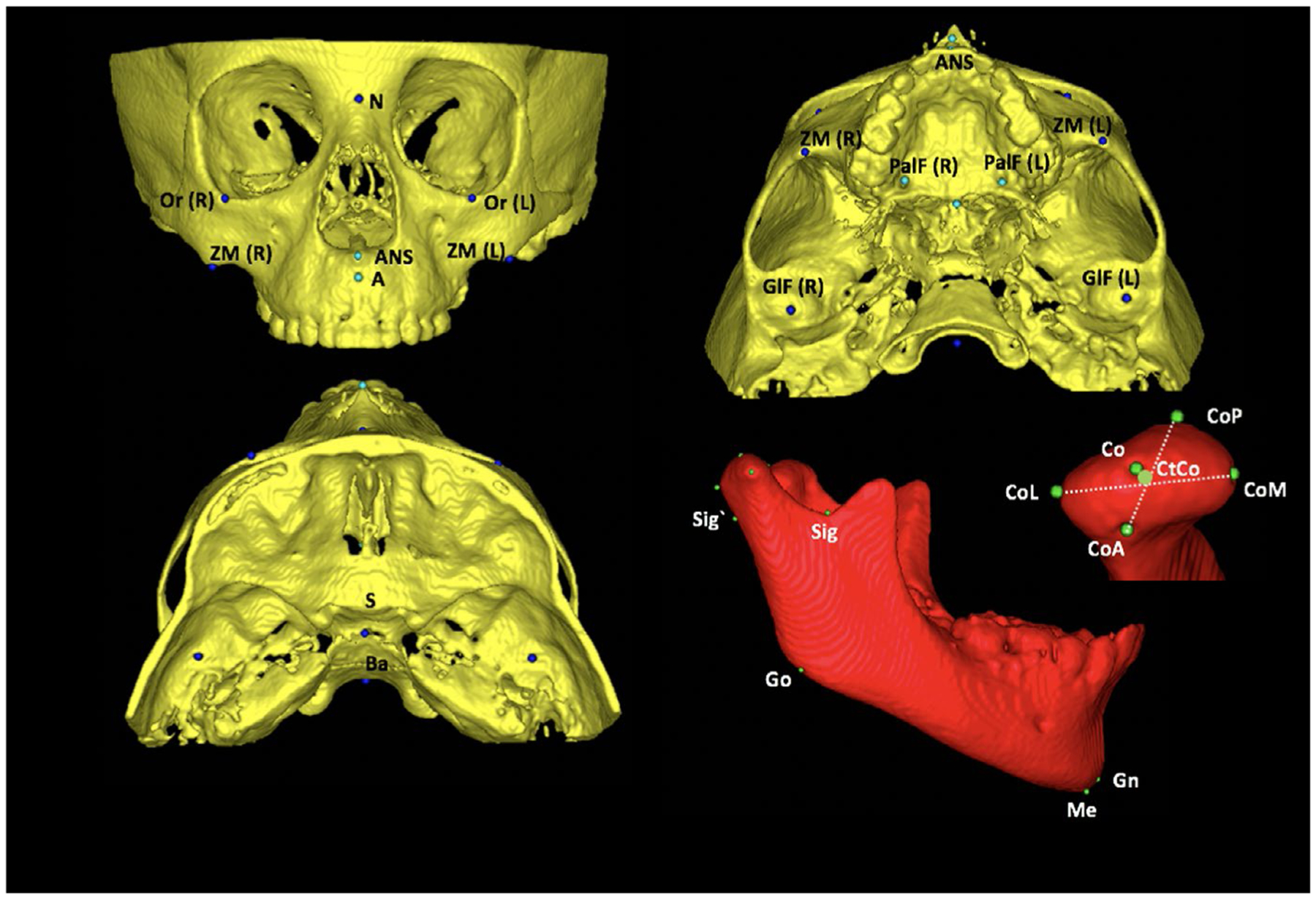
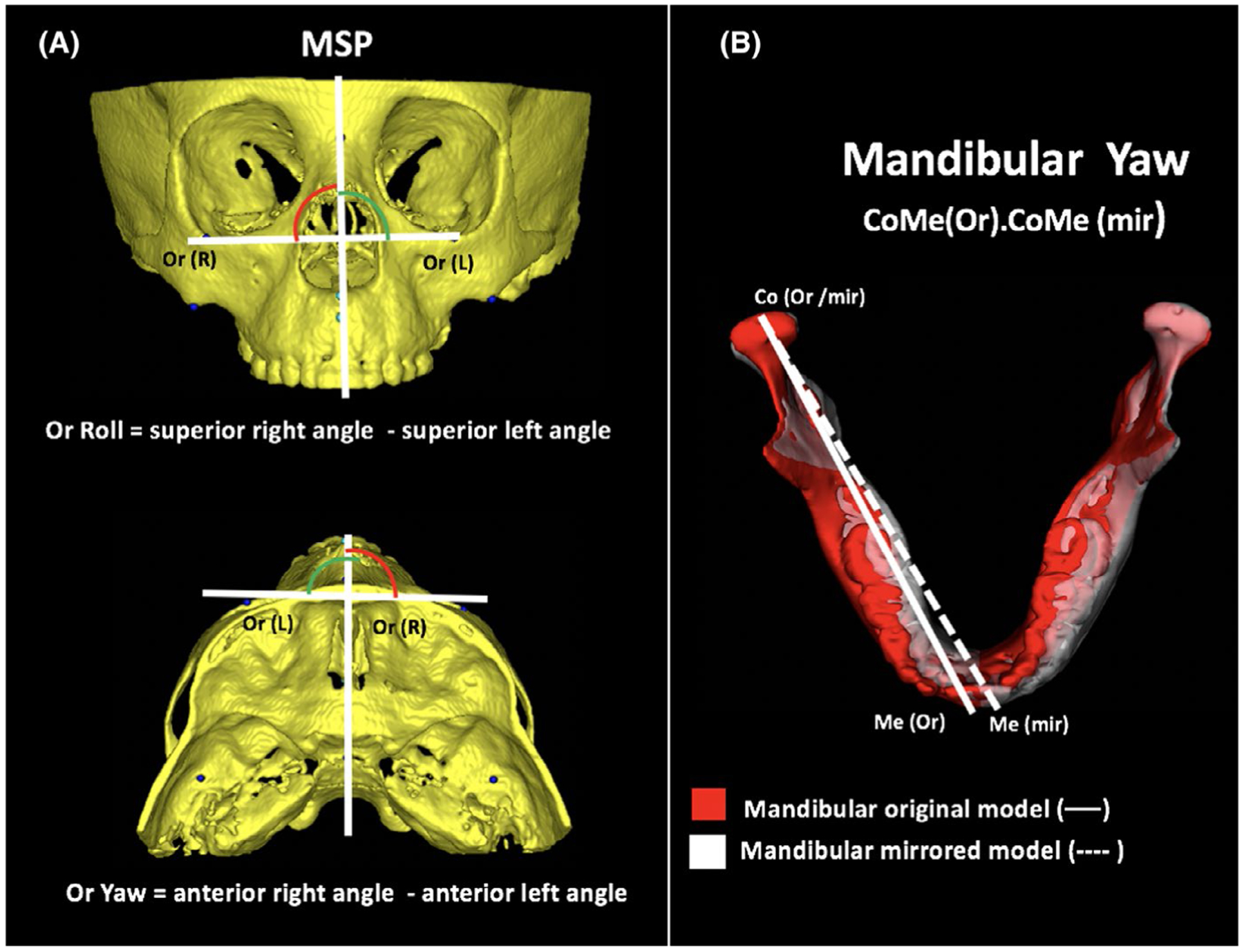
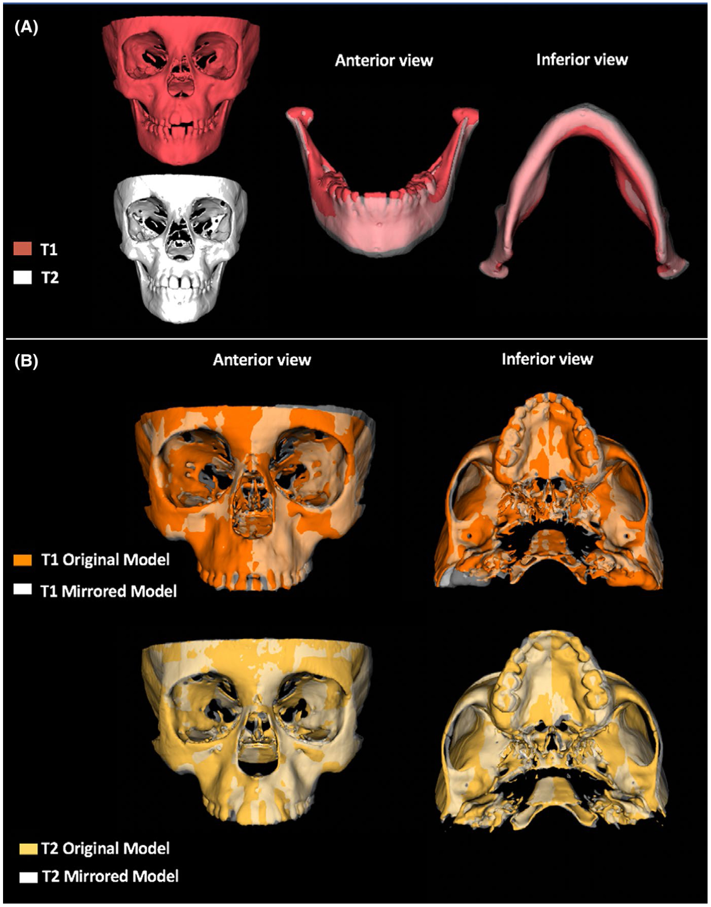
Material and methods: This prospective study consisted of 2 groups: 25 children with functional UPC (6.77 ± 1.5 years) and 26 children without UPC (7.41 ± 1.31 years). Linear and angular measurements were obtained from zygomatic, maxilla, glenoid fossa and mandible, using original and mirrored 3D overlapped models. All right and left side comparisons in both groups and intergroups asymmetries were compared using MANOVA and t test for independent samples, respectively, statistically significant at P < .05.
Results: The UPC group showed no side differences, but mandibular horizontal rotation at T1, and this asymmetry was improved in T2. The non-UPC group showed at baseline significant lateral asymmetry in orbitale, position of palatine foramen, respectively, in average 2.95 mm and 1.16 mm, and 0.49 mm of average asymmetry in condylar height. The glenoid fossa was symmetric in both groups at T1 and T2.
Conclusions: Children with transverse maxillary deficiency showed slight morphological asymmetry, located in the mandible position in cases of UPC, and in the orbital and maxillary regions in cases without UPC. One year after RME, patients improved their craniofacial asymmetry, with significant changes in the mandible and correction of the mandibular rotation in patients who presented UPC.
Keywords: cone beam computed tomography; craniofacial abnormalities; crossbite; facial asymmetry; maxillary expansion.
© 2020 John Wiley & Sons A/S. Published by John Wiley & Sons Ltd.
Conflict of interest statement
CONFLIC T OF INTEREST
All authors have nothing to disclose.
Figures
Similar articles
-
Evaluation of mandibular changes after rapid maxillary expansion: a CBCT study in youngsters with unilateral posterior crossbite using a surface-to-surface matching technique.Clin Oral Investig. 2021 Apr;25(4):1775-1785. doi: 10.1007/s00784-020-03480-5. Epub 2020 Aug 2. Clin Oral Investig. 2021. PMID: 32743674
-
Cone beam computed tomographic evaluation of the changes in condylar position in growing patients with unilateral posterior crossbite undergoing rapid maxillary expansion followed by fixed orthodontic therapy.Eur Arch Paediatr Dent. 2021 Oct;22(5):959-967. doi: 10.1007/s40368-021-00628-z. Epub 2021 May 5. Eur Arch Paediatr Dent. 2021. PMID: 33950475
-
Mandibular transverse dentoalveolar and skeletal changes associated with lip bumper and rapid maxillary expander: A cone-beam computed tomography study.Am J Orthod Dentofacial Orthop. 2023 Mar;163(3):407-425. doi: 10.1016/j.ajodo.2021.12.026. Epub 2022 Dec 12. Am J Orthod Dentofacial Orthop. 2023. PMID: 36517377
-
Is there an asymmetry of the condylar and coronoid processes of the mandible in individuals with unilateral crossbite?Angle Orthod. 2019 May;89(3):464-469. doi: 10.2319/052518-398.1. Epub 2018 Dec 28. Angle Orthod. 2019. PMID: 30644758 Free PMC article.
-
Maxillary Expansion and Its Effects on Circummaxillary Structures: A Review.Cureus. 2023 Jan 13;15(1):e33755. doi: 10.7759/cureus.33755. eCollection 2023 Jan. Cureus. 2023. PMID: 36793826 Free PMC article. Review.
Cited by
-
Very early orthodontic treatment: when, why and how?Dental Press J Orthod. 2022 Jun 10;27(2):e22spe2. doi: 10.1590/2177-6709.27.2.e22spe2. eCollection 2022. Dental Press J Orthod. 2022. PMID: 35703618 Free PMC article.
-
Comparison of Mirroring and Overlapping Analysis and Three-Dimensional Soft Tissue Spatial Angle Wireframe Template in Evaluating Facial Asymmetry.Bioengineering (Basel). 2025 Jan 16;12(1):79. doi: 10.3390/bioengineering12010079. Bioengineering (Basel). 2025. PMID: 39851353 Free PMC article.
-
Prevalence of mandibular asymmetry in different skeletal sagittal patterns.Angle Orthod. 2022 Jan 1;92(1):118-126. doi: 10.2319/040921-292.1. Angle Orthod. 2022. PMID: 34546287 Free PMC article.
-
Analysis of maxillary asymmetry before and after treatment of functional posterior cross-bite: a retrospective study using 3D imaging system and deviation analysis.Prog Orthod. 2023 Dec 11;24(1):41. doi: 10.1186/s40510-023-00494-z. Prog Orthod. 2023. PMID: 38072875 Free PMC article.
-
Effects on the Facial Growth of Rapid Palatal Expansion in Growing Patients Affected by Juvenile Idiopathic Arthritis with Monolateral Involvement of the Temporomandibular Joints: A Case-Control Study on Posteroanterior and Lateral Cephalograms.J Clin Med. 2020 Apr 18;9(4):1159. doi: 10.3390/jcm9041159. J Clin Med. 2020. PMID: 32325675 Free PMC article.
References
-
- Proffit WR, Fields HW Jr. Contemporary orthodontics, 5th edn St. Louis, MO: Mosby; 2012:768.
-
- Melsen B, Stensgaard K, Pedersen J. Sucking habits and their influence on swallowing pattern and prevalence of malocclusion. Europ J Orthod. 1979;1:271–280. - PubMed
-
- Adenoids L-A. Their effect on mode of breathing and nasal airflow and their relationship to characteristics of the facial skeleton and the dentition. Acta Otolaryngol Suppl. 1970;265:1–132. - PubMed
-
- Thilander B, Lennartsoon B. A study of children with unilateral crossbite, treated and untreated in deciduous dentition. J Orofac Orthop. 2002;63:371–383. - PubMed
-
- Andrade AS, Gameiro GH, Derossi M, Gavião MB. Posterior cross-bite and functional changes.A systematic review. Angle Orthod. 2009;79:380–386. - PubMed
