. 2020 May 15;201(10):1249-1262.
doi: 10.1164/rccm.201908-1513OC.

Hyperoxia Injury in the Developing Lung Is Mediated by Mesenchymal Expression of Wnt5A

Affiliations

Hyperoxia Injury in the Developing Lung Is Mediated by Mesenchymal Expression of Wnt5A

Jennifer M S Sucre et al. Am J Respir Crit Care Med. .

Abstract

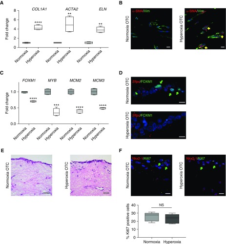
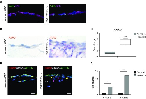
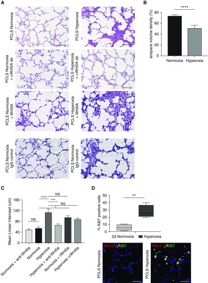
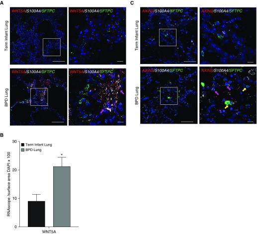
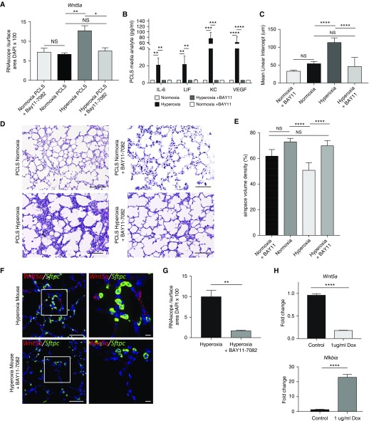
Rationale: Bronchopulmonary dysplasia (BPD) is a leading complication of preterm birth that affects infants born in the saccular stage of lung development at <32 weeks of gestation. Although the mechanisms driving BPD remain uncertain, exposure to hyperoxia is thought to contribute to disease pathogenesis.Objectives: To determine the effects of hyperoxia on epithelial-mesenchymal interactions and to define the mediators of activated Wnt/β-catenin signaling after hyperoxia injury.Methods: Three hyperoxia models were used: A three-dimensional organotypic coculture using primary human lung cells, precision-cut lung slices (PCLS), and a murine in vivo hyperoxia model. Comparisons of normoxia- and hyperoxia-exposed samples were made by real-time quantitative PCR, RNA in situ hybridization, quantitative confocal microscopy, and lung morphometry.Measurements and Main Results: Examination of an array of Wnt ligands in the three-dimensional organotypic coculture revealed increased mesenchymal expression of WNT5A. Inhibition of Wnt5A abrogated the BPD transcriptomic phenotype induced by hyperoxia. In the PCLS model, Wnt5A inhibition improved alveolarization following hyperoxia exposure, and treatment with recombinant Wnt5a reproduced features of the BPD phenotype in PCLS cultured in normoxic conditions. Chemical inhibition of NF-κB with BAY11-7082 reduced Wnt5a expression in the PCLS hyperoxia model and in vivo mouse hyperoxia model, with improved alveolarization in the PCLS model.Conclusions: Increased mesenchymal Wnt5A during saccular-stage hyperoxia injury contributes to the impaired alveolarization and septal thickening observed in BPD. Precise targeting of Wnt5A may represent a potential therapeutic strategy for the treatment of BPD.

Keywords: Wnt; bronchopulmonary dysplasia; hyperoxia; lung injury.


Figures

Figure 1.
Figure 1.
Three-dimensional organotypic cocultures (3D-OTC) replicate the bronchopulmonary dysplasia transcriptomic phenotype. (A) With hyperoxia exposure, 3D-OTC comprised of primary fetal lung mesenchymal cells and type 2 alveolar epithelial cells have increased expression of genes associated with fibrosis with hyperoxia exposure, shown as mean ± SD. **P < 0.01 and ****P < 0.0001. (B) Immunofluorescence (IF) shows increased α-SMA (α-smooth muscle actin) (red) in fibroblasts stained for vimentin (Vim) (green) in hyperoxia 3D-OTC (yellow arrows indicate double-positive cells). Scale bars, 10 μm. (C) With hyperoxia exposure, 3D-OTC have decreased expression of genes associated with alveolarization when compared with normoxia-cultured controls, shown as mean ± SD. ***P < 0.001 and ****P < 0.0001. (D) IF for FOXM1 (green) and RNA in situ hybridization for SPC gene Sftpc showed decreased nuclear FOXM1 in hyperoxia 3D-OTC (green arrows indicate FOXM1+ nuclei). Scale bars, 10 μm. (E) Hematoxylin and eosin staining of 3D-OTC cultured in normoxia and hyperoxia. Scale bars, 25 μm. (F) IF for mitotic marker Ki67 (green) and epithelial marker Nkx2.1 (red) shows no significant difference in proliferation between 3D-OTCs cultured in normoxia and hyperoxia, with quantification expressed as percent Ki67-positive cells and determined by counting 36 high-powered fields per group (green arrows indicate Ki67+ cells). N = 5 biological replicates, with 3 technical replicates used for each sample. NS = nonsignificant.
Figure 2.
Figure 2.
Hyperoxia exposure of three-dimensional organotypic cocultures (3D-OTC) stimulates active Wnt/β-catenin signaling. (A) Immunofluorescence for p-β-cateninY489 (nuclear β-catenin phosphorylated at tyrosine-489) (green), an established marker of Wnt activation, and SPB (surfactant protein-B) (purple) shows increased nuclear p-β-cateninY489 in hyperoxia-exposed OTC when compared with normoxic-cultured controls. Scale bars, 10 μm. (B) RNA in situ hybridization (RNA ISH) shows increased expression of Wnt target gene AXIN2 with hyperoxia exposure. Scale bars, 10 μm. (C) Real-time quantitative PCR (qPCR) comparing hyperoxia-exposed 3D-OTC with normoxia-cultured OTC shows increased expression of AXIN2. ***P < 0.01. (D) Multiplex RNA ISH showing increased expression of AXIN2 (red) in hyperoxia 3D-OTC relative to normoxia controls. AXIN2 is expressed in epithelial cells coexpressing SFTPC in green (yellow arrows) and mesenchymal cells expressing fibroblast marker S100A4 in white (magenta arrows). Scale bars, 10 μm. (E) MLE-15 cells were combined with human lung fibroblasts to make 3D-OTC, which were exposed to hyperoxia. Real-time quantitative PCR was performed with species-specific probes for AXIN2/Axin2. *P < 0.05 and **P< 0.01. Data are representative of three biological replicates for each condition, with three technical replicates for each sample used for qPCR.
Figure 3.
Figure 3.
Hyperoxia exposure of three-dimensional organotypic cocultures (3D-OTC) results in mesenchymal expression of Wnt5A. (A) Wnt pathway array shows relative expression of Wnt ligands in 3D-OTC cultured in hyperoxia and normoxia conditions, normalized to normoxia. Expression was considered to be 0 if cycle threshold value was >35. *P< 0.001. (B) Confirmation of increased expression of WNT5A by quantitative PCR (qPCR) with TaqMan probes. ****P< 0.001. (C) RNA in situ hybridization demonstrating increased expression of WNT5A (red) with hyperoxia exposure. Scale bars, 10 μm. (D) Multiplex RNA in situ hybridization shows coexpression of WNT5A (red) by mesenchymal cells expressing fibroblast marker S100A4 (green), with yellow arrows indicating cells coexpressing both genes. Scale bars, 10 μm. (E) MLE-15 cells were combined with human lung fibroblasts to make 3D-OTC, which were exposed to hyperoxia. Real-time quantitative PCR was performed with species-specific probes, with only increased expression of human WNT5A. Cycle threshold was >35. ***P < 0.001. (F) Addition of rWnt5A (recombinant Wnt5A) protein to OTC partially replicates the transcriptomic response to hyperoxia, with significantly increased expression of COL1A1 and ACTA2, with decreased expression of FOXM1 and MCM2 in normoxia. Addition of rWNT5a had no significant effect of the expression of MYB, ELN, and MCM3. Added rWnt5A to hyperoxia 3D-OTC further decreased expression of FOXM1 and MCM2. **P < 0.01. (G) Treatment of hyperoxia-exposed cocultures with anti-Wnt5A neutralizing antibody (aWnt5A) during hyperoxia exposure partially attenuated the transcriptomic response to hyperoxia, with decreased expression of ACTA2, COL1A1, and increased expression of alveologenesis-associated genes FOXM1 and MCM2. There were no significant differences between hyperoxia-exposed OTC and hyperoxia-exposed OTC treated with IgG isotype control antibody, or between normoxia OTC and normoxia OTC exposed to aWnt5A. **P < 0.01. Data are representative of three biological replicates for each condition, with three technical replicates for each sample used for qPCR. ND = not detected; NS = nonsignificant.
Figure 4.
Figure 4.
Wnt5A mediates septal thickening and impaired alveolarization observed in hyperoxia exposure. (A) Hematoxylin and eosin staining of precision-cut lung slices (PCLS) from PN4 mouse exposed to normoxia and hyperoxia, with or without added anti-Wnt5A neutralizing antibody (ab), recombinant Wnt5a, or IgG isotype control. Scale bars, 100 μm. (B) Airspace volume density (%) of PCLS cultured in normoxia relative to hyperoxia. ****P < 0.0001 by Welch’s t test. (C) Mean linear intercept of PCLS cultured in normoxia and hyperoxia, with and without anti-Wnt5A antibody and with and without recombinant Wnt5A.****P < 0.0001, ***P < 0.001, and *P < 0.05 by two-way ANOVA and secondary analysis by Tukey’s test for multiple comparisons. (D) Immunofluorescence for mitotic marker Ki67 (green) and epithelial marker Nkx2.1 (red) shows increased proliferation in PCLS in both Nkx2.1 positive and negative cells cultured in hyperoxia. **P < 0.01. Scale bars, 10 μm. Quantification of Ki67+ cells was determined by counting 36 high powered fields per group (green arrows indicate Ki67+ cells). Data are representative of ≥3 biological replicates for each condition, with 4 technical replicates for histology. NS = nonsignificant.
Figure 5.
Figure 5.
(A) Hematoxylin and eosin staining of neonatal mouse lungs after 14 days of exposure to normoxia or hyperoxia shows increased alveolar airspaces. Scale bars, 100 μm. (B) Multiplex RNA in situ hybridization (RNA ISH) showing increased expression of Wnt5a (red) coexpressed in cells also expressing fibroblast marker S100a4 (white), with no expression of Wnt5a in cells expressing Sftpc; magenta arrow indicates cells coexpressing S100a4 and Wnt5a. Scale bars, 10 μm. (C) Multiplex RNA ISH demonstrating expression of Wnt target gene Axin2 in both Wnt5A-expressing cells (magenta arrow) and in adjacent Sftpc-expressing alveolar type 2 cells (yellow arrows). Scale bars, 10 μm. (D) Quantification of RNA ISH normalized to cell number shows significantly increased expression of both Wnt5a and Axin2 in response to hyperoxia. ***P < 0.001 and ****P < 0.0001. White boxes in B and C denote area of inset. Data are representative of ≥3 biological replicates for each condition, with 3 technical replicates for histology.
Figure 6.
Figure 6.
The pattern of mesenchymal expression of WNT5A is replicated in the lungs of human infants with bronchopulmonary dysplasia (BPD). (A) RNA in situ hybridization (RNA ISH) demonstrates increased expression of WNT5A (red) in lungs of infants who died with BPD (at >40 wk corrected gestational age at the time of death) when compared with term infants who died from nonrespiratory causes, with colocalization of WNT5A expression with expression of fibroblast marker S100A4 (white) indicated by magenta arrows, and no expression of WNT5A in cells coexpressing SFTPC. Scale bars, 100 μm. (B) Quantification of RNA ISH normalized to nuclear surface area shows significantly increased expression of WNT5A by BPD lung tissue when compared with term infant controls. *P < 0.05. (C) RNA ISH demonstrates increased expression of AXIN2 (red) in cells coexpressing S100A4 (magenta arrows) in lungs of infants who died with BPD (at >40 wk corrected gestational age at the time of death) when compared with term infants who died from nonrespiratory causes, with colocalization of WNT5A expression with expression of fibroblast marker S100A4 (white) indicated by magenta arrows, and coexpression of AXIN2 in cells coexpressing SFTPC (green) indicated by yellow arrows. Scale bars, 100 μm. White boxes in A and C denote inset area shown in higher magnification on the right. These data are representative of four infants who died with BPD at corrected gestational age 45–65 weeks and three term infant controls who died from nonrespiratory causes.
Figure 7.
Figure 7.
Wnt5a expression in the setting of hyperoxia injury is mediated by NF-κB (nuclear factor-κB) in the developing lung. (A) Quantification of Wnt5a expression normalized to cell number for each condition, analyzed by two-way ANOVA and secondary analysis by Tukey’s test for multiple comparisons. N = 4, *P < 0.05 and **P < 0.01. (B) Cytokine analysis on tissue lysate samples from precision-cut lung slices (PCLS) from P4 lungs cultured in normoxia, hyperoxia, normoxia + BAY11–7082, and hyperoxia + BAY11–7082 (n = 4 mice per group). **P < 0.01, ***P < 0.001, and ****P < 0.0001 by two-way ANOVA with secondary analysis by Tukey’s test for multiple comparisons. (C) Mean linear intercept of PCLS cultured in normoxia or hyperoxia, with or without BAY11–7082, analyzed by two-way ANOVA and secondary analysis by Tukey’s test for multiple comparisons. ****P < 0.0001. (D) Hematoxylin and eosin staining of PCLS cultured in normoxia or hyperoxia, with or without BAY11–7082. Scale bars, 100 μm. (E) Airspace volume density of PCLS cultured in normoxia and hyperoxia, with or without BAY11–7082. ****P < 0.0001. (F) Multiplex RNA in situ hybridization comparing expression of Wnt5A (red) in hyperoxia-exposed pups treated with BAY11–7082 on P12–13 and untreated hyperoxia pups; Sftpc expression is in green. Scale bars, 10 μm. (G) Quantification of RNA in situ hybridization normalized to nuclear surface area shows significantly decreased expression of Wnt5a in hyperoxia-exposed pups treated with BAY11–7082 when compared with untreated hyperoxia controls. **P < 0.01. (H) Expression of inducible dominant-negative inhibitor (with doxycycline treatment) of NF-κB signaling in mouse lung fibroblasts showed decrease in Wnt5a expression by real-time quantitative PCR (qPCR) when NF-κB signaling was inhibited. ****P < 0.0001. qPCR confirmed expression of IκBα gene Nfkbia with treatment of doxycycline. ****P < 0.0001. Data are representative of ≥4 biological replicates for each condition, with 3 technical replicates for histology and 4 replicates used for qPCR. Dox = treated with doxycycline; NS = nonsignificant (P > 0.05).

Comment in

References

    1. Baraldi E, Filippone M. Chronic lung disease after premature birth. N Engl J Med. 2007;357:1946–1955. - PubMed
    1. Surate Solaligue DE, Rodríguez-Castillo JA, Ahlbrecht K, Morty RE. Recent advances in our understanding of the mechanisms of late lung development and bronchopulmonary dysplasia. Am J Physiol Lung Cell Mol Physiol. 2017;313:L1101–L1153. - PubMed
    1. Zhang M, Shi J, Huang Y, Lai L. Expression of canonical WNT/β-CATENIN signaling components in the developing human lung. BMC Dev Biol. 2012;12:21. - PMC - PubMed
    1. Frank DB, Peng T, Zepp JA, Snitow M, Vincent TL, Penkala IJ, et al. Emergence of a wave of Wnt signaling that regulates lung alveologenesis by controlling epithelial self-renewal and differentiation. Cell Reports. 2016;17:2312–2325. - PMC - PubMed
    1. Baarsma HA, Königshoff M. ‘WNT-er is coming’: WNT signalling in chronic lung diseases. Thorax. 2017;72:746–759. - PMC - PubMed

Publication types

MeSH terms