. 2020 Mar 1;123(3):1247-1264.
doi: 10.1152/jn.00278.2019. Epub 2020 Feb 5.

Object and place information processing by CA1 hippocampal neurons of C57BL/6J mice

Affiliations

Object and place information processing by CA1 hippocampal neurons of C57BL/6J mice

Herborg N Ásgeirsdóttir et al. J Neurophysiol. .

Abstract

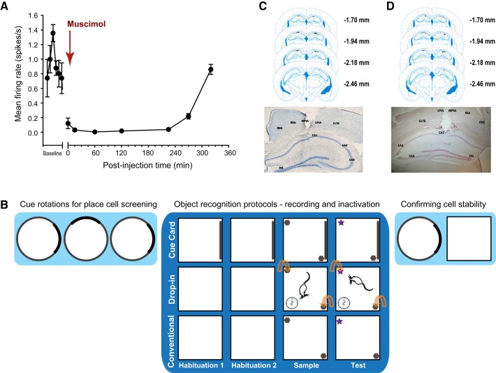
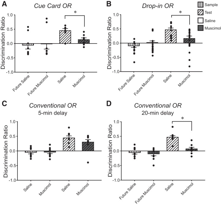
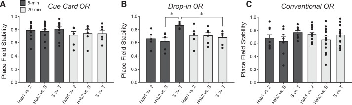
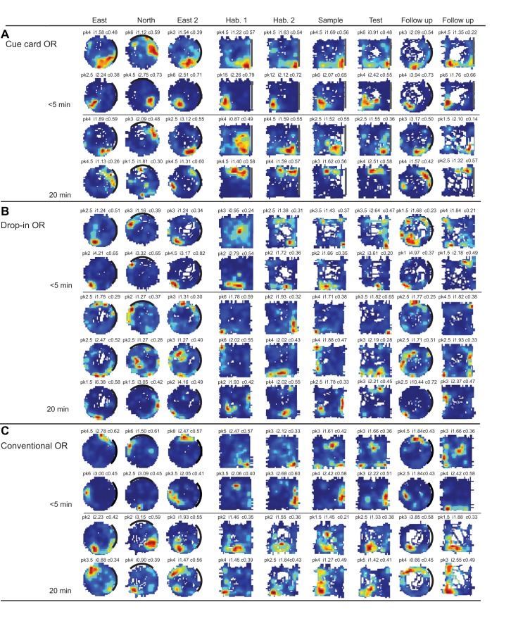
Medial and lateral entorhinal cortices convey spatial/contextual and item/object information to the hippocampus, respectively. Whether the distinct inputs are integrated as one cognitive map by hippocampal neurons to represent location and the objects therein, or whether they remain as parallel outputs, to be integrated in a downstream region, remains unclear. Principal, or complex spike bursting, neurons of hippocampus exhibit location-specific firing, and it is likely that the activity of "place cells" supports spatial memory/navigation in rodents. Consistent with cognitive map theory, the activity of CA1 hippocampal neurons is also critical for nonspatial memory, such as object recognition. However, the degree to which CA1 neuronal activity represents the associations of object-context or object-in-place memory is not well understood. Here, the contributions of mouse CA1 neuronal activity to object recognition memory and the emergence of object-place conjunctive representations were tested using in vivo recordings and functional inactivation. Independent of arena configuration, CA1 place fields were stable throughout testing and object-place representations were not identified in CA1, although the number of fields per cell increased during object sessions, and few object-related firing CA1 neurons (nonplace) were recorded. The results of the inactivation studies confirmed the significant contribution of CA1 neuronal activity to object recognition memory when a delay of 20 min, but not 5 min, was imposed between encoding and retrieval. Together, our results confirm the delay-dependent contribution of the CA1 region to object memory and suggest that object information is processed in parallel with the ongoing spatial mapping function that is a hallmark of hippocampal memory.NEW & NOTEWORTHY We developed variations of the object recognition task to examine the contribution of mouse CA1 neuronal activity to object memory and the degree to which object-context conjunctive representations are formed during object training. Our results indicate that, within the CA1 region, object information is processed in a parallel but delay-dependent manner, with ongoing spatial mapping.

Keywords: CA1; contextual information; memory; muscimol; object recognition.


Conflict of interest statement

No conflicts of interest, financial or otherwise, are declared by the authors.

Figures

Fig. 1.
Fig. 1.
A: mean ± SE normalized firing frequency of CA1 hippocampal neurons before, during and at multiple time points after a local infusion of muscimol. Time 0 = the start of the infusion of muscimol. B: schematic depicting the in vivo recording and functional inactivation protocols. The darker blue background highlights the specific sessions for the three different object recognition (OR) protocols. Top: Cue Card OR, white square arena with dark cue card on one wall throughout all sessions. Middle: Drop-In OR, white square arena (no polarizing cue) where the mouse explored the empty arena for 2 min, after which the objects were placed into the arena in the positions indicated by the curved arrows. Bottom: conventional OR, same white square arena as in Drop-In OR, except that objects were present from the beginning upon mice entering the arena for the object sessions. Mice who received bilateral intrahippocampal infusion of saline or muscimol (i.e., inactivation studies) completed only the sessions in the darker blue section of the figure, while mice implanted with microdrive/tetrode arrays (i.e., in vivo recording studies) completed the lighter blue cue card rotations before, and immediately after, behavioral testing in the OR protocols (while place cells were recorded), to confirm cell stability and unit isolation. C, top: representative placement of the unilateral tetrode arrays implanted over the CA1 region of the right dorsal hippocampus for all mice (black dots). Bottom: representative photomicrograph of tetrode tip location in the dorsal hippocampus. D, top: representative bilateral intrahippocampal infusion sites within the CA1 region of the dorsal hippocampus for the Cue Card OR experiment (black dots). These sites are representative of the infusion locations for all of the inactivation experiments. Bottom: characteristic photomicrograph of the intrahippocampal microinfusion site into the CA1. Abbreviations in photomicrographs in C and D: CA1, CA2, and CA3, respective pyramidal cell fields of the hippocampus; DG, dentate gyrus; LPtA, lateral parietal association cortex; MPtA, medial parietal association cortex; RSA, retrosplenial agranular cortex; RSG, retrosplenial granular cortex; S1TR, primary somatosensory cortex trunk region.
Fig. 2.
Fig. 2.
AD: mean ± SE discrimination ratios for object sessions in the Cue Card object recognition (OR), Drop-In OR, and Conventional OR protocols (5-min and 20-min delays). White and gray bars of each graph represent the vehicle- and muscimol-treated groups, respectively. Within the bars, vertical lines represent object discrimination during sample, while diagonal lines indicate object discrimination during the test. Object exploration during sample was equivalent across the treatment groups and mice did not discriminate between the two identical sample session objects. Significant differences between groups were found for the test (all, except Conventional OR protocol, 5-min delay), and these are designated with an asterisk. *P < 0.05 versus vehicle group. Each mouse received bilateral 0.335-µl microinfusion of vehicle or muscimol (1 µg/µl) immediately after acquiring sample exploration criterion (see materials and methods), or in the 5-min delay of the Conventional OR protocol, the treatments were administered 20-min before sample.
Fig. 3.
Fig. 3.
AC: mean ± SE place × firing rate map correlation (0–1) calculated between two adjacent recording sessions for Cue Card OR, Drop-In OR, and Conventional OR protocols, respectively. Calculated correlations between Habituation 1 and Habituation 2 sessions is represented by “Hab1 versus 2”; correlation between Habituation 2 and sample is represented by “Hab 2 versus S”; and the correlation between sample and test object sessions is represented by “S versus T.” Darker colored bars indicate a 5-min delay between recording sessions whereas lighter colored bars represent a 20-min delay between adjacent sessions. *P < 0.01. OR, object recognition.
Fig. 4.
Fig. 4.
Representative place × firing rate maps of individual CA1 place cells recorded during each of the three object recognition (OR) protocols. For each map, firing rate is represented by the color-coded pixels, with red referring to the location(s) where the cell discharged at its highest rate, and blue referring to the visited locations where the cell was silent. White pixels represent areas of the arena that were unvisited by the given mouse. Each row represents the location-specific firing properties of a given CA1 neuron recorded across the nine 10-min sessions of a respective protocol. The label above the top row of maps identifies the respective recording sessions. The numbers on the left and above each place × firing rate map represent the average in-field firing rate for that cell (pk), information content (i), and spatial coherence (c). A: representative place × firing rate maps from two mice tested in the Cue Card OR protocol with an intersession delay of 5-min and 20-min, respectively. B: same as in A, but for two mice tested in the Drop-In OR protocol. C: same as in A, but for two mice tested in the Conventional OR protocol. Representative place × firing maps for “object cells” can be found in Supplemental Fig. S1 at https://doi.org/10.6084/m9.figshare.11482281.
Fig. 5.
Fig. 5.
A: spatial coherence across the three protocols at both delays calculated from place fields during sample and test. *P < 0.01. B: spatial information content of place fields across protocols at both delays obtained from sample and test.
Fig. 6.
Fig. 6.
A: linear regression analysis illustrates the positive relationship between the normalized discrimination ratio and the normalized place field stability averaged for each mouse across object recognition protocols. B: regression analysis and representative plot demonstrate the positive relationship between the normalized discrimination ratio and the normalized place field stability averaged over each mouse by delay.

References

    1. Ainge JA, Heron-Maxwell C, Theofilas P, Wright P, de Hoz L, Wood ER. The role of the hippocampus in object recognition in rats: examination of the influence of task parameters and lesion size. Behav Brain Res 167: 183–195, 2006. doi:10.1016/j.bbr.2005.09.005. - DOI - PubMed
    1. Amaral DG, Witter MP. Hippocampal formation. In: The Rat Nervous System, edited by Paxinos G. San Diego, CA: Academic Press Inc, 1995, p. 443–493.
    1. Baker KB, Kim JJ. Effects of stress and hippocampal NMDA receptor antagonism on recognition memory in rats. Learn Mem 9: 58–65, 2002. doi:10.1101/lm.46102. - DOI - PMC - PubMed
    1. Bettis T, Jacobs LF. Sex differences in object recognition are modulated by object similarity. Behav Brain Res 233: 288–292, 2012. doi:10.1016/j.bbr.2012.04.028. - DOI - PubMed
    1. Bonnevie T, Dunn B, Fyhn M, Hafting T, Derdikman D, Kubie JL, Roudi Y, Moser EI, Moser MB. Grid cells require excitatory drive from the hippocampus. Nat Neurosci 16: 309–317, 2013. doi:10.1038/nn.3311. - DOI - PubMed

Publication types

MeSH terms

LinkOut - more resources