Review
. 2020 May 15;21(10):1387-1396.
doi: 10.1002/cbic.202000017. Epub 2020 Mar 2.

Hijacking Translation Initiation for Synthetic Biology

Affiliations
Review

Hijacking Translation Initiation for Synthetic Biology

Jeffery M Tharp et al. Chembiochem. .

Abstract

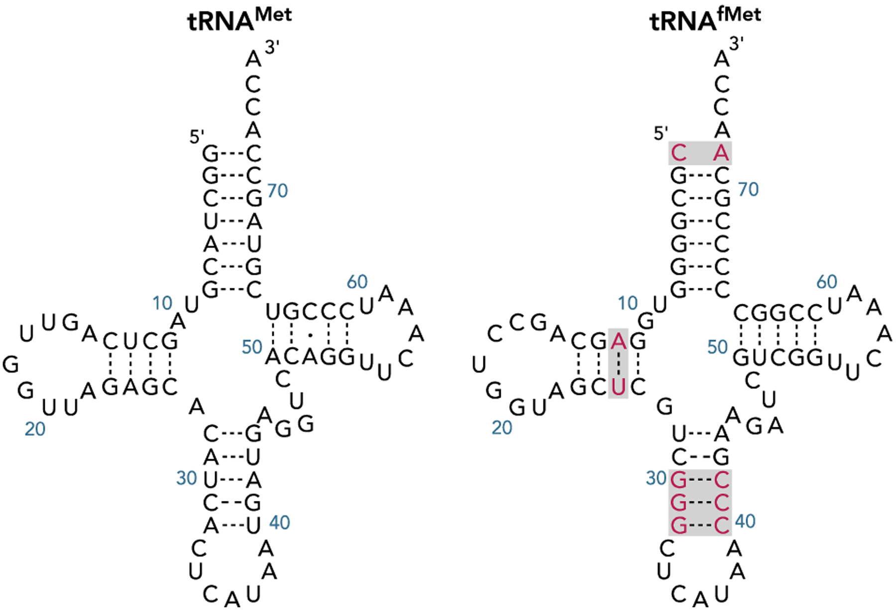
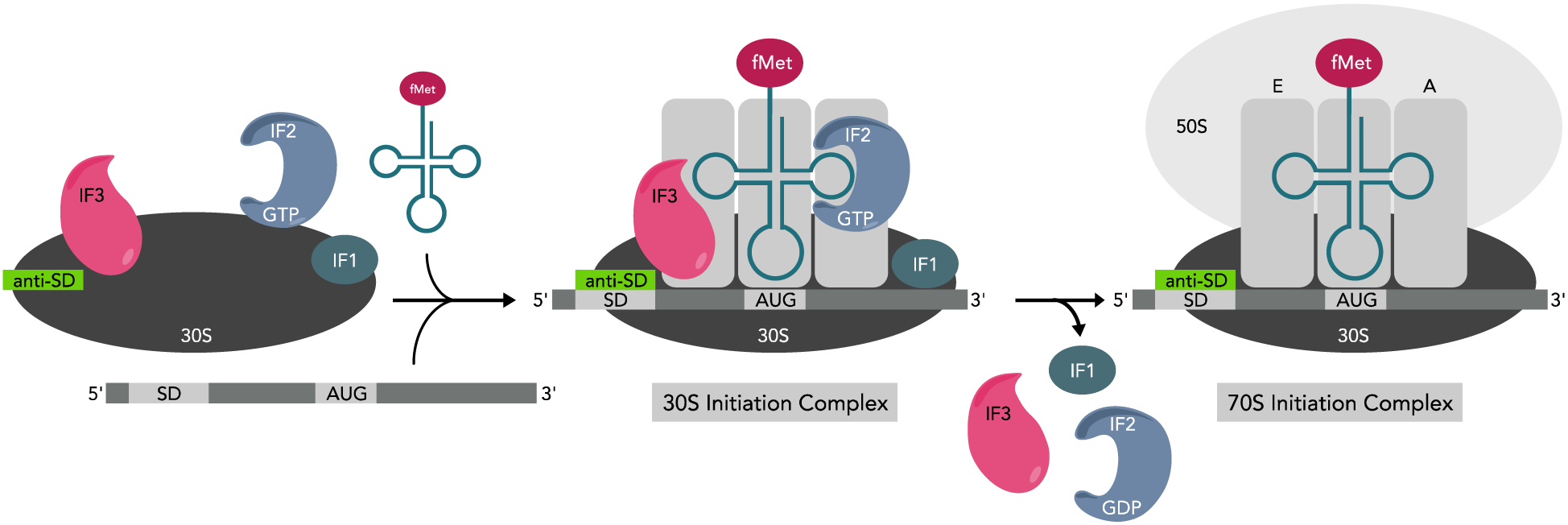
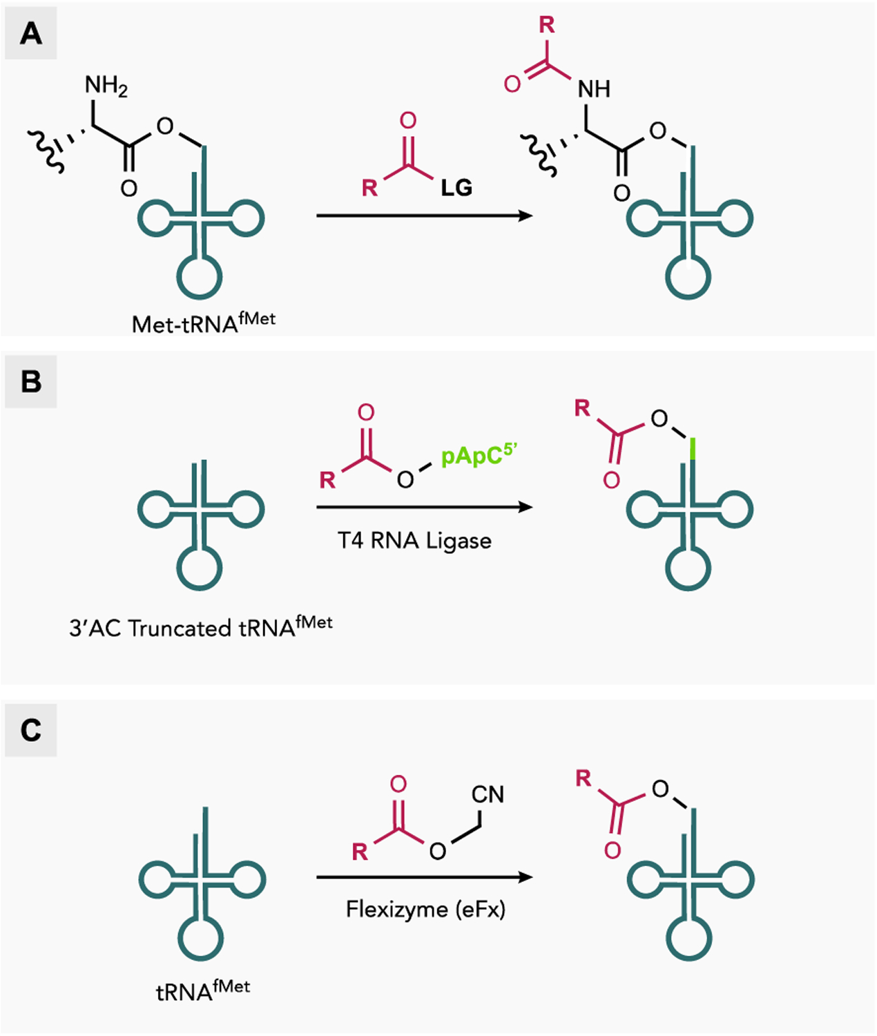
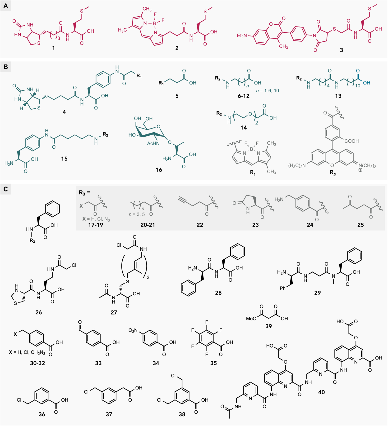
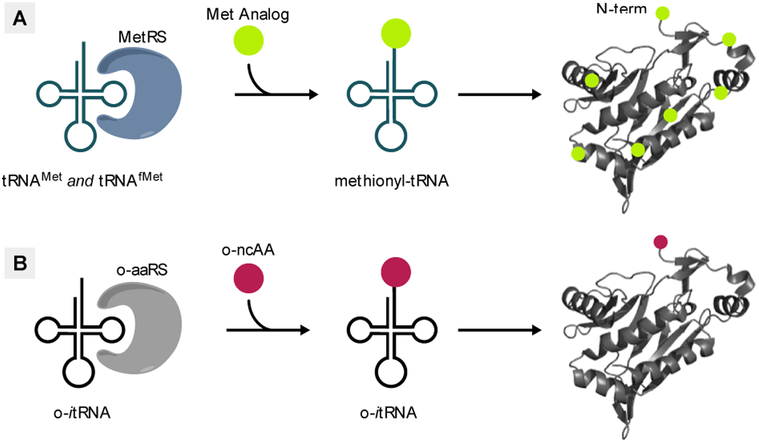
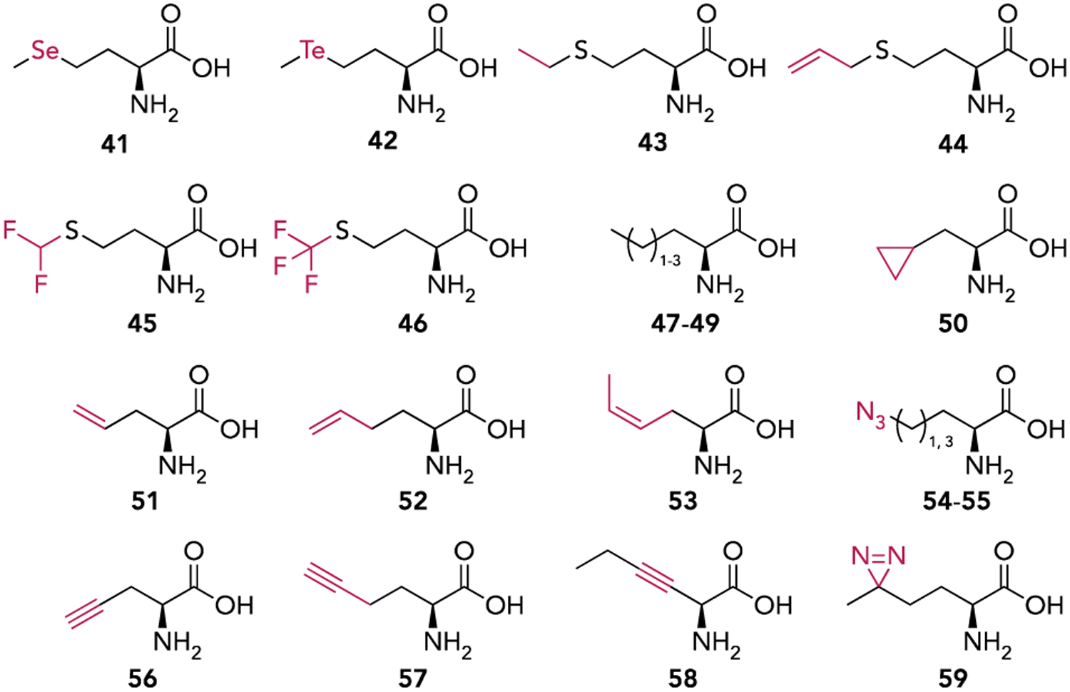
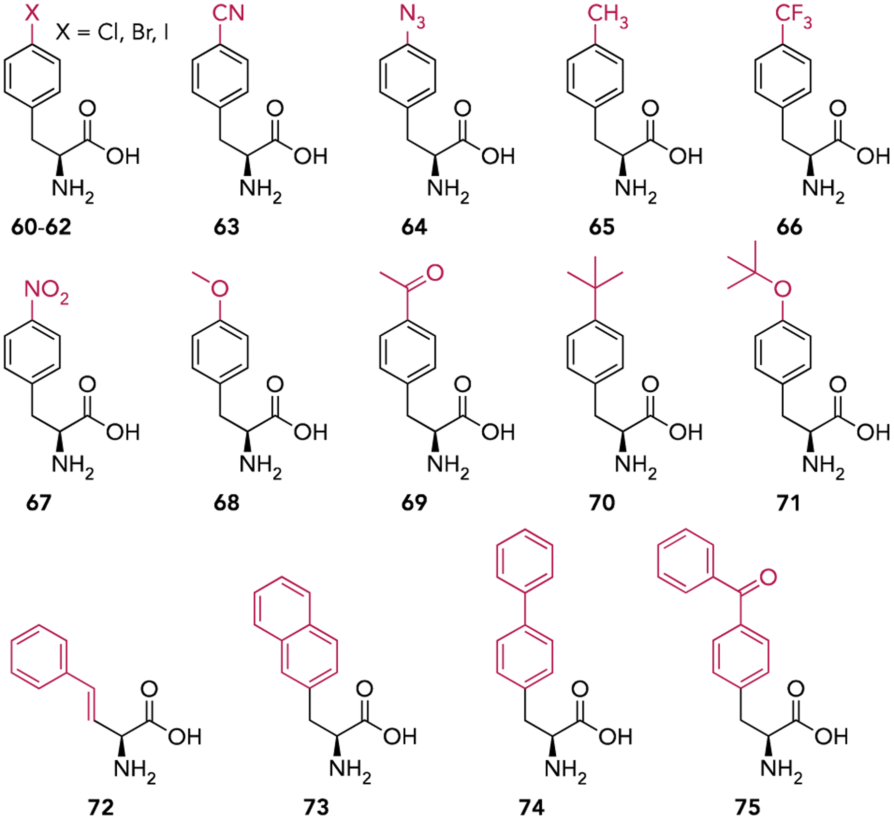
Genetic code expansion (GCE) has revolutionized the field of protein chemistry. Over the past several decades more than 150 different noncanonical amino acids (ncAAs) have been co-translationally installed into proteins within various host organisms. The vast majority of these ncAAs have been incorporated between the start and stop codons within an open reading frame. This requires that the ncAA be able to form a peptide bond at the α-amine, limiting the types of molecules that can be genetically encoded. In contrast, the α-amine of the initiating amino acid is not required for peptide bond formation. Therefore, including the initiator position in GCE allows for co-translational insertion of more diverse molecules that are modified, or completely lacking an α-amine. This review explores various methods which have been used to initiate protein synthesis with diverse molecules both in vitro and in vivo.

Keywords: chemical biology; genetic code expansion; noncanonical amino acids; synthetic biology; translation initiation.


Figures

Figure 1.
Figure 1.
The cloverleaf structures of E. coli elongator (tRNAMet) and initiator (tRNAfMet) methionine tRNA. Nucleotides in tRNAfMet that contribute to its ability to initiate are highlighted.
Figure 2.
Figure 2.
Model of translation initiation. The first step in initiation is binding of IF3 and IF2, followed by IF1, mRNA, and fMet-tRNAfMet to the 30S ribosomal subunit giving way to the 30S pre-initiation complex. Initiation factor-mediated anticodon-codon pairing between mRNA and fMet-tRNAfMet gives rise to a more stable 30S initiation complex. 50S ribosomal subunit binding stimulates GTP hydrolysis by IF2 resulting in a conformational change and initiation factor dissociation, giving rise the translationally competent 70S initiation complex.
Figure 3.
Figure 3.
Strategies for generating mis-acylated tRNAfMet for non-canonical initiation in vitro. (A) Aminoacylation by MetRS followed by chemical modification of the α-amine. (B) Enzymatic ligation of a synthetic 2’,(3’)-O-acyl-pCpA to a truncated tRNAfMet transcript. (C) Flexizyme-catalyzed acylation.
Figure 4.
Figure 4.
Representative carboxylic acids used to initiate translation in vitro. Mis-acylated tRNAfMets for in vitro translation were prepared by acylation with MetRS followed by chemical modification of the α-amine (A), enzymatic ligation of synthetic 2’,(3’)-O-acyl-pCpA to a 3’-truncated tRNAfMet (B), or flexizyme catalyzed acylation (C).
Figure 5.
Figure 5.
Methods for genetic code expansion. (A) Residue-specific incorporation uses endogenous aaRS/tRNA pairs to incorporate ncAAs that are structural analogs of canonical amino acids. This method replaces all instances of a canonical amino acid with the ncAA. (B) Site-specific incorporation uses orthogonal aaRS/tRNA pairs to incorporate ncAAs that are not recognized by endogenous aaRSs. This method allows for incorporation of ncAAs at defined sites within the protein.
Figure 6.
Figure 6.
The structures of methionine analogs used to initiate translation in vivo via the residue-specific incorporation technique.
Figure 7.
Figure 7.
The structures of non-canonical amino acids used to initiate translation in vivo via site-specific incorporation.

References

    1. Agostini F, Völler JS, Koksch B, Acevedo-Rocha CG, Kubyshkin V, Budisa N, Angew. Chem. Int. Ed 2017, 56, 9680–9703; - PubMed
    2. Drienovská I, Mayer C, Dulson C, Roelfes G, Nat. Chem 2018, 10, 946–952; - PubMed
    3. Chin JW, Nature 2017, 550, 53–60. - PubMed
    1. Laursen BS, Sørensen HP, Mortensen KK, Sperling-Petersen HU, Microbiol. Mol. Biol. Rev 2005, 69, 101–123. - PMC - PubMed
    1. Gualerzi CO, Pon CL, Cell Mol. Life Sci 2015, 72, 4341–4367; - PMC - PubMed
    2. Rodnina MV, Cold Spring Harb. Perspect. Biol 2018, 10, a032664. - PMC - PubMed
    1. RajBhandary UL, J. Bacteriol 1994, 176, 547–552. - PMC - PubMed
    1. Varshney U, Lee CP, RajBhandary UL, Proc. Natl. Acad. Sci. USA 1993, 90, 2305–2309. - PMC - PubMed

Publication types

LinkOut - more resources