. 2020 Mar 19;77(6):1251-1264.e9.
doi: 10.1016/j.molcel.2020.01.014. Epub 2020 Feb 4.

Partitioning of MLX-Family Transcription Factors to Lipid Droplets Regulates Metabolic Gene Expression

Affiliations

Partitioning of MLX-Family Transcription Factors to Lipid Droplets Regulates Metabolic Gene Expression

Niklas Mejhert et al. Mol Cell. .

Abstract

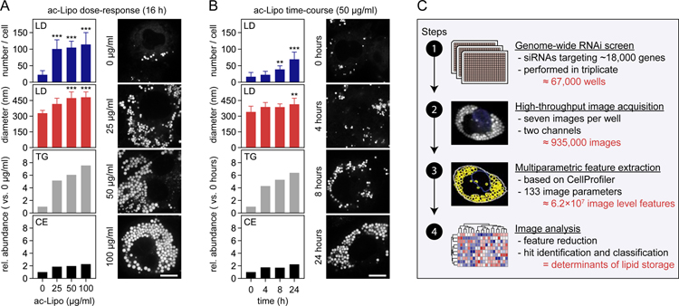
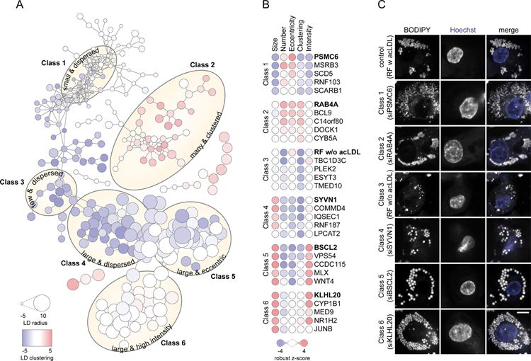
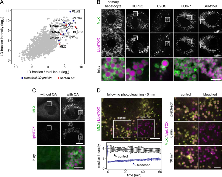
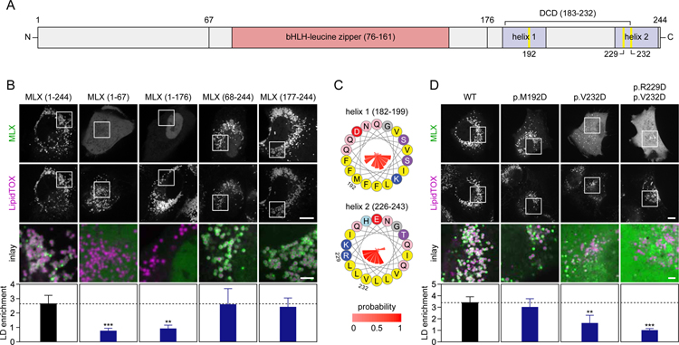
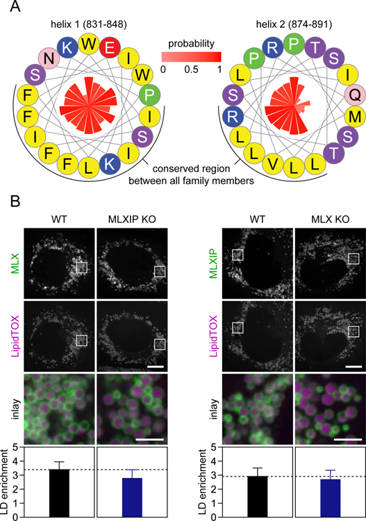
Lipid droplets (LDs) store lipids for energy and are central to cellular lipid homeostasis. The mechanisms coordinating lipid storage in LDs with cellular metabolism are unclear but relevant to obesity-related diseases. Here we utilized genome-wide screening to identify genes that modulate lipid storage in macrophages, a cell type involved in metabolic diseases. Among ∼550 identified screen hits is MLX, a basic helix-loop-helix leucine-zipper transcription factor that regulates metabolic processes. We show that MLX and glucose-sensing family members MLXIP/MondoA and MLXIPL/ChREBP bind LDs via C-terminal amphipathic helices. When LDs accumulate in cells, these transcription factors bind to LDs, reducing their availability for transcriptional activity and attenuating the response to glucose. Conversely, the absence of LDs results in hyperactivation of MLX target genes. Our findings uncover a paradigm for a lipid storage response in which binding of MLX transcription factors to LD surfaces adjusts the expression of metabolic genes to lipid storage levels.

Keywords: ChREBP; MLX; MLXIP; MLXIPL; MondoA; MondoB; glucose; lipid droplets; metabolism; transcription factor.


Conflict of interest statement

Declaration of Interests The authors declare no competing interests.

Figures

Figure 1.
Figure 1.. Systematic Identification of Regulators of Lipid Storage in Human Macrophages
(A–B) Induction of lipid storage in macrophages by acetylated apolipoprotein B-containing lipoproteins. Macrophages were incubated in the absence/presence of ac-Lipo (0–100 µg/mL) for 0–24 hours followed by assessment of LD formation using BODIPY staining and quantification of lipid composition by TLC. Results from one representative experiment are shown for TLC analyses and quantifications of LDs were based on multiple cells (n=8–19). Results were evaluated using one-way non-parametric ANOVA (Kruskal-Wallis followed by Dunn’s multiple comparisons test). Scale bar, 10 µm. (C) Overview of the experimental and computational design of the study. Based on four major steps, regulators of lipid storage in macrophages were identified. Abbreviations: ac-Lipo, acetylated apolipoprotein B-containing lipoprotein; CE, cholesterol ester; TG, triacylglycerol.
Figure 2.
Figure 2.. Genetic Determinants of Lipid Storage Belong to Six Major Classes
(A) RNAi screen hits cluster into six major classes. Based on pair-wise similarities derived from Spearman’s rank correlation, RNAi screen hits were interconnected by edges into major classes as indicated by yellow ellipses. Each node represents a hit and its size and color are proportional to the robust z-score of the hit for LD radius and LD clustering, respectively. Data are also available as an interactive tree view file, see Data S1. (B–C) Representative hits for classes 1–6. (B) For each class, five hits are visualized as nodes where the color of each circle is proportional to the robust z-score of the hit. (C) Confocal images displaying LDs (stained by BODIPY) and nuclei (stained by Hoechst) of a representative hit for each class. Scale bar, 5 µm.
Figure 3.
Figure 3.. Transcription Factor MLX Is an LD Protein
(A) Identification of macrophage LD proteins in THP-1 macrophages. Proteins localized to LDs in THP-1 macrophages incubated with oleic acid (0.5 mM) for 6 hours were identified by mass spectrometry. For each protein, intensities in whole-cell lysate and LD fractions were compared. Known LD proteins were used to calculate a fold-change cut-off (for LD enrichment) based on the 99% confidence interval of their distribution (lower boundary is indicated by the dashed line). Results from one representative experiment are shown. (B) MLX localizes to LDs in multiple cell types. Representative confocal images of mouse primary hepatocytes, HEPG2, U2OS, COS-7, and SUM159 cells transfected with GFP-tagged MLX. Cells were incubated with oleic acid (0.5 mM) for 1 day, stained with LipidTOX Deep Red and thereafter imaged. Scale bars, 10 µm and 2.5 µm (inlay). (C) Endogenously tagged MLX targets to LDs. MLX was endogenously tagged C-terminally with EGFP using CRISPR/Cas9-mediated engineering in SUM159 cells. Representative images of the localization of MLX in live cells incubated in the presence or absence of oleic acid (0.5 mM) for 1 day are shown. LDs were stained prior to imaging using LipidTOX Deep Red. Scale bars, 10 µm and 2.5 µm (inlay). (D) Stable binding of MLX to LDs. After transfection with EGFP-tagged MLX, oleation (0.5 mM) for 1 day and staining of LDs with LipidTOX Deep Red, LD-binding properties of MLX were tested using fluorescence recovery after photobleaching. Representative examples of a cell and inlay images are shown in the upper left and right panels, respectively. Recovery kinetics of MLX was quantified from three independent experiments as highlighted in the lower left panel. Scale bars, 5 µm and 2.5 µm (inlay). Abbreviation: OA, oleic acid.
Figure 4.
Figure 4.. MLX Binds LDs Through a C-Terminal Amphipathic Helix
(A) Overview of MLX domains and expression constructs. (B) The C-terminal domain of MLX is sufficient and required for LD binding. The ability of truncated forms of MLX tagged with EGFP to target to LDs was evaluated in SUM159 cells. Cells were transfected with the indicated constructs and thereafter incubated with oleic acid (0.5 mM) for 1 day. Prior to imaging, LDs were stained with LipidTOX Deep Red. Representative images from one experiment are shown, and results were quantified (n=10–12 cells per construct) using CellProfiler. Results were evaluated using one-way non-parametric ANOVA (Kruskal-Wallis followed by Dunn’s multiple comparisons test). Scale bars, 10 µm and 2.5 µm (inlay). (C) Two C-terminal amphipathic helices are present in MLX. Helical wheels displaying amphipathic helices present in MLX. Amino acid properties and positions are indicated by colors and numbers, respectively, and conservation (expressed as probability %) is indicated by the inner bars. (D) Point-mutations in the most C-terminal amphipathic helix of MLX abolish LD binding. The experiment was performed exactly as described in Panel (B) with the exception that cells were transfected with full-length constructs containing point-mutations in the amphipathic helices of MLX. Results were evaluated using one-way non-parametric ANOVA (Kruskal-Wallis followed by Dunn’s multiple comparisons test). Scale bars, 10 µm and 2.5 µm (inlay). Abbreviations: bHLH, basic helix-loop-helix; DCD, dimerization and cytoplasmic localization domain.
Figure 5.
Figure 5.. MLX and MLXIP Bind LDs Independently of Each Other
(A) Two amphipathic helices, similar to those found in MLX, are present in the C-terminus of MLXIP. Helical wheels displaying amphipathic helices present in MLXIP. Amino acid properties are indicated by colors, and conservation (expressed as probability %) is indicated by the inner bars. (B) MLX:MLXIP heterocomplex formation is not required for LD targeting of either protein. SUM159 cells were transfected with EGFP-tagged MLX or MLXIP, incubated with oleic acid (0.5 mM) for 1 day, stained with LipidTOX Deep Red and imaged. Left panel; localization of MLX in MLXIP KO cells. Right panel; localization of MLXIP in MLX KO cells. Representative images from one experiment are shown and results were quantified (n=7–12 cells per construct and cell type) using CellProfiler. Results were evaluated using one-way non-parametric ANOVA (Kruskal-Wallis followed by Dunn’s multiple comparisons test). Scale bars, 10 µm and 2.5 µm (inlay).
Figure 6.
Figure 6.. LD Binding Modulates MLX:MLXIP Transcriptional Activity
(A–B) LD binding sequesters MLX away from the nuclei. Effects of alterations in lipid storage and glucose on endogenous MLX localization were determined in SUM159 cells by confocal microscopy. (A) In the presence or absence of oleic acid (0.5 mM), cells were starved from glucose for 2 days and thereafter incubated for 1 day with 0 or 25 mM glucose-containing media. (B) Cells were starved from glucose for 2 days and thereafter incubated overnight with DMSO or DGAT1/DGAT2 inhibitors as well as glucose (25 mM) and oleic acid (0.5 mM). Prior to live cell imaging, LDs and nuclei were stained with LipidTOX Deep Red and Hoechst, respectively. For panels A-B, representative images from one experiment are shown, and results were quantified (n=19–25 cells per condition) by CellProfiler. Results were evaluated using two-way ANOVA followed by Tukey-Kramer test. Scale bars, 10 µm and 2.5 µm (inlay). (C) TXNIP mRNA levels are upregulated by glucose. Genes regulated by glucose were identified in SUM159 cells using RNA sequencing. Prior to lysis, cells were starved from glucose for 2 days and thereafter incubated with or without glucose (25 mM) for 6 hours. Results are based on three replicates per condition and were evaluated using DESeq2. (D–E) Glucose-mediated induction of TXNIP mRNA requires MLX and MLXIP. Messenger RNA levels of TXNIP were measured by qPCR in WT, MLXIP (D), and MLX (E) knockout cells. The cells were starved from glucose for 2 days and thereafter incubated in the presence/absence of glucose (25 mM) for 1 day. (E) As indicated in the panel, MLX-EGFP was stably expressed in MLX knockout cells from the AAVS1 locus (add-back). Results from one representative experiment containing three replicates are shown. (F–G) LDs regulate MLX:MLXIP activity. TXNIP mRNA levels were measured in SUM159 cells with or without LDs. (F) Cells were starved from glucose in the presence or absence of oleic acid (0.5 mM) for one day and thereafter washed and starved from glucose for another 12 hours. After this, cells were incubated in with or without glucose (25 mM) and oleic acid (0.5 mM) for 1 day. (G) Cells were starved from glucose for 2.5 days and thereafter incubated with or without glucose (25 mM) in the presence of DMSO or DGAT1/DGAT2 inhibitors overnight. Results are based on two independent experiments, each containing three replicates and were evaluated using two-way ANOVA followed by Tukey-Kramer test. (H) LDs regulate MLX promoter binding. TXNIP promoter binding by endogenously EGFP-tagged MLX was measured in SUM159 cells. Cells were starved from glucose for 1.5 days and thereafter incubated OA-containing media with or without glucose (25 mM) in the presence of DMSO or DGAT1/DGAT2 inhibitors for 12 hours. Results from three replicates are shown using an anti-MLX antibody (left panel) or an anti-GFP antibody (right panel). Abbreviations: DGATi, DGAT1/2 inhibition; KO, knockout; OA, oleic acid.
Figure 7.
Figure 7.. LDs modulate MLX:MLXIPL Gene Regulation in Human Macrophages
(A–B) MLX controls a specific set of putative lipid storage response genes. (A) Genes depending on MLX for induction after glucose stimulus were identified by RNA sequencing. Control and MLX-depleted THP-1 macrophages were starved from glucose for 2 days and thereafter incubated in the presence or absence of glucose (25 mM) for 1 day. Known targets of MLX (TXNIP and ARRDC4) and genes displaying a similar LD phenotype as MLX in the RNAi screen (r>0.5) are highlighted. Results are based on three replicates per condition and were evaluated using DESeq2. (B) Heatmap and distribution chart of the effects of lipid storage induction by ac-Lipo on putative target gene expression. Counts from the RNA sequencing were scaled prior to clustering (left panel) and the relative position among all measured genes highlighted (right panel). Results are based on two replicates per condition. (C) MLX binds specifically to TG-containing LDs. Based on fractionated THP-1 cells, protein and lipid composition of isolated LDs induced by incubations with OA (0.5 mM) or ac-LDL (25 µg/mL) for 2 days were determined using western blotting and TLC, respectively. (D–E) LDs regulate MLX activity. TXNIP promoter binding of MLX and TXNIP mRNA levels were measured in THP-1 macrophages. Cells were starved from glucose for 1.5 days and thereafter incubated OA-containing media with or without glucose (10 mM in panel D and 25 mM in panel E) in the presence of DMSO or DGAT1/DGAT2 inhibitors for 12–16 hours. Results are based on one (D) or two (E) independent experiments, each containing three replicates and (E) were evaluated using two-way ANOVA followed by Tukey-Kramer test. (F–G) Alterations in MLX:MLXIPL activity control 2-DG uptake. Uptake of [3H] 2-Deoxy-D-glucose in THP-1 macrophages was determined in cells (F) starved from glucose for 1 day and thereafter incubated with or without glucose in the presence of DMSO or 10 µM SBI-477 for 1 day and (G) incubated in varying concentrations of glucose in the presence or absence of OA (0.5 mM) or ac-LDL (25 µg/mL) for 2 days. Results are based on three (F) or two (G) independent experiments, each containing three replicates and were evaluated using two-way ANOVA followed by Tukey-Kramer test. Abbreviations: 2-DG, 2-deoxyglucose; ac-LDL, acetylated low-density lipoprotein; ac-Lipo, acetylated apolipoprotein B-containing lipoprotein; CE, cholesterol ester; comp., composition; OA, oleic acid; TG, triacylglycerol; untr., untreated.

References

    1. Abdul-Wahed A, Guilmeau S, and Postic C (2017). Sweet Sixteenth for ChREBP: Established Roles and Future Goals. Cell Metab 26, 324–341. - PubMed
    1. Accad M, Smith SJ, Newland DL, Sanan DA, King LE, Linton MF, Fazio S, and Farese RV (2000). Massive xanthomatosis and altered composition of atherosclerotic lesions in hyperlipidemic mice lacking acyl CoA:cholesterol acyltransferase 1. J. Clin. Invest 105, 711–719. - PMC - PubMed
    1. Ahn B, Soundarapandian MM, Sessions H, Peddibhotla S, Roth GP, Li J-L, Sugarman E, Koo A, Malany S, Wang M, et al. (2016). MondoA coordinately regulates skeletal myocyte lipid homeostasis and insulin signaling. J. Clin. Invest 126, 3567–3579. - PMC - PubMed
    1. Altschul SF, Gish W, Miller W, Myers EW, and Lipman DJ (1990). Basic local alignment search tool. J. Mol. Biol 215, 403–410. - PubMed
    1. Basu SK, Goldstein JL, Anderson GW, and Brown MS (1976). Degradation of cationized low density lipoprotein and regulation of cholesterol metabolism in homozygous familial hypercholesterolemia fibroblasts. Proc. Natl. Acad. Sci. U. S. A 73, 3178–3182. - PMC - PubMed

Publication types

MeSH terms