

The Laboratory Diagnosis of Neisseria gonorrhoeae: Current Testing and Future Demands
- PMID: 32024032
- PMCID: PMC7169389
- DOI: 10.3390/pathogens9020091
The Laboratory Diagnosis of Neisseria gonorrhoeae: Current Testing and Future Demands
Abstract
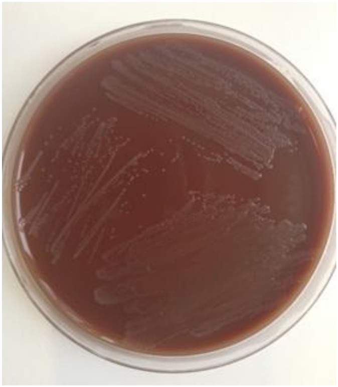
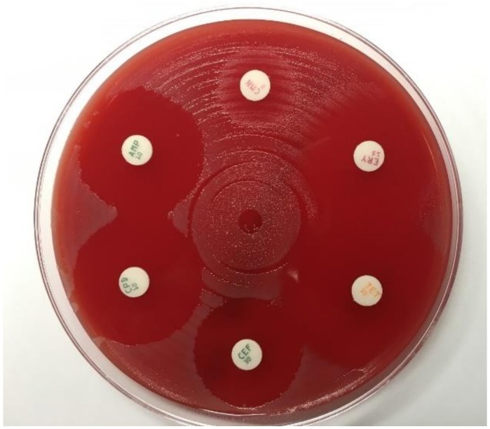
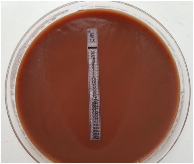
The ideal laboratory test to detect Neisseria gonorrhoeae (Ng) should be sensitive, specific, easy to use, rapid, and affordable and should provide information about susceptibility to antimicrobial drugs. Currently, such a test is not available and presumably will not be in the near future. Thus, diagnosis of gonococcal infections presently includes application of different techniques to address these requirements. Microscopy may produce rapid results but lacks sensitivity in many cases (except symptomatic urogenital infections in males). Highest sensitivity to detect Ng was shown for nucleic acid amplification technologies (NAATs), which, however, are less specific than culture. In addition, comprehensive analysis of antibiotic resistance is accomplished only by in vitro antimicrobial susceptibility testing of cultured isolates. As a light at the end of the tunnel, new developments of molecular techniques and microfluidic systems represent promising opportunities to design point-of-care tests for rapid detection of Ng with high sensitivity and specificity, and there is reason to hope that such tests may also provide antimicrobial resistance data in the future.
Keywords: NAAT; antimicrobial resistance; culture; diagnostic; gonorrhea; microfluidic; microscopy; point-of-care test.
Conflict of interest statement
The authors declare no conflict of interest.
Figures

References
-
- European Centre for Disease Prevention and Control . Gonorrhoea—Annual Epidemiological Report for 2017. European Centre for Disease Prevention and Control; Stockholm, Sweden: 2019.
-
- Department of Health and Human Services . Sexually Transmitted Disease Surveillance 2018. Centers for Disease Control and Prevention; Atlanta, GE, USA: 2019. - DOI
-
- Fifer H., Saunders J., Soni S., Sadiq T., FitzGerald M. British Association for Sexual Health and HIV Web Site; 2019. [(accessed on 30 January 2020)]. National Guideline for the Management of Infection with Neisseria gonorrhoeae. Available online: https://www.bashhguidelines.org/media/1208/gc-2019.pdf. - PubMed
-
- Dudareva-Vizule S., Haar K., Sailer A., Wisplinghoff H., Wisplinghoff F., Marcus U., PARIS Study Group Prevalence of pharyngeal and rectal Chlamydia trachomatis and Neisseria gonorrhoeae infections among men who have sex with men in Germany. Sex Transm. Infect. 2014;90:46–51. doi: 10.1136/sextrans-2012-050929. - DOI - PubMed
Publication types
LinkOut - more resources
Full Text Sources
Other Literature Sources
Research Materials
