. 2020 Feb 1;11(2):158.
doi: 10.3390/mi11020158.

Nano-in-Micro Dual Delivery Platform for Chronic Wound Healing Applications

Affiliations

Nano-in-Micro Dual Delivery Platform for Chronic Wound Healing Applications

Jana Zarubova et al. Micromachines (Basel). .

Abstract

Here, we developed a combinatorial delivery platform for chronic wound healing applications. A microfluidic system was utilized to form a series of biopolymer-based microparticles with enhanced affinity to encapsulate and deliver vascular endothelial growth factor (VEGF). Presence of heparin into the structure can significantly increase the encapsulation efficiency up to 95% and lower the release rate of encapsulated VEGF. Our in vitro results demonstrated that sustained release of VEGF from microparticles can promote capillary network formation and sprouting of endothelial cells in 2D and 3D microenvironments. These engineered microparticles can also encapsulate antibiotic-loaded nanoparticles to offer a dual delivery system able to fight bacterial infection while promoting angiogenesis. We believe this highly tunable drug delivery platform can be used alone or in combination with other wound care products to improve the wound healing process and promote tissue regeneration.

Keywords: angiogenesis; antibacterial properties; chronic wounds; drug delivery; microfluidics; microparticles; nanoparticles.


Conflict of interest statement

The authors declare no conflict of interest.

Figures

Figure 1
Figure 1
Formation of alginate-based microparticles. (A) Schematic of microfluidics platform used to control formation of microparticles. Hydrophobic coated glass microfluidic droplet junction chip with 100 µm diameter used in this study. (B) Controlling flow rates was used to tune the size of alginate-based microparticles. The presented data are expressed as average ± SD (number of independent experiments; n = 3; for each independent experiment more than 40 microparticles were measured). (C) Bright-field image of calcium crosslinked alginate-heparin microparticles at water phase/oil phase flow ratio of 0.33 (oil flow rate: 12 µL/min; water flow rate: 4 µL/min).
Figure 2
Figure 2
The presence of heparin increased vascular endothelial growth factor (VEGF) binding and prolongs its release. (A) Schematic of microparticles (16 µm in diameter) were used to encapsulate therapeutic proteins (here VEGF). (B) VEGF binding efficiency of alginate and alginate-heparin microparticles at various initial concentrations of VEGF after 16 h incubation at 4 °C. (C) VEGF release kinetic of microfluidic-synthesized microparticles prepared from alginate and alginate-heparin in phosphate buffered saline (PBS) at 37 °C. (D) Calculated diffusion coefficients of VEGF from microparticles and (E) the cumulative amount of released VEGF after 24 h. The presented data are expressed as an average ± SD; number of independent experiments, n = 3; number of samples per experiment, s = 5). The lines in the graphs are guide for the eye. *, **, and *** indicate significant difference of p < 0.05, p < 0.01, and p < 0.001, respectively, as evaluated by one-way ANOVA.
Figure 3
Figure 3
2D capillary network formation. Capillary-like network formation by endothelial cells on growth factor-reduced Matrigel affected by addition of microparticles releasing VEGF-A. (A) Morphology of vascular networks after 24 h and (B) evaluation with the Angiogenesis Analyzer ImageJ plugin; red dots indicate junctions, blue lines indicate loops, scale bar: 300 µm. MP: microparticles, MP-VEGF: microparticles releasing VEGF-A (50 ng/mL initial loading), MP-VEGF-H: microparticles releasing high concentration of VEGF-A (100 ng/mL initial loading). (C) Total vascular tube length. (D) Number of junctions. (E) Number of loops, ‡ indicates significant difference comparing to samples containing microparticles alone (p < 0.05) evaluated by one-way ANOVA.
Figure 4
Figure 4
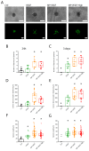
VEGF release accelerate spheroids sprouting in 3D. (A) The effect of microparticles releasing VEGF-A on endothelial cell sprouting from spheroids in 3D microenvironments. Morphology of endothelial cell sprouts in bright-field and fluorescence microscopy images, scale bar: 100 µm. Ctrl: HUVEC spheroids grown in EGM-2 medium, VEGF: HUVEC spheroids stimulated with 50 ng/mL VEGF-A, MP-VEGF: microparticles releasing VEGF-A (50 ng/mL initial loading), MP-VEGF-H: microparticles releasing high concentration of VEGF-A (100 ng/mL initial loading). Number of sprouts per spheroid after (B) 24 h and (C) 3 days. Cumulative sprout length after (D) 24 h and (E) 3 days. Length of individual sprouts after (F) 24 h and (G) 3 days. * indicates significant difference comparing to Ctrl (p < 0.001) evaluated by one-way ANOVA.
Figure 5
Figure 5
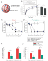
Evaluation of gentamicin release. (A) Gentamicin-loaded poly(lactic-co-glycolic) acid (PLGA) nanoparticles (NPs) encapsulated in alginate-heparin microparticles. Estimated amount of released gentamicin after 24 h. (B) Cumulative release profiles of gentamicin from free PLGA NPs and from microparticle-encapsulated PLGA NPs. Cumulative release profiles of co-encapsulated VEGF from alginate-heparin microparticles are also shown here. Analysis of the ability of gentamicin PLGA NPs to inhibit the growth of (C) Escherichia coli and (D) Pseudomonas aeruginosa as determined by measurement of absorbance at 600 nm. Estimated (E) half-maximal inhibitory (IC50) and (F) minimum inhibitory concentrations (MIC) values for free and PLGA-encapsulated PLGA NPs (average ± SD, n = 3). * and ** indicate significant difference of p < 0.05 and p < 0.01, respectively, as evaluated by one-way ANOVA. The lines in the graphs are a guide for the eye.

References

    1. Menke N.B., Ward K.R., Witten T.M., Bonchev D.G., Diegelmann R.F. Impaired wound healing. Clin. Dermatol. 2007;25:19–25. doi: 10.1016/j.clindermatol.2006.12.005. - DOI - PubMed
    1. Guo S., Dipietro L.A. Factors affecting wound healing. J. Dent. Res. 2010;89:219–229. doi: 10.1177/0022034509359125. - DOI - PMC - PubMed
    1. Gosain A., DiPietro L.A. Aging and wound healing. World J. Surg. 2004;28:321–326. doi: 10.1007/s00268-003-7397-6. - DOI - PubMed
    1. Patel S., Srivastava S., Singh M.R., Singh D. Mechanistic insight into diabetic wounds: Pathogenesis, molecular targets and treatment strategies to pace wound healing. Biomed. Pharmacother. 2019;112:108615. doi: 10.1016/j.biopha.2019.108615. - DOI - PubMed
    1. Mihai M.M., Preda M., Lungu I., Gestal M.C., Popa M.I., Holban A.M. Nanocoatings for Chronic Wound Repair-Modulation of Microbial Colonization and Biofilm Formation. Int. J. Mol. Sci. 2018;19:1179. doi: 10.3390/ijms19041179. - DOI - PMC - PubMed