

Human Mesenchymal Stem Cells-mediated Transcriptomic Regulation of Leukemic Cells in Delivering Anti-tumorigenic Effects
- PMID: 32024378
- PMCID: PMC7444238
- DOI: 10.1177/0963689719885077
Human Mesenchymal Stem Cells-mediated Transcriptomic Regulation of Leukemic Cells in Delivering Anti-tumorigenic Effects
Abstract
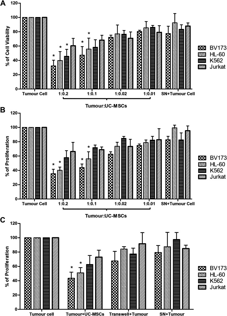
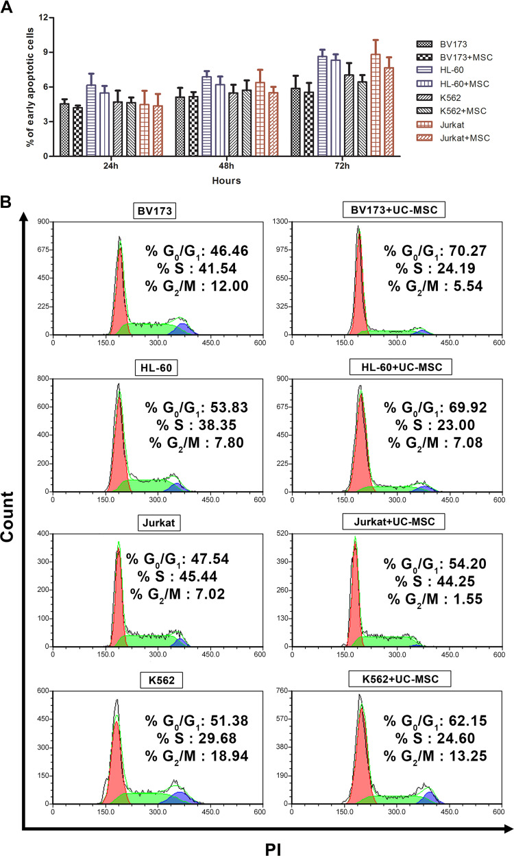
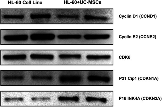
Treatment of leukemia has become much difficult because of resistance to the existing anticancer therapies. This has thus expedited the search for alternativ therapies, and one of these is the exploitation of mesenchymal stem cells (MSCs) towards control of tumor cells. The present study investigated the effect of human umbilical cord-derived MSCs (UC-MSCs) on the proliferation of leukemic cells and gauged the transcriptomic modulation and the signaling pathways potentially affected by UC-MSCs. The inhibition of growth of leukemic tumor cell lines was assessed by proliferation assays, apoptosis and cell cycle analysis. BV173 and HL-60 cells were further analyzed using microarray gene expression profiling. The microarray results were validated by RT-qPCR and western blot assay for the corresponding expression of genes and proteins. The UC-MSCs attenuated leukemic cell viability and proliferation in a dose-dependent manner without inducing apoptosis. Cell cycle analysis revealed that the growth of tumor cells was arrested at the G0/G1 phase. The microarray results identified that HL-60 and BV173 share 35 differentially expressed genes (DEGs) (same expression direction) in the presence of UC-MSCs. In silico analysis of these selected DEGs indicated a significant influence in the cell cycle and cell cycle-related biological processes and signaling pathways. Among these, the expression of DBF4, MDM2, CCNE2, CDK6, CDKN1A, and CDKN2A was implicated in six different signaling pathways that play a pivotal role in the anti-tumorigenic activity exerted by UC-MSCs. The UC-MSCs perturbate the cell cycle process of leukemic cells via dysregulation of tumor suppressor and oncogene expression.
Keywords: cell cycle; gene expression profiling; leukemia; mesenchymal stem cells.
Conflict of interest statement
Figures
References
-
- Colby-Graham MF, Chordas C. The childhood leukemias. J Pediatr Nurs. 2003;18(2):87–95. - PubMed
-
- Kurtova AV, Balakrishnan K, Chen R, Ding W, Schnabl S, Quiroga MP, Sivina M, Wierda WG, Estrov Z, Keating MJ, Shehata M, et al. Diverse marrow stromal cells protect CLL cells from spontaneous and drug-induced apoptosis: development of a reliable and reproducible system to assess stromal cell adhesion-mediated drug resistance. Blood. 2009;114(20):4441–4450. - PMC - PubMed
-
- Jiang Y, Jahagirdar BN, Reinhardt RL, Schwartz RE, Keene CD, Ortiz-Gonzalez XR, Reyes M, Lenvik T, Lund T, Blackstad M, Du J, et al. Pluripotency of mesenchymal stem cells derived from adult marrow. Nature. 2002;418(6893):41–49. - PubMed
Publication types
MeSH terms
LinkOut - more resources
Full Text Sources
Medical
Molecular Biology Databases
Research Materials
Miscellaneous
