

Multi-template matching: a versatile tool for object-localization in microscopy images
- PMID: 32024462
- PMCID: PMC7003318
- DOI: 10.1186/s12859-020-3363-7
Multi-template matching: a versatile tool for object-localization in microscopy images
Erratum in
-
Correction to: Multi-template matching: a versatile tool for object-localization in microscopy images.BMC Bioinformatics. 2022 Jan 4;23(1):3. doi: 10.1186/s12859-021-04524-7. BMC Bioinformatics. 2022. PMID: 34983368 Free PMC article. No abstract available.
Abstract
Background: The localization of objects of interest is a key initial step in most image analysis workflows. For biomedical image data, classical image-segmentation methods like thresholding or edge detection are typically used. While those methods perform well for labelled objects, they are reaching a limit when samples are poorly contrasted with the background, or when only parts of larger structures should be detected. Furthermore, the development of such pipelines requires substantial engineering of analysis workflows and often results in case-specific solutions. Therefore, we propose a new straightforward and generic approach for object-localization by template matching that utilizes multiple template images to improve the detection capacity.
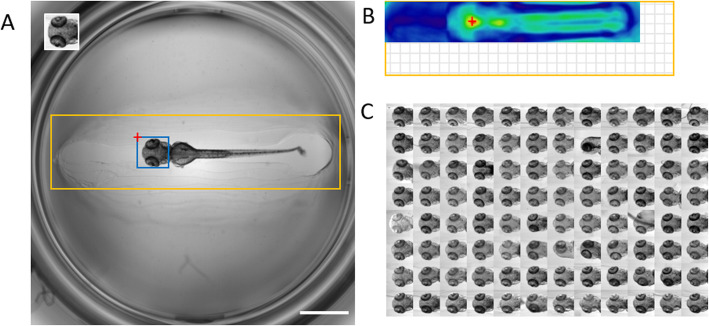
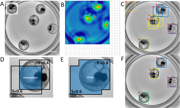
Results: We provide a new implementation of template matching that offers higher detection capacity than single template approach, by enabling the detection of multiple template images. To provide an easy-to-use method for the automatic localization of objects of interest in microscopy images, we implemented multi-template matching as a Fiji plugin, a KNIME workflow and a python package. We demonstrate its application for the localization of entire, partial and multiple biological objects in zebrafish and medaka high-content screening datasets. The Fiji plugin can be installed by activating the Multi-Template-Matching and IJ-OpenCV update sites. The KNIME workflow is available on nodepit and KNIME Hub. Source codes and documentations are available on GitHub (https://github.com/multi-template-matching).
Conclusion: The novel multi-template matching is a simple yet powerful object-localization algorithm, that requires no data-pre-processing or annotation. Our implementation can be used out-of-the-box by non-expert users for any type of 2D-image. It is compatible with a large variety of applications including, for instance, analysis of large-scale datasets originating from automated microscopy, detection and tracking of objects in time-lapse assays, or as a general image-analysis step in any custom processing pipelines. Using different templates corresponding to distinct object categories, the tool can also be used for classification of the detected regions.
Keywords: Classification; Fiji; KNIME; Medaka; Object-localization; Object-recognition; OpenCV; Pattern recognition; Template matching; Zebrafish.
Conflict of interest statement
Both authors are employees of ACQUIFER, a division of DITABIS Digital Biomedical Imaging Systems AG.
Figures
Similar articles
-
Fiji plugins for qualitative image annotations: routine analysis and application to image classification.F1000Res. 2020 Oct 15;9:1248. doi: 10.12688/f1000research.26872.2. eCollection 2020. F1000Res. 2020. PMID: 33841801 Free PMC article.
-
SHERPA: an image segmentation and outline feature extraction tool for diatoms and other objects.BMC Bioinformatics. 2014 Jun 25;15:218. doi: 10.1186/1471-2105-15-218. BMC Bioinformatics. 2014. PMID: 24964954 Free PMC article.
-
BigDataProcessor2: a free and open-source Fiji plugin for inspection and processing of TB sized image data.Bioinformatics. 2021 Sep 29;37(18):3079-3081. doi: 10.1093/bioinformatics/btab106. Bioinformatics. 2021. PMID: 33594413 Free PMC article.
-
Workflows for microscopy image analysis and cellular phenotyping.J Biotechnol. 2017 Nov 10;261:70-75. doi: 10.1016/j.jbiotec.2017.07.019. Epub 2017 Jul 27. J Biotechnol. 2017. PMID: 28757289 Review.
-
Live cell fluorescence microscopy-an end-to-end workflow for high-throughput image and data analysis.Biol Methods Protoc. 2024 Oct 11;9(1):bpae075. doi: 10.1093/biomethods/bpae075. eCollection 2024. Biol Methods Protoc. 2024. PMID: 39484095 Free PMC article. Review.
Cited by
-
Cell state-specific cytoplasmic density controls spindle architecture and scaling.Nat Cell Biol. 2025 Jun;27(6):959-971. doi: 10.1038/s41556-025-01678-x. Epub 2025 Jun 13. Nat Cell Biol. 2025. PMID: 40514430 Free PMC article.
-
Latent Space Search-Based Adaptive Template Generation for Enhanced Object Detection in Bin-Picking Applications.Sensors (Basel). 2024 Sep 19;24(18):6050. doi: 10.3390/s24186050. Sensors (Basel). 2024. PMID: 39338795 Free PMC article.
-
Dysregulation of N-terminal acetylation causes cardiac arrhythmia and cardiomyopathy.Res Sq [Preprint]. 2024 Jul 19:rs.3.rs-3398860. doi: 10.21203/rs.3.rs-3398860/v1. Res Sq. 2024. Update in: Nat Commun. 2025 Apr 16;16(1):3604. doi: 10.1038/s41467-025-58539-2. PMID: 39070617 Free PMC article. Updated. Preprint.
-
Dysregulation of N-terminal acetylation causes cardiac arrhythmia and cardiomyopathy.Nat Commun. 2025 Apr 16;16(1):3604. doi: 10.1038/s41467-025-58539-2. Nat Commun. 2025. PMID: 40234403 Free PMC article.
-
Efficient and reproducible generation of human iPSC-derived cardiomyocytes using a stirred bioreactor.bioRxiv [Preprint]. 2024 Feb 28:2024.02.24.581789. doi: 10.1101/2024.02.24.581789. bioRxiv. 2024. Update in: Nat Commun. 2024 Jul 15;15(1):5929. doi: 10.1038/s41467-024-50224-0. PMID: 38464269 Free PMC article. Updated. Preprint.
References
-
- Marcato D, et al. An automated and high-throughput photomotor response platform for chemical screens. 2015. pp. 7728–7731. - PubMed
MeSH terms
Grants and funding
LinkOut - more resources
Full Text Sources
