Review
. 2020 Mar 31;94(8):e02120-19.
doi: 10.1128/JVI.02120-19. Print 2020 Mar 31.

Bridging the Gap: Virus Long-Distance Spread via Tunneling Nanotubes

Affiliations
Review

Bridging the Gap: Virus Long-Distance Spread via Tunneling Nanotubes

Robert J J Jansens et al. J Virol. .

Abstract

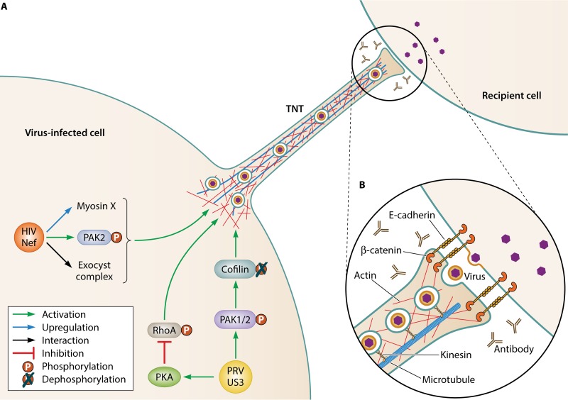
Tunneling nanotubes (TNTs) are actin-based intercellular conduits that connect distant cells and allow intercellular transfer of molecular information, including genetic information, proteins, lipids, and even organelles. Besides providing a means of intercellular communication, TNTs may also be hijacked by pathogens, particularly viruses, to facilitate their spread. Viruses of many different families, including retroviruses, herpesviruses, orthomyxoviruses, and several others have been reported to trigger the formation of TNTs or TNT-like structures in infected cells and use these structures to efficiently spread to uninfected cells. In the current review, we give an overview of the information that is currently available on viruses and TNT-like structures, and we discuss some of the standing questions in this field.

Keywords: TNT; antibodies; intercellular; spread; tunneling nanotubes; virus.


Figures

FIG 1
FIG 1
(A) Schematic representation of two of the best characterized models of virus-induced TNT formation: Nef-induced TNT formation in HIV-infected macrophages and US3-induced TNT formation in PRV-infected epithelial cells. Both types of TNTs have been shown to carry virus particles in vesicles (see, for example, references and 43). Some of the molecular players that have been shown to be involved in these two types of TNT formation are indicated. More detailed information on their role is provided in the present study. (B) An inset image shows a schematic representation of what is known about the contact area between a PRV US3-induced TNT and a recipient cell based on electron microscopy and confocal microscopy analyses (based on reference 17).

References

    1. van Niel G, D’Angelo G, Raposo G. 2018. Shedding light on the cell biology of extracellular vesicles. Nat Rev Mol Cell Biol 19:213–228. doi:10.1038/nrm.2017.125. - DOI - PubMed
    1. Rustom A, Saffrich R, Markovic I, Walther P, Gerdes H-H. 2004. Nanotubular highways for intercellular organelle transport. Science 303:1007–1010. doi:10.1126/science.1093133. - DOI - PubMed
    1. Biran A, Perelmutter M, Gal H, Burton DGA, Ovadya Y, Vadai E, Geiger T, Krizhanovsky V. 2015. Senescent cells communicate via intercellular protein transfer. Genes Dev 29:791–802. doi:10.1101/gad.259341.115. - DOI - PMC - PubMed
    1. Watkins SC, Salter RD. 2005. Functional connectivity between immune cells mediated by tunneling nanotubules. Immunity 23:309–318. doi:10.1016/j.immuni.2005.08.009. - DOI - PubMed
    1. Thayanithy V, Dickson EL, Steer C, Subramanian S, Lou E. 2014. Tumor-stromal cross talk: direct cell-to-cell transfer of oncogenic microRNAs via tunneling nanotubes. Transl Res 164:359–365. doi:10.1016/j.trsl.2014.05.011. - DOI - PMC - PubMed

Publication types

LinkOut - more resources