Comparative Study
. 2020 Feb 5;7(2):e674.
doi: 10.1212/NXI.0000000000000674. Print 2020 Mar 5.

International multicenter examination of MOG antibody assays

Affiliations
Comparative Study

International multicenter examination of MOG antibody assays

Markus Reindl et al. Neurol Neuroimmunol Neuroinflamm. .

Erratum in

Abstract

Objective: To compare the reproducibility of 11 antibody assays for immunoglobulin (Ig) G and IgM myelin oligodendrocyte glycoprotein antibodies (MOG-IgG and MOG-IgM) from 5 international centers.

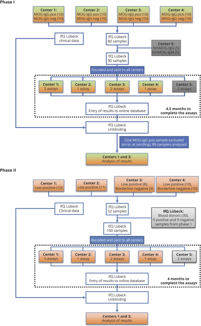
Methods: The following samples were analyzed: MOG-IgG clearly positive sera (n = 39), MOG-IgG low positive sera (n = 39), borderline negative sera (n = 13), clearly negative sera (n = 40), and healthy blood donors (n = 30). As technical controls, 18 replicates (9 MOG-IgG positive and 9 negative) were included. All samples and controls were recoded, aliquoted, and distributed to the 5 testing centers, which performed the following antibody assays: 5 live and 1 fixed immunofluorescence cell-based assays (CBA-IF, 5 MOG-IgG, and 1 MOG-IgM), 3 live flow cytometry cell-based assays (CBA-FACS, all MOG-IgG), and 2 ELISAs (both MOG-IgG).

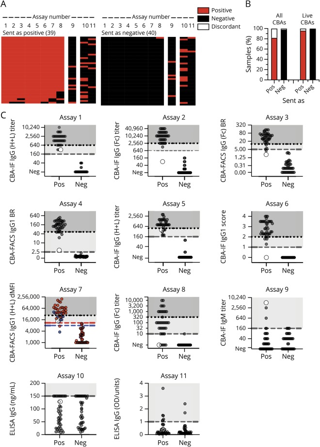
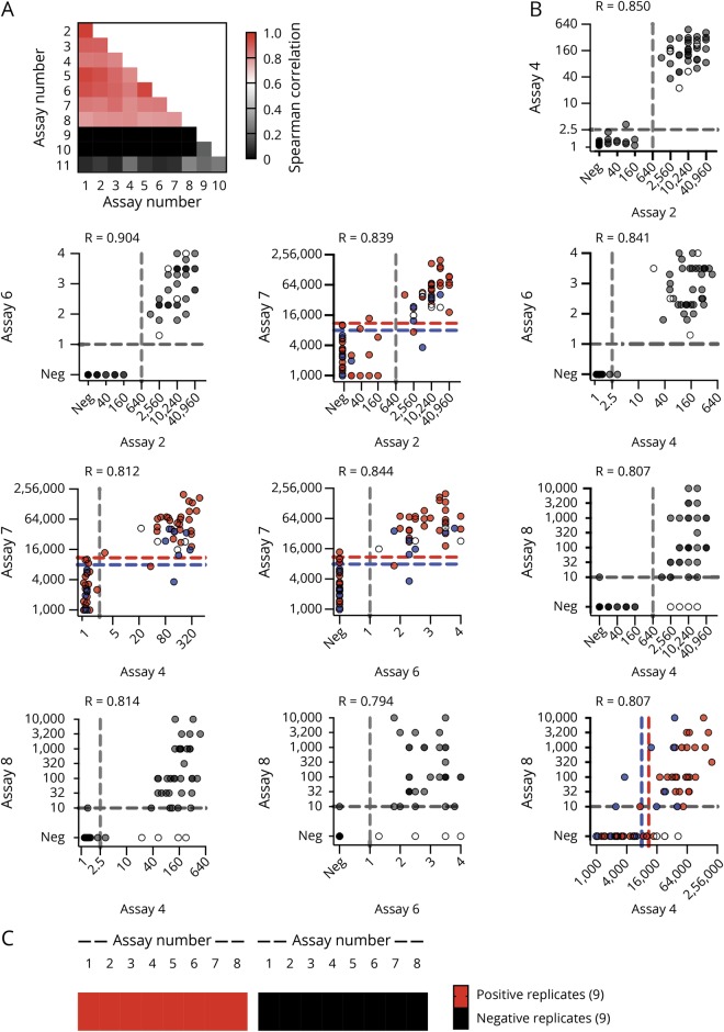
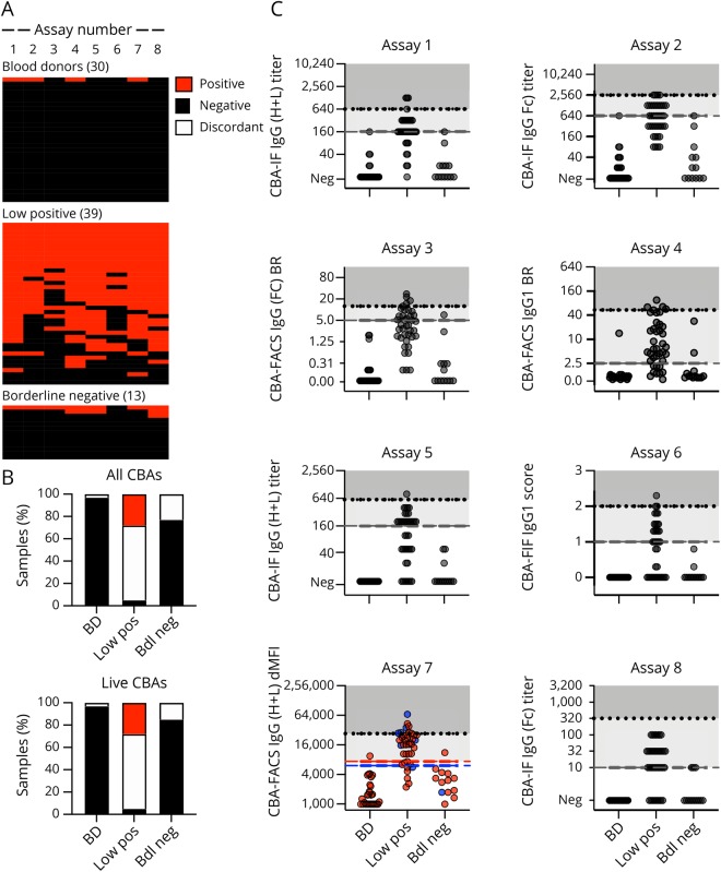
Results: We found excellent agreement (96%) between the live CBAs for MOG-IgG for samples previously identified as clearly positive or negative from 4 different national testing centers. The agreement was lower with fixed CBA-IF (90%), and the ELISA showed no concordance with CBAs for detection of human MOG-IgG. All CBAs showed excellent interassay reproducibility. The agreement of MOG-IgG CBAs for borderline negative (77%) and particularly low positive (33%) samples was less good. Finally, most samples from healthy blood donors (97%) were negative for MOG-IgG in all CBAs.

Conclusions: Live MOG-IgG CBAs showed excellent agreement for high positive and negative samples at 3 international testing centers. Low positive samples were more frequently discordant than in a similar comparison of aquaporin-4 antibody assays. Further research is needed to improve international standardization for clinical care.


Figures

Figure 1
Figure 1. Flowchart showing phases I and II of this study
Center 1 (Innsbruck) performed 5 assays (live CBA-IF MOG-IgG (H + L), live CBA-IF MOG-IgG(Fc), live CBA-FACS MOG-IgG(Fc), live CBA-IF MOG-IgM, and ELISA MOG-IgG); center 2 (Mayo Clinic) performed 1 assay (live CBA-FACS MOG-IgG1); center 3 (Oxford) performed 2 assays (live CBA-IF MOG-IgG (H + L) and live CBA-IF MOG-IgG1); center 4 (Sydney) performed 1 assay (live CBA-FACS MOG-IgG (H + L)), which was repeated twice; center 5 (Euroimmun) performed 2 assays (fixed CBA-IF MOG-IgG(Fc) and ELISA MOG-IgG(Fc)). CBA = cell-based assay; FACS = fluorescence-activated cell sorting; IF = immunofluorescence; IfQ = Institute for Quality Assurance; Ig = immunoglobulin; MOG = myelin oligodendrocyte glycoprotein.
Figure 2
Figure 2. Agreement of MOG-Ab assays on clear positive and negative samples
(A) Heatmap of the qualitative results for samples sent as clearly positive (n = 39) or negative (n = 40). Each column is an individual assay (1–8 MOG-IgG CBAs, 9 MOG-IgM CBAs, and 10–11 MOG-IgG ELISAs), and each row is an individual serum sample. Results are based on qualitative results; negative samples are black, and positive samples are red. The samples are shown according to their serostatus sent by the individual centers. (B) Agreement of MOG-IgG CBAs according to the samples sent (pos = positive, neg = negative). Results (in % of all samples) are grouped according to their agreement in all 8 CBAs or in the 7 live CBAs (red: positive in all CBAs, black: negative in all CBAs, white: discordant). (C) Quantitative results for all assays. The cutoff values for all assays except assay Nr. 7 are indicated by the dashed gray lines. For assay Nr. 7, cutoff levels for pediatric samples (blue dots) are indicated by the blue dashed line, and cutoff levels for adult samples (red dots) are indicated by the red dashed line. The quantitative range of each assay result for its probability to be seropositive in all live CBAs is indicated by the dotted line and shaded in darker gray (100% probability), whereas the range of discordant samples is shaded in light gray. A single sample with high IgM titer 1:5120 and low positive in the IgG (H + L) and on IgG1 but not in another IgG1 and the IgG(Fc) assays is indicated by the larger white dot. BR = binding ratio; CBA = cell-based assay; dMFI = delta mean fluorescence intensity; Ig = immunoglobulin; MOG = myelin oligodendrocyte glycoprotein.
Figure 3
Figure 3. Correlation of MOG-IgG assays and reproducibility of assay results
(A) Heatmap of Spearman correlation coefficients for all correlations. (B) Correlation of illustrative live (Nr. 2, 4, 6, and 7) and fixed CBAs (Nr. 8): Nr. 2 (center 1): CBA-IF IgG(Fc) titer (1), Nr. 4 (center 2): CBA-FACS IgG1 binding ratio, Nr. 6 (center 3): CBA-IF IgG1 binding score, Nr. 7 (center 4): CBA-FACS IgG (H + L) delta mean fluorescence intensity, Nr, 8 (center 5): CBA-IF IgG(Fc) titer (1). The cutoff values for all assays except assay Nr. 7 are indicated by the dashed gray lines. For assay Nr. 7, cutoff levels for pediatric samples (blue dots) are indicated by the blue dashed line, and cutoff levels for adult samples (red dots) are indicated by the red dashed line. Samples that are positive in live CBAs but not the fixed CBA (assay Nr. 8) are indicated by the white dots. (C) Qualitative results for 9 positive and 9 negative samples from phase I retested in a blinded way by all assays in phase II. CBA = cell-based assay; IF = immunofluorescence; Ig = immunoglobulin; MOG = myelin oligodendrocyte glycoprotein.
Figure 4
Figure 4. Agreement of MOG-Ab assays on low positive and borderline negative samples and blood donors
Qualitative and quantitative results of all MOG-IgG CBAs for blood donors (BD, n = 30), low positive (n = 39), and borderline negative (n = 40) samples. (A) Qualitative results according to the serostatus sent. Each column is an individual MOG-IgG CBA, and each row is an individual serum sample. Negative samples are black, and positive samples are red. The samples are shown according to their serostatus sent by the individual centers. (B) Agreement of assay results for IgG CBAs (assays 1–8). Sample are grouped according to their agreement in all 8 CBAs or in the 7 live CBAs (red: positive in all CBAs, black: negative in all CBAs, white: discordant). (C) Quantitative results for the 8 MOG-IgG CBA-IF and CBA-FACS assays. The cutoff values for all assays except assay Nr. 7 are indicated by the dashed gray lines. For assay Nr. 7, cutoff levels for pediatric samples (blue dots) are indicated by the blue dashed line, and cutoff levels for adult samples (red dots) are indicated by the red dashed line. The quantitative range of each assay result for its probability to be seropositive in all live CBAs is indicated by the dotted line and shaded in darker gray (100% probability), whereas the range of discordant samples is shaded in light gray. BD = blood donor; bdl neg = borderline negative values just below the individual cutoff levels; CBA = cell-based assay; FACS = fluorescence-activated cell sorting; IF = immunofluorescence; Ig = immunoglobulin; low pos = positive values just above the individual cutoff levels; MOG = myelin oligodendrocyte glycoprotein.

References

    1. Reindl M, Waters P. Myelin oligodendrocyte glycoprotein antibodies in neurological disease. Nat Rev Neurol 2019;15:89–102. - PubMed
    1. Mader S, Gredler V, Schanda K, et al. . Complement activating antibodies to myelin oligodendrocyte glycoprotein in neuromyelitis optica and related disorders. J Neuroinflammation 2011;8:184. - PMC - PubMed
    1. Waters P, Woodhall M, O'Connor KC, et al. . MOG cell-based assay detects non-MS patients with inflammatory neurologic disease. Neurol Neuroimmunol Neuroinflamm 2015;2:e89 doi: 10.1212/NXI.0000000000000089. - DOI - PMC - PubMed
    1. Waters PJ, Komorowski L, Woodhall M, et al. . A multicenter comparison of MOG-IgG cell-based assays. Neurology 2019;92:e1250–e1255. - PMC - PubMed
    1. Di Pauli F, Mader S, Rostasy K, et al. . Temporal dynamics of anti-MOG antibodies in CNS demyelinating diseases. Clin Immunol 2011;138:247–254. - PubMed

Publication types

MeSH terms