. 2020 Feb 5;12(529):eaaw6143.
doi: 10.1126/scitranslmed.aaw6143.

Osteoclast-mediated bone resorption is controlled by a compensatory network of secreted and membrane-tethered metalloproteinases

Affiliations

Osteoclast-mediated bone resorption is controlled by a compensatory network of secreted and membrane-tethered metalloproteinases

Lingxin Zhu et al. Sci Transl Med. .

Abstract

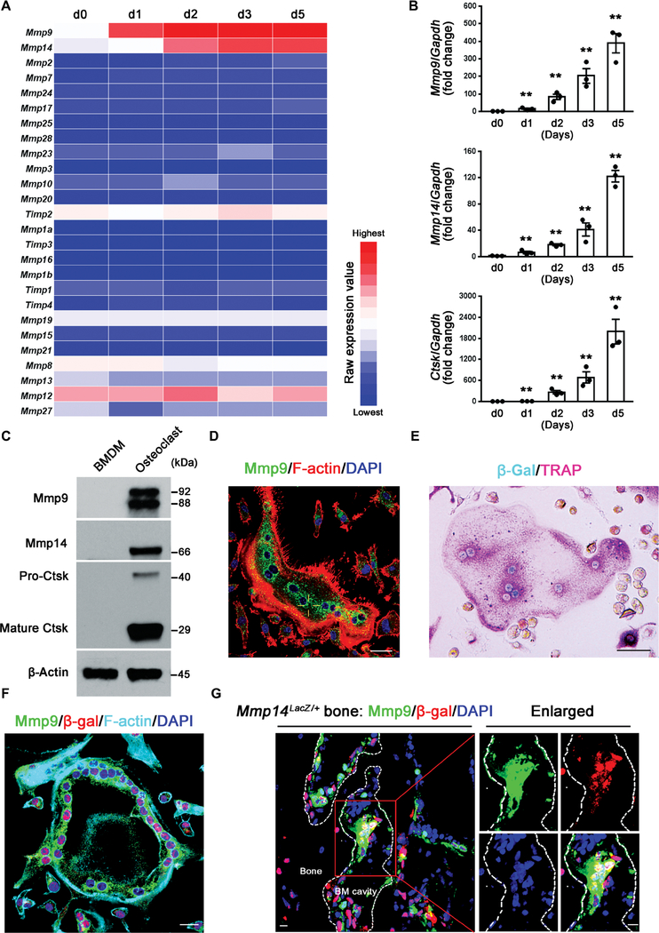
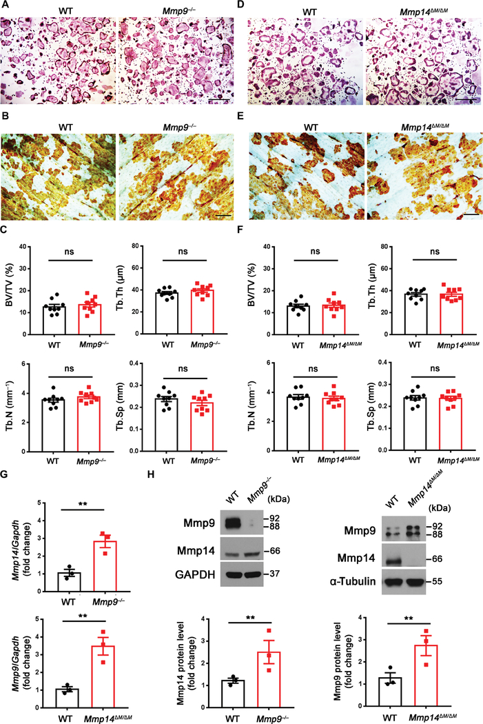
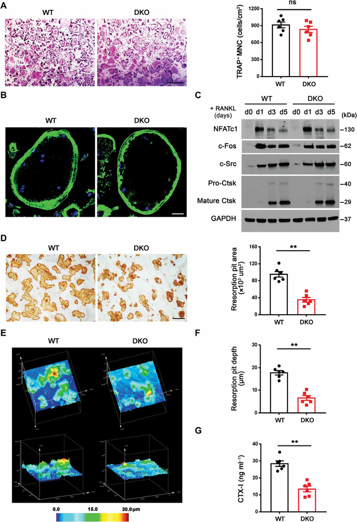
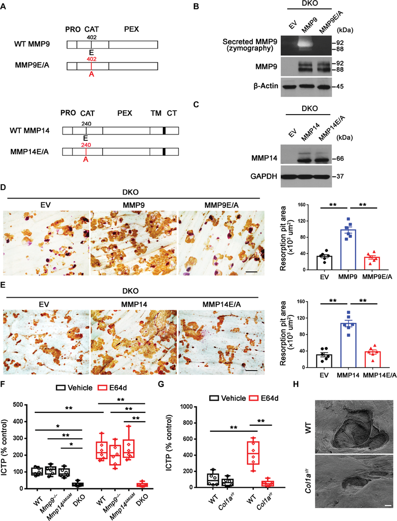
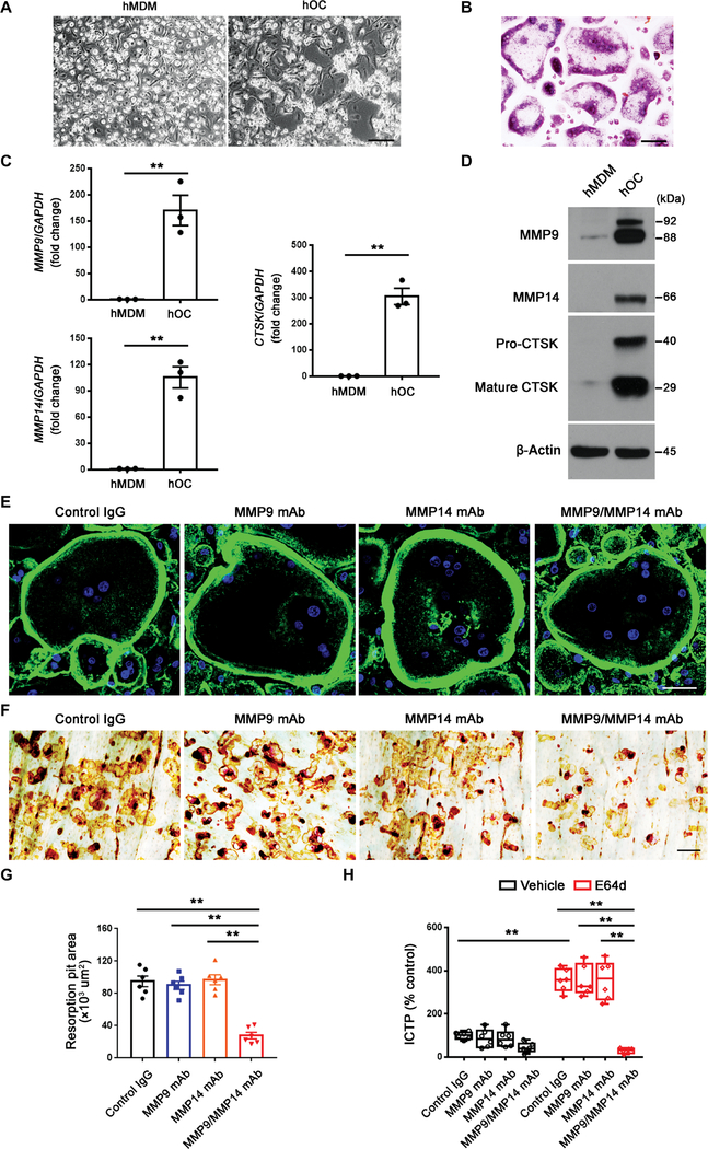
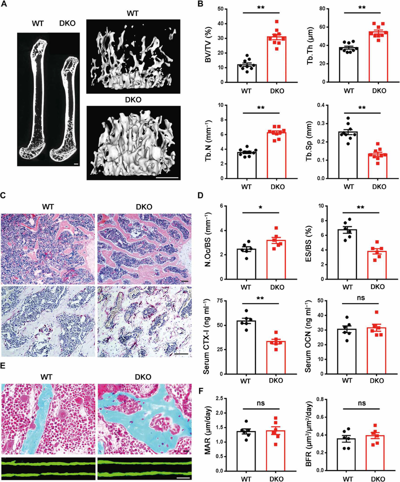
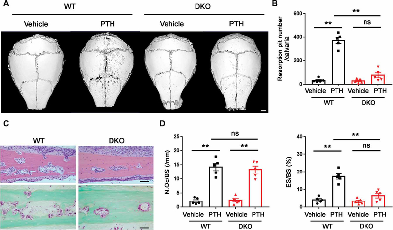
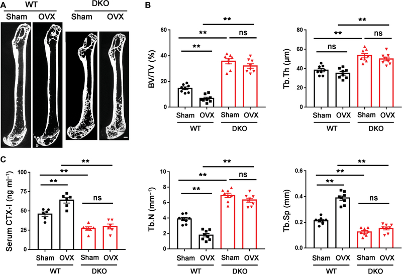
Osteoclasts actively remodel both the mineral and proteinaceous components of bone during normal growth and development as well as pathologic states ranging from osteoporosis to bone metastasis. The cysteine proteinase cathepsin K confers osteoclasts with potent type I collagenolytic activity; however, cathepsin K-null mice, as well as cathepsin K-mutant humans, continue to remodel bone and degrade collagen by as-yet-undefined effectors. Here, we identify a cathepsin K-independent collagenolytic system in osteoclasts that is composed of a functionally redundant network of the secreted matrix metalloproteinase MMP9 and the membrane-anchored matrix metalloproteinase MMP14. Unexpectedly, whereas deleting either of the proteinases individually leaves bone resorption intact, dual targeting of Mmp9 and Mmp14 inhibited the resorptive activity of mouse osteoclasts in vitro and in vivo and human osteoclasts in vitro. In vivo, Mmp9/Mmp14 conditional double-knockout mice exhibited marked increases in bone density and displayed a highly protected status against either parathyroid hormone- or ovariectomy-induced pathologic bone loss. Together, these studies characterize a collagenolytic system operative in mouse and human osteoclasts and identify the MMP9/MMP14 axis as a potential target for therapeutic interventions for bone-wasting disease states.


Conflict of interest statement

Competing interests: The authors declare that they have no competing interests.

Figures

Fig. 1.
Fig. 1.. Osteoclasts selectively express Mmp9 and Mmp14 in vitro and in vivo.
(A) Transcriptional profile of MMPs and TIMPs [tissue inhibitors of metalloproteinases; (16, 17)] during M-CSF/RANKL-induced differentiation of BMDMs to osteoclasts from days 0 to 5 (d0 to d5) (color bar, raw expression value). (B) Relative mRNA expression of Mmp9, Mmp14, and Ctsk during the differentiation from BMDMs to osteoclasts as a function of time in culture (n = 3). (C) Western blot of Mmp9, Mmp14, and Ctsk expression in BMDMs and mature osteoclasts. The Mmp9 doublet represents the glycosylated and nonglycosylated proforms of the proteinase (16, 17). (D) Mmp9 (green) and F-actin (red) immunofluorescence of wild-type BMDM-derived osteoclasts. Scale bar, 20 μm. DAPI, 4′,6-diamidino-2-phenylindole. (E) β-gal activity (cyan) and TRAP (pink) staining of osteoclasts differentiated from Mmp14LacZ/+ BMDMs. Scale bar, 50 μm. (F) Mmp9 (green), β-gal (red), and F-actin (cyan) immunofluorescence of osteoclasts differentiated from Mmp14LacZ/+ BMDMs. Scale bar, 20 μm. (G) Mmp9 (green) and β-gal (red) immunofluorescence of a femur section from a Mmp14LacZ/+ mouse. The MNCs associated with the surface of the bone marrow cavity (BM cavity) that are shown in the red box are further enlarged in the panels to the right. Scale bars, 10 μm. All results are representative of data generated in at least three independent experiments. **P < 0.01. Error bars are means ± SEM. Data were analyzed using one-way analysis of variance (ANOVA) with Bonferroni correction.
Fig. 2.
Fig. 2.. Mmp9−/− and myeloid-specific Mmp14 conditional knockout mice display normal osteoclast activity in vitro and in vivo.
(A) BMDMs were isolated from wild-type (WT) or Mmp9−/− mice, cultured on plastic substrata with M-CSF and RANKL for 5 days, and stained with TRAP. Scale bar, 500 μm. (B) Wild-type or Mmp9−/− BMDMs were cultured atop bone slices and induced into osteoclasts for 6 days. After cell removal, resorption pits were visualized by WGA-DAB staining. Scale bar, 100 μm. (C) Quantification of bone volume/tissue volume (BV/TV), trabecular thickness (Tb.Th), trabecular number (Tb.N), and trabecular separation (Tb.Sp) as determined by μCT of 5-month-old wild-type and Mmp9−/− male mice (n = 9). (D) BMDMs were isolated from wild-type or Mmp14ΔM/ΔM mice, cultured atop plastic substrata with M-CSF and RANKL for 5 days, and stained with TRAP. Scale bar, 500 μm. (E) Wild-type or Mmp14ΔM/ΔM BMDMs were cultured atop bovine bone slices and induced into osteoclasts for 6 days. Cells were removed and resorption pits visualized by WGA-DAB staining. Scale bar, 100 μm. (F) Quantification of BV/TV, Tb.Th, Tb.N, and Tb.Sp as determined by μCT of 5-month-old wild-type and Mmp14ΔM/ΔM male mice (n = 9). (G) Relative mRNA expression of Mmp14 or Mmp9 in osteoclasts differentiated from Mmp9−/− or Mmp14ΔM/ΔM BMDMs (n = 3). (H) Western blot and quantification of Mmp9 and Mmp14 expression in osteoclasts differentiated from Mmp9−/− or Mmp14ΔM/ΔM BMDMs (n = 3). All results are representative of data generated in at least three independent experiments. ns, not significant. **P < 0.01. Error bars are means ± SEM. All data were analyzed using unpaired Student’s t test. GAPDH, glyceraldehyde-3-phosphate dehydrogenase.
Fig. 3.
Fig. 3.. Impaired bone-resorbing activity of DKO osteoclasts in vitro.
(A) BMDMs were isolated from wild-type or DKO mice, cultured on plastic substrata with M-CSF and RANKL for 5 days, and stained with TRAP, and the number of TRAP+ MNCs was determined (n = 6). Scale bar, 500 μm. (B) Phalloidin (green) staining of F-actin in wild-type or DKO osteoclasts cultured on glass. Scale bar, 20 μm. (C) NFATc1, c-Fos, c-Src, and Ctsk expression as assessed by Western blot in BMDMs during osteoclast differentiation. (D) After a 6-day culture period, wild-type or DKO osteoclasts were removed from bone slices, resorption pits were visualized by WGA-DAB staining, and resorption pit area was quantified (n = 6). Scale bar, 100 μm. (E and F) Resorption pits were three-dimensionally reconstructed by reflective confocal laser scanning microscope (E) in tandem with (F) quantification of resorption pit depth (n = 6). Color bar, pit depth. (G) Supernatant CTX-I was determined using ELISA (n = 6). All results are representative of data generated in at least three independent experiments. **P < 0.01. Error bars are means ± SEM. All data were analyzed using unpaired Student’s t test.
Fig. 4.
Fig. 4.. Mmp9/Mmp14 catalytic activity codetermines osteoclast-mediated bone resorption by proteolyzing bone type I collagen.
(A) A schematic diagram of full-length human MMP9 and MMP14 and their respective catalytically inactive mutants depicting the pro (PRO), catalytic (CAT), hemopexin (HPX), transmembrane (TM) domains, and cytosolic tail (CT). (B) DKO BMDMs were transduced with lentiviral vectors expressing full-length MMP9, an MMP9E/A mutant, or an empty control (EV) and differentiated into osteoclasts. Supernatant and cell lysate were collected for gelatin zymography and MMP9 immunoblots, respectively. (C) DKO BMDMs were transduced with lentiviral vectors expressing full-length MMP14, MMP14E/ A, or an empty control (EV) and differentiated into osteoclasts. Cell lysates were collected for MMP14 immunoblotting. (D) MMP9 or MMP9E/A-transduced BMDMs were induced into osteoclasts and cultured atop bone slices for 3 days. Osteoclasts were removed, resorption pits were visualized by WGA-DAB staining, and resorption pit area was quantified (n = 6). Scale bar, 100 μm. (E) MMP14 or MMP14E/A-transduced BMDMs were induced into osteoclasts and cultured atop bone slices as described in (D). Resorption pits were visualized by WGA-DAB staining, and resorption pit area was quantified (n = 6). Scale bar, 100 μm. (F) Osteoclasts differentiated from wild-type, Mmp9−/−, Mmp14ΔM/ΔM, or DKO BMDMs were cultured atop decalcified cortical bone slices with or without E64d (20 μM) for 3 days, and supernatants were collected for ICTP ELISA (n = 6). (G) Wild-type osteoclasts were cultured atop normal or Col1ar/r mutant bone with or without E64d for 3 days, and supernatants were collected for ICTP ELISA (n = 6). (H) Resorption pits generated on normal or Col1ar/r bone by wild-type osteoclasts ex vivo were imaged by scanning electron microscopy. Scale bar, 10 μm. All results are representative of data generated in at least three independent experiments. *P < 0.05, **P < 0.01. Error bars are means ± SEM. Data were analyzed using one-way ANOVA (D and E) or two-way ANOVA (F and G) with Bonferroni correction.
Fig. 5.
Fig. 5.. Dual MMP9/MMP14 activity regulates human osteoclast-mediated bone resorption.
(A) Phase contrast images of human monocyte-derived macrophages (hMDMs) and human osteoclasts (hOCs) differentiated from human CD14+ monocytes. Scale bar, 200 μm. (B) TRAP staining of human osteoclasts. Scale bar, 100 μm. (C) Relative mRNA expression of MMP9, MMP14, and CTSK in hMDM and mature hOC (n = 3). (D) MMP9, MMP14, and CTSK protein expression as assessed by Western blot was determined in hMDMs and mature hOCs. (E) Human osteoclasts were cultured on glass in the presence or absence of an MMP9 function-blocking mAb and MMP14 function-blocking antibody (DX-2400), and the cells were stained with fluorescein isothiocyanate–phalloidin. Scale bar, 50 μm. IgG, immunoglobulin G. (F and G) Human osteoclasts were cultured atop bone slices for 6 days in the presence or absence of either the MMP9 function-blocking mAb or DX-2400. Osteoclasts were removed from the bone slices, resorption pits were visualized by (F) WGA-DAB staining, and (G) resorption pit area was quantified (n = 6). Scale bar, 100 μm. (H) Human osteoclasts were cultured atop cortical bone slices for 6 days in the presence or absence of the MMP9 or MMP14 blocking antibodies with or without E64d, and the supernatants were collected for ICTP ELISA (n = 6). All results are representative of data generated in at least three independent experiments. **P < 0.01. Error bars are means ± SEM. Data were analyzed using unpaired Student’s t test (C), one-way ANOVA (G), or two-way ANOVA (H) with Bonferroni correction.
Fig. 6.
Fig. 6.. DKO mice exhibit an osteopetrotic phenotype with decreased osteoclast activity.
(A) Representative μCT of sagittal sections of femurs with three-dimensional (3D) reconstruction of the distal femur trabeculae of 5-month-old wild-type and DKO male mice is shown. Scale bars, 500 μm. (B) Quantification of BV/TV, Tb.Th, Tb.N, and Tb.Sp as determined by μCT in 5-month-old wild-type and DKO male mice (n = 9). (C) Hematoxylin and eosin (H&E) and TRAP staining of the distal femurs of 5-month-old male wild-type and DKO mice. Scale bars, 100 μm. (D) Quantification of osteoclast number per bone surface (N.Oc/BS) and eroded surface per bone surface (ES/BS) and serum CTX-I and OCN in 5-month-old wild-type and DKO male mice are shown (n = 6). (E) Golden’s trichrome staining of the distal femurs of 5-month-old male wild-type and DKO mice was performed along with double calcein bone labeling and confocal imaging to assess bone formation. Scale bars, 10 μm. (F) Quantification of MAR and BFR as assessed in 5-month-old male wild-type and DKO mice (n = 6). *P < 0.05, **P < 0.01. Error bars are means ± SEM. All data were analyzed using unpaired Student’s t test.
Fig. 7.
Fig. 7.. PTH-induced calvarial bone erosion is alleviated in DKO mice.
(A) Representative μCT of 3D reconstructed images of calvaria from 3-month-old male wild-type or DKO mice injected with PTH or vehicle control. Scale bar, 1000 μm. (B) Quantification of resorption pit number per calvaria of 3-month-old male wild-type or DKO mice injected with PTH or vehicle control (n = 5). (C) H&E and TRAP staining of calvaria from 3-month-old male wild-type or DKO mice injected with PTH. Scale bars, 50 μm. (D) Quantification of N.Oc/BS and ES/BS of calvaria from 3-month-old male wild-type or DKO mice injected with PTH or vehicle control (n = 5). **P < 0.01. Error bars are means ± SEM. All data were analyzed using two-way ANOVA with Bonferroni correction.
Fig. 8.
Fig. 8.. DKO mice are protected from OVX-induced osteoporosis.
(A) Representative μCT sagittal sections of femur recovered from 5-month-old female wild-type or DKO mice that underwent either sham or OVX surgery at 3 months of age. Scale bar, 500 μm. (B) BV/TV, Tb.Th, Tb.N, and Tb.Sp were determined by μCT of femurs from sham or OVX surgery mice (n = 8). (C) Serum CTX-I of sham or OVX surgery mice determined using ELISA (n = 8). **P < 0.01. Error bars are means ± SEM. All data were analyzed using two-way ANOVA with Bonferroni correction.

References

    1. Zaidi M, Skeletal remodeling in health and disease. Nat. Med 13, 791–801 (2007). - PubMed
    1. Teitelbaum SL, Ross FP, Genetic regulation of osteoclast development and function. Nat. Rev. Genet 4, 638–649 (2003). - PubMed
    1. Boyle WJ, Simonet WS, Lacey DL, Osteoclast differentiation and activation. Nature 423, 337–342 (2003). - PubMed
    1. Takayanagi H, Osteoimmunology: Shared mechanisms and crosstalk between the immune and bone systems. Nat. Rev. Immunol 7, 292–304 (2007). - PubMed
    1. Rachner TD, Khosla S, Hofbauer LC, Osteoporosis: Now and the future. Lancet 377, 1276–1287 (2011). - PMC - PubMed

Publication types