. 2020 Feb 6;12(1):5.
doi: 10.1038/s41368-019-0072-z.

Temporomandibular joint damage in K/BxN arthritic mice

Affiliations

Temporomandibular joint damage in K/BxN arthritic mice

Sabine Kuchler-Bopp et al. Int J Oral Sci. .

Abstract

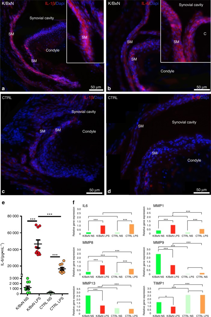
Rheumatoid arthritis (RA) is an autoimmune disease affecting 1% of the world population and is characterized by chronic inflammation of the joints sometimes accompanied by extra-articular manifestations. K/BxN mice, originally described in 1996 as a model of polyarthritis, exhibit knee joint alterations. The aim of this study was to describe temporomandibular joint (TMJ) inflammation and damage in these mice. We used relevant imaging modalities, such as micro-magnetic resonance imaging (μMRI) and micro-computed tomography (μCT), as well as histology and immunofluorescence techniques to detect TMJ alterations in this mouse model. Histology and immunofluorescence for Col-I, Col-II, and aggrecan showed cartilage damage in the TMJ of K/BxN animals, which was also evidenced by μCT but was less pronounced than that seen in the knee joints. μMRI observations suggested an increased volume of the upper articular cavity, an indicator of an inflammatory process. Fibroblast-like synoviocytes (FLSs) isolated from the TMJ of K/BxN mice secreted inflammatory cytokines (IL-6 and IL-1β) and expressed degradative mediators such as matrix metalloproteinases (MMPs). K/BxN mice represent an attractive model for describing and investigating spontaneous damage to the TMJ, a painful disorder in humans with an etiology that is still poorly understood.


Conflict of interest statement

The authors declare no competing interests.

Figures

Fig. 1
Fig. 1
Histological and morphological analyses of K/BxN and control TMJs. Histology of the temporomandibular joint (TMJ) of an 8-month-old K/BxN mouse (ac) and a control mouse (df) after alcian blue staining (a, b, d, e) and Safranin O staining (c, f). g, j Micro-computed tomography (μCT) sections of the TMJ of an 8-month-old K/BxN mouse (g) and a control mouse (j). (h, i, k, l) 3D reconstructions of the condyles of an 8-month-old K/BxN mouse (h, i) and a control mouse (k, l). B, bone; D, disc; FC, fibrocartilage; FL, fibrous layer; PL, proliferative layer
Fig. 2
Fig. 2
Immunolocalization of specific cartilage and bone proteins in K/BxN and control TMJs. Immunofluorescence for Collagen I (a, b, e, f), Collagen II (c, g), Aggrecan (d, h), BSPII (i, j), Osteopontin and CD31 (k, l), and RUNX2 (mo) in cryostat sections of a K/BxN TMJ (ad, i, k, m, n) and a control TMJ (eh, j, l, o)
Fig. 3
Fig. 3
Comparison of synovial fluid volume of K/BxN and control TMJs. Magnetic resonance imaging (MRI) and 3D reconstructions of the TMJ of a K/BxN mouse (a) and a control mouse (b). c Volume measurement of the upper articular cavity of seven K/BxN mice and three control mice. The orange square represents the K/BxN mouse shown in a, and the green circle represents the control mouse shown in b
Fig. 4
Fig. 4
Characterization of fibroblast-like synoviocytes (FLS) from K/BxN and control TMJs. Characterization of fibroblast-like synoviocytes (FLSs) from K/BxN TMJs (a, c, e, g) and control TMJs (b, d, f, h) by using hematoxylin–eosin staining (a, b) and immunofluorescence for vimentin (c, d), phalloidin and TEM1 (e, f), and CD90 and fibronectin (g, h). Nuclei were stained with 4′,6-diamidino-2-phenylindole (DAPI)
Fig. 5
Fig. 5
Expression of inflammatory cytokines and matrix metalloproteinases in K/BxN and control FLS. Immunofluorescence for IL-1β (a, c) and IL-6 (b, d) in sagittal sections of the TMJ of K/BxN mice (a, b) and control mice (c, d). (e) Quantification of IL-6 (pg·mL1) in the culture medium of unstimulated (NS) K/BxN and control TMJ FLSs and those stimulated with LPS for 24 h. (f) RT-qPCR experiments to monitor Il-6, Mmp1, 8, 9, and 13 and Timp1 expression in K/BxN and control FLSs treated with or without LPS for 24 h. The values are the means ± SEMs; *P < 0.01, ***P < 0.000 1

References

    1. Perschbacher, S. Oral Radiology: Principles and Interpretation (eds Pharoah, MJ & White, SC) (Mosby-Year Book Inc., 2014).
    1. van Bellinghen X, et al. Temporomandibular joint regenerative medicine. Int. J. Mol. Sci. 2018;19:E446. doi: 10.3390/ijms19020446. - DOI - PMC - PubMed
    1. Bartok B, Firestein GS. Fibroblast-like synoviocytes: key effector cells in rheumatoid arthritis. Immunol. Rev. 2010;233:233–55. doi: 10.1111/j.0105-2896.2009.00859.x. - DOI - PMC - PubMed
    1. Te Veldhuis EC, Te Veldhuis AH, Koudstaal MJ. Treatment management of children with juvenile idiopathic arthritis with temporomandibular joint involvement: a systematic review. Oral Surg. Oral Med. Oral Pathol. Oral Radiol. 2014;117:581–589.e582. doi: 10.1016/j.oooo.2014.01.226. - DOI - PubMed
    1. Keller H, et al. Is early TMJ involvement in children with juvenile idiopathic arthritis clinically detectable? Clinical examination of the TMJ in comparison with contrast enhanced MRI in patients with juvenile idiopathic arthritis. Pediatr. Rheumatol. Online J. 2015;13:56. doi: 10.1186/s12969-015-0056-2. - DOI - PMC - PubMed

Publication types

MeSH terms

Substances