. 2020 Feb 5;11(1):709.
doi: 10.1038/s41467-020-14610-8.

Genetic influence is linked to cortical morphology in category-selective areas of visual cortex

Affiliations

Genetic influence is linked to cortical morphology in category-selective areas of visual cortex

Nooshin Abbasi et al. Nat Commun. .

Abstract

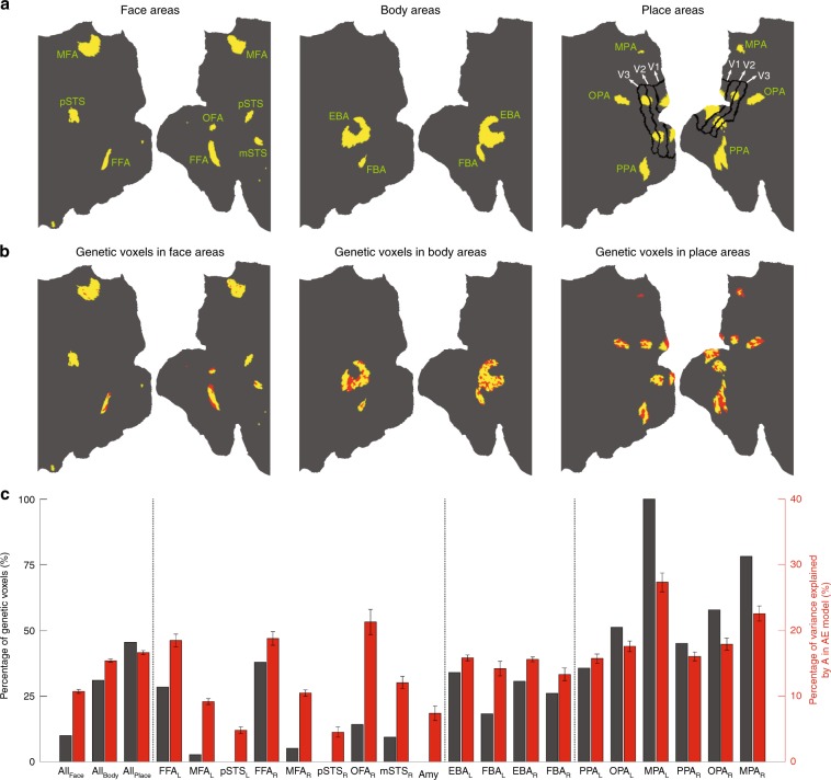
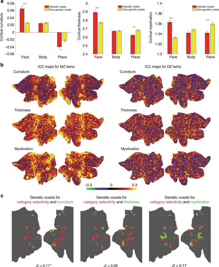
Human visual cortex contains discrete areas that respond selectively to specific object categories such as faces, bodies, and places. A long-standing question is whether these areas are shaped by genetic or environmental factors. To address this question, here we analyzed functional MRI data from an unprecedented number (n = 424) of monozygotic (MZ) and dizygotic (DZ) twins. Category-selective maps were more identical in MZ than DZ twins. Within each category-selective area, distinct subregions showed significant genetic influence. Structural MRI analysis revealed that the 'genetic voxels' were predominantly located in regions with higher cortical curvature (gyral crowns in face areas and sulcal fundi in place areas). Moreover, we found that cortex was thicker and more myelinated in genetic voxels of face areas, while it was thinner and less myelinated in genetic voxels of place areas. This double dissociation suggests a differential development of face and place areas in cerebral cortex.


Conflict of interest statement

The authors declare no competing interests.

Figures

Fig. 1
Fig. 1. Correlation between category-selective activations in MZ and DZ twins.
a For each category, ICC maps in MZ and DZ twins are displayed on a 2D flat patch of left and right hemispheres (left and right maps in each subpanel). b Left panel: A schematic diagram depicting the procedure for computing Pearson’s r correlation. In each twin pair, the activation patterns (z maps) of one twin and the co-twin were correlated. In the activation vector, data from two hemispheres were concatenated. Right panels: Distribution of correlation coefficient values for all MZ and DZ twin pairs in face-, body-, and place-selective maps. c Mean correlation was calculated separately for each cortical lobe (O occipital, P parietal, T temporal, F frontal). Lobar parcellation was based on PALS-B12 atlas of human cerebral cortex (http://brainvis.wustl.edu/wiki/index.php/Caret:Atlases). Error bars represent one standard error of the mean, here and in the other figures. *p < 0.05, **p < 0.005, ***p < 0.0005; Bonferroni-corrected pooled t test for three comparisons in panel (b) and 12 comparisons in panel (c).
Fig. 2
Fig. 2. Genetic analysis in category-selective areas.
a Face, body, and place areas are displayed on a 2D flat patch of left and right hemispheres (left and right maps in each subpanel; see text for the full name of areas). The face-selective area in the posterior cingulate cortex was named MFA (medial face area) here. The areal borders of V1/V2/V3 were estimated based on a probabilistic map of retinotopic areas in a group of 12 subjects. Place-related activations in V1/V2/V3, which avoided the V1/V2 border, were localized in/near regions representing the mid-peripheral visual field. For each category, there were 913 category-selective voxels. b Voxels with a significant genetic effect (FDR-adjusted p < 0.05 based on 913 comparisons) are marked red in the face, body, and place maps. For these voxels, the average A value was ~25%. c Region-of-interest analysis demonstrating descriptive statistics for the genetic effect in category-selective networks (all areas together) and in individual category-selective areas. For this analysis, EBA and FBA in the right hemisphere, which were conjoined in the map, were separated at their junction. The effect of A varied significantly (p < 0.0005; one-way ANOVA) across networks, across face areas, and across place areas.
Fig. 3
Fig. 3. Relationship between genetic effect in category-selective areas and structural/morphological properties of cortical gray matter.
a In face, body, and place areas, mean cortical curvature, mean cortical thickness, and mean cortical myelination were computed across genetic and nongenetic voxels. Place-selective voxels in V1/V2/V3 were excluded in this analysis. Regions with positive curvature corresponded to cortical gyri, and regions with negative curvature corresponded to cortical sulci. *p < 0.05, **p < 0.005, ***p < 0.0005; Bonferroni-corrected pooled t test for three comparisons between genetic and nongenetic voxels in each plot. b ICC maps for curvature, thickness, and myelination in MZ and DZ twins, displayed on a 2D flat patch of left and right hemispheres. c Using the same procedure for estimating the heritability of category selectivity (see Methods), the heritability of curvature, thickness, and myelination was estimated in each category-selective voxel. The maps show voxels with genetic influence on category selectivity (red), cortical structure (green), or both (yellow). The degree of overlap between the binary genetic influence maps was quantified using the Jaccard Index (JI). JI was low in all cases. In the case of curvature, it was slightly and significantly above the expected JI under independence (p < 0.05; bootstrap test). In the case of thickness and myelination, the difference was not significant (p > 0.05; bootstrap test).

References

    1. Grill-Spector K, Weiner KS. The functional architecture of the ventral temporal cortex and its role in categorization. Nat. Rev. Neurosci. 2014;15:536–548. doi: 10.1038/nrn3747. - DOI - PMC - PubMed
    1. Kanwisher N. Functional specificity in the human brain: a window into the functional architecture of the mind. Proc. Natl Acad. Sci. USA. 2010;107:11163–11170. doi: 10.1073/pnas.1005062107. - DOI - PMC - PubMed
    1. Deen B, et al. Organization of high-level visual cortex in human infants. Nat. Commun. 2017;8:13995. doi: 10.1038/ncomms13995. - DOI - PMC - PubMed
    1. Livingstone MS, et al. Development of the macaque face-patch system. Nat. Commun. 2017;8:14897. doi: 10.1038/ncomms14897. - DOI - PMC - PubMed
    1. Arcaro MJ, Schade PF, Vincent JL, Ponce CR, Livingstone MS. Seeing faces is necessary for face-domain formation. Nat. Neurosci. 2017;20:1404–1412. doi: 10.1038/nn.4635. - DOI - PMC - PubMed

Publication types