

Cellular uptake of magnetic nanoparticles imaged and quantified by magnetic particle imaging
- PMID: 32024926
- PMCID: PMC7002802
- DOI: 10.1038/s41598-020-58853-3
Cellular uptake of magnetic nanoparticles imaged and quantified by magnetic particle imaging
Abstract
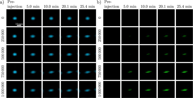
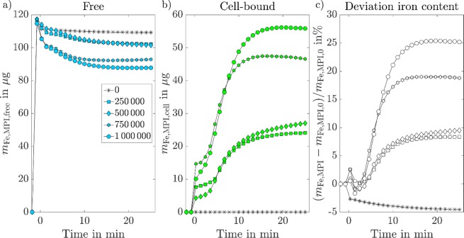
Magnetic particle imaging (MPI) is a non-invasive, non-ionizing imaging technique for the visualization and quantification of magnetic nanoparticles (MNPs). The technique is especially suitable for cell imaging as it offers zero background contribution from the surrounding tissue, high sensitivity, and good spatial and temporal resolutions. Previous studies have demonstrated that the dynamic magnetic behaviour of MNPs changes during cellular binding and internalization. In this study, we demonstrate how this information is encoded in the MPI imaging signal. Through MPI imaging we are able to discriminate between free and cell-bound MNPs in reconstructed images. This technique was used to image and quantify the changes that occur in-vitro when free MNPs come into contact with cells and undergo cellular-uptake over time. The quantitative MPI results were verified by colorimetric measurements of the iron content. The results showed a mean relative difference between the MPI results and the reference method of 23.8% for the quantification of cell-bound MNPs. With this technique, the uptake of MNPs in cells can be imaged and quantified directly from the first MNP cell contact, providing information on the dynamics of cellular uptake.
Conflict of interest statement
The authors declare no competing interests.
Figures

References
-
- Wells James, Kazakova Olga, Posth Oliver, Steinhoff Uwe, Petronis Sarunas, Bogart Lara K, Southern Paul, Pankhurst Quentin, Johansson Christer. Standardisation of magnetic nanoparticles in liquid suspension. Journal of Physics D: Applied Physics. 2017;50(38):383003. doi: 10.1088/1361-6463/aa7fa5. - DOI
-
- Rahmer J, Antonelli A, Sfara C, Tiemann B, Gleich B, Magnani M, Weizenecker J, Borgert J. Nanoparticle encapsulation in red blood cells enables blood-pool magnetic particle imaging hours after injection. Physics in Medicine and Biology. 2013;58(12):3965–3977. doi: 10.1088/0031-9155/58/12/3965. - DOI - PubMed
-
- Sedlacik Jan, Frölich Andreas, Spallek Johanna, Forkert Nils D., Faizy Tobias D., Werner Franziska, Knopp Tobias, Krause Dieter, Fiehler Jens, Buhk Jan-Hendrik. Magnetic Particle Imaging for High Temporal Resolution Assessment of Aneurysm Hemodynamics. PLOS ONE. 2016;11(8):e0160097. doi: 10.1371/journal.pone.0160097. - DOI - PMC - PubMed
Publication types
MeSH terms
Substances
LinkOut - more resources
Full Text Sources
