. 2020 Feb 5;10(1):1894.
doi: 10.1038/s41598-020-58726-9.

Identification and characterization of novel filament-forming proteins in cyanobacteria

Affiliations

Identification and characterization of novel filament-forming proteins in cyanobacteria

Benjamin L Springstein et al. Sci Rep. .

Abstract

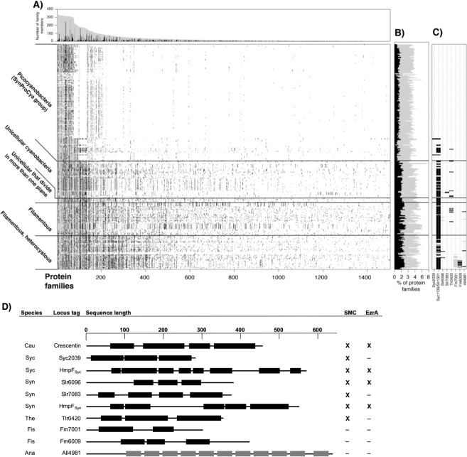
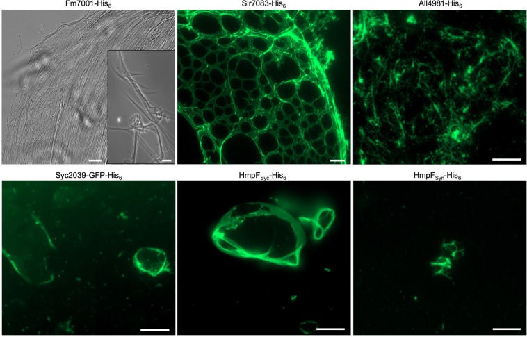
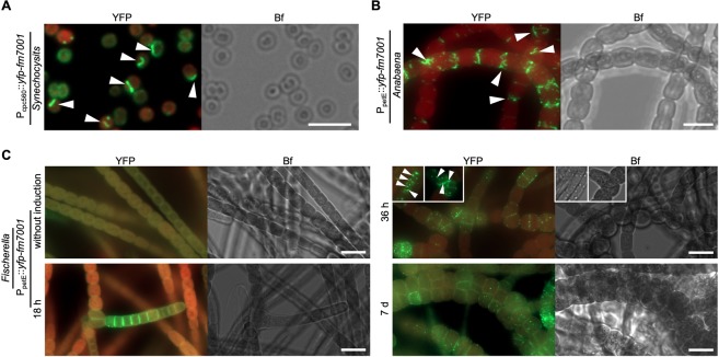
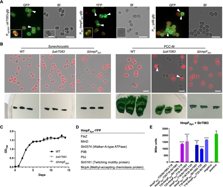
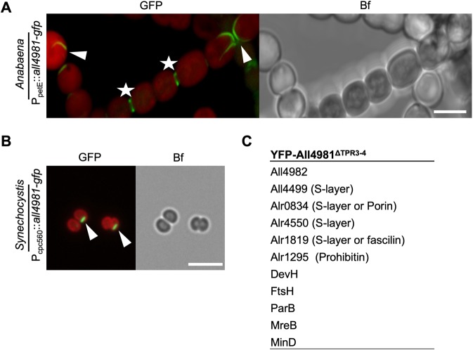
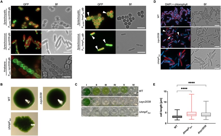
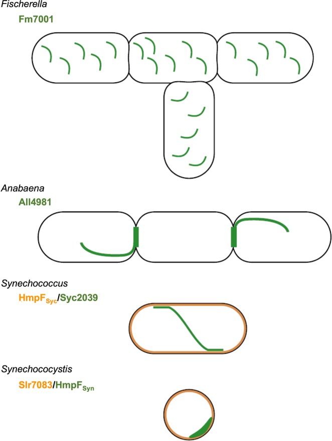
Filament-forming proteins in bacteria function in stabilization and localization of proteinaceous complexes and replicons; hence they are instrumental for myriad cellular processes such as cell division and growth. Here we present two novel filament-forming proteins in cyanobacteria. Surveying cyanobacterial genomes for coiled-coil-rich proteins (CCRPs) that are predicted as putative filament-forming proteins, we observed a higher proportion of CCRPs in filamentous cyanobacteria in comparison to unicellular cyanobacteria. Using our predictions, we identified nine protein families with putative intermediate filament (IF) properties. Polymerization assays revealed four proteins that formed polymers in vitro and three proteins that formed polymers in vivo. Fm7001 from Fischerella muscicola PCC 7414 polymerized in vitro and formed filaments in vivo in several organisms. Additionally, we identified a tetratricopeptide repeat protein - All4981 - in Anabaena sp. PCC 7120 that polymerized into filaments in vitro and in vivo. All4981 interacts with known cytoskeletal proteins and is indispensable for Anabaena viability. Although it did not form filaments in vitro, Syc2039 from Synechococcus elongatus PCC 7942 assembled into filaments in vivo and a Δsyc2039 mutant was characterized by an impaired cytokinesis. Our results expand the repertoire of known prokaryotic filament-forming CCRPs and demonstrate that cyanobacterial CCRPs are involved in cell morphology, motility, cytokinesis and colony integrity.


Conflict of interest statement

The authors declare no competing interests.

Figures

Figure 1
Figure 1
Distribution of CCRP protein families within cyanobacteria. (A) Lines in the presence/absence matrix designate cyanobacterial genomes; each column shows a protein family. Gray dots designate any homologous protein in the same protein family and black dots represent CCRP members. Protein families are sorted according to the number of members. Protein family size and the number of CCRP members are presented in a bar graph above. (B) The proportion of protein families containing CCRPs (gray) and CCRP proteins (black) in each genome. (C) Presence/absence pattern of CCRP candidate protein families. Only protein families with at least three members predicted to be CCRPs are shown. (D) Domain prediction of CCRP candidates. Scale on top is given in amino acid residues. Amino acid sequences in coiled-coil conformation are depicted by black bars with non-coiled-coil sequences represented by black lines. Tetratricopeptide repeats (TPR), also predicted by the COILS algorithm, are shown as grey bars. Proteins are given as cyanobase locus tags. Fm7001 and Fm6009 correspond to NCBI accession numbers WP_016868005.1 and WP_020476706, respectively. Abbreviations: Cau: C. crescentus; Syc: Synechococcus, Syn: Synechocystis; Ana: Anabaena; The: Thermosynechococcus elongatus BP-1; Fis: Fischerella. Cyanobacterial CCRPs had conserved domains present in prokaryotic IF-like CCRPs and eukaryotic IF proteins (Supplementary Table 1). Presence of a structural maintenance of chromosomes (SMC) domain or structural similarities to the cell division protein EzrA are marked with “X”, absence is indicated with “”. Full list is given in Supplementary Table 1.
Figure 2
Figure 2
Cyanobacterial CCRPs assemble into diverse filament-like structures in vitro. Bright field and epifluorescence micrographs of filament-like structures formed by purified and renatured Fm7001-His6 (0.7 mg ml−1), Slr7083-His6 (1 mg ml−1), All4981-His6 (0.5 mg ml−1), Syc2039-GFP-His6 (0.3 mg ml−1), HmpFSyc-His6 (0.5 mg ml−1) and HmpFSyn-His6 (0.5 mg ml−1). Proteins were dialyzed into 2 mM Tris-HCl, 4.5 M urea pH 7.5 (Fm7001), HLB (Slr7083), PLB (All4981, HmpFSyc, HmpFSyn) or BG11 (Syc2039). Renatured proteins were either directly analyzed by bright field microscopy (Fm7001) or stained with an excess of NHS-Fluorescein and analyzed by epifluorescence microscopy. The NHS-Fluorescein dye binds primary amines and is thus incompatible with urea, which is why Fm7001 filament-like structures were visualized by bright field microscopy. Scale bars: 10 µm or (Fm7001 inlay and Slr7083) 20 µm.
Figure 3
Figure 3
Host-independency for Fm7001 in vivo filamentation. Merged GFP fluorescence and chlorophyll autofluorescence (red) and bright field micrographs of (A) Synechocystis, (B) Anabaena or (C) Fischerella cells expressing YFP-Fm7001. Cells were either grown in (A,B) BG11 or (C) BG11 without copper and then induced with 0.5 µM CuSO4. (C) Micrographs were taken before induction of yfp-fm7001 expression (without induction) and 18 h, 36 h or 7 d post induction. White triangles point to selected YFP-Fm7001 filament-like strings within the cells. Notably, unlike in Anabaena and Fischerella, Fm7001-GFP induced a swollen morphotype in E. coli and a subpopulation of Synechocystis cells (Supplementary Fig. 1E). (B) Maximum intensity projection of a Z-stack. Scale bars: (A,B) 5 µm, (C) 10 µm.
Figure 4
Figure 4
Slr7083 and HmpFSyn are involved in twitching motility in Synechocystis. (A) Merged GFP fluorescence and chlorophyll autofluorescence (red) and bright field micrographs of Synechocystis cells expressing, Slr7083-GFP, HmpFSyn-YFP or PilB-GFP from Pcpc560 (Slr7083) or Ptrc (HmpFSyn, PilB). Expression of PilB-GFP in PCC-M resulted in the same localization pattern (data not shown). White triangles indicate focal spots and crescent-like formations. Scale bars: 5 μm. (B) Merged bright field and chlorophyll autofluorescence micrographs of motile and non-motile Synechocystis WT, Δslr7083 and ΔhmpFSyn mutant cells. Below, motility tests of three single colonies from indicated cells streaked on BG11 plates and illuminated from only one direction are shown. (C) Growth curve of Synechocystis WT, Δslr7083 and ΔhmpFSyn mutant strains grown in quadruples at standard growth conditions. OD750 values were recorded once a day for 15 d. Error bars show the standard deviation (n = 4). (D) Excerpt of interacting proteins of interest from mass spectrometry analysis of anti-GFP co-immunoprecipitations of Synechocystis cells expressing HmpFSyn-YFP from Ptrc. (E) Beta-galactosidase assays of E. coli cells co-expressing indicated translational fusion constructs of all possible pair-wise combinations of Slr7083 with HmpFSyn grown for 1 d at 30 °C. Quantity values are given in Miller Units per milligram LacZ of the mean results from three independent colonies. Error bars indicate standard deviations (n = 3). Neg: pKNT25 plasmid carrying hmpFSyn co-transformed with empty pUT18C. Pos: Zip/Zip control. Values indicated with * are significantly different from the negative control. *P < 0.05, **P < 0.01, ***P < 0.001, ****P < 0.0001 (Dunnett’s multiple comparison test and one-way ANOVA).
Figure 5
Figure 5
All4981 forms cell-traversing filament-like structures in cyanobacteria. (A,B) GFP fluorescence and merged GFP fluorescence and chlorophyll autofluorescence (red) and bright field micrographs of (A) Anabaena and (B) Synechocystis cells expressing All4981-GFP. Anabaena cells were grown in BG110 and Synechocystis cells were grown in BG11. (A) Maximum intensity projections of a Z-stack. White triangles indicate selected filament-like strings traversing through the cells. White arrows point to spindle-like All4981-GFP structures. White stars mark septal formations between two neighboring cells. Scale bars: 5 µm. (C) Excerpt of interacting proteins of interest from mass spectrometry analysis of anti-GFP co-immunoprecipitations of Anabaena cells expressing YFP-All4981ΔTPR3–4 from PpetE.
Figure 6
Figure 6
Synechococcus CCRPs affect cytokinesis and cellular integrity. (A) Merged GFP fluorescence and chlorophyll autofluorescence (red) and bright field micrographs of Synechocystis, Synechococcus and Anabaena cells expressing Syc2039-GFP or HmpFSyc-GFP from Pcpc560, PpetE or Ptrc. Synechocystis cells were grown in BG11, Anabaena cells were grown in BG110 supplemented with 0.25 µM CuSO4 for 1 day, and Synechococcus cells were grown on BG11 plates supplemented with 0.01 mM (Syc2039) or 1 mM (HmpFSyc) IPTG. Micrographs of Synechococcus and Anabaena cells expressing Syc2039-GFP are maximum intensity projections of a Z-stack. White triangles indicate HmpFSyc-GFP spots. Attempts to translationally fuse a YFP-tag to the N-terminus of Syc2039 were unsuccessful, possibly due to the transmembrane domain predicted to the Syc2039 N-terminus (Supplementary Table 1). (B) Colony formation of Synechococcus WT and mutant strains on BG11 plates. (C) Cell viability of Synechococcus WT and mutant strains grown in (I) BG11 or BG11 supplemented with (II) 5 mM glucose, (III) 200 mM glucose, (IV) 2 mM NH4Cl, (V) 200 mM maltose or (VI) 500 mM NaCl. (D) Merged DAPI fluorescence and chlorophyll autofluorescence (red) and bright field micrographs of Synechococcus WT and mutant strains grown on BG11 plates and stained with 10 μg ml−1 DAPI. White triangles indicate non-dividing cells revealing inhomogeneous DNA placement. (E) Cell length of Synechococcus WT (n = 648), non-segregated ΔhmpFSyc (n = 417) and Δsyc2039 (n = 711) mutant cells. Values indicated with * are significantly different from the WT. ****P < 0.0001 (one-way ANOVA, using Turkey’s multiple comparison test). Scale bars: 5 µm.
Figure 7
Figure 7
Cyanobacterial CCRP systems. Schematic models for the in vivo localization of cyanobacterial CCRPs in their respective hosts. Fm7001 forms filament-like strings in Fischerella. In Anabaena, All4981 assembles into pole-arising filament-like structures that traverse through the cell or forms septal-localized bridge-like formations. Syc2039, either independently of other Synechococcus proteins, or in direct cooperation with other putative filamentous proteins, forms long and sometimes helical strings that are often aligned with or in close proximity to the cell periphery. In Synechococcus, HmpFSyc likely forms a protective proteinaceous layer below the cytoplasmic membrane. In Synechocystis, HmpFSyn forms crescent-like structures while Slr7083 seemingly underlies the cytoplasmic membrane. Both localization types were also observed for PilB, suggesting a cooperative function.

References

    1. Rippka R, Stanier RY, Deruelles J, Herdman M, Waterbury JB. Generic Assignments, Strain Histories and Properties of Pure Cultures of Cyanobacteria. Microbiology. 1979;111:1–61. doi: 10.1099/00221287-111-1-1. - DOI
    1. Kumar K, Mella-Herrera RA, Golden JW. Cyanobacterial heterocysts. Cold Spring Harb. Perspect. Biol. 2010;2:1–19. doi: 10.1101/cshperspect.a000315. - DOI - PMC - PubMed
    1. Singh SP, Montgomery BL. Determining cell shape: adaptive regulation of cyanobacterial cellular differentiation and morphology. Trends Microbiol. 2011;19:278–285. doi: 10.1016/j.tim.2011.03.001. - DOI - PubMed
    1. Wilk L, et al. Outer membrane continuity and septosome formation between vegetative cells in the filaments of Anabaena sp. PCC 7120. Cell. Microbiol. 2011;13:1744–1754. doi: 10.1111/j.1462-5822.2011.01655.x. - DOI - PubMed
    1. Herrero A, Stavans J, Flores E. The multicellular nature of filamentous heterocyst-forming cyanobacteria. FEMS Microbiol. Rev. 2016;40:831–854. doi: 10.1093/femsre/fuw029. - DOI - PubMed

Publication types

MeSH terms

Supplementary concepts