Review
. 2020 Apr;20(4):218-232.
doi: 10.1038/s41568-019-0235-4. Epub 2020 Feb 5.

CD8+ T cell states in human cancer: insights from single-cell analysis

Affiliations
Review

CD8+ T cell states in human cancer: insights from single-cell analysis

Anne M van der Leun et al. Nat Rev Cancer. 2020 Apr.

Abstract

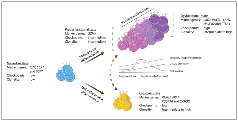
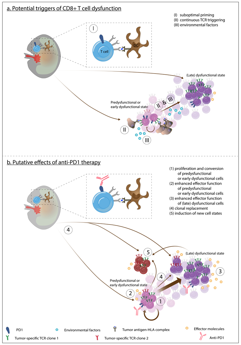
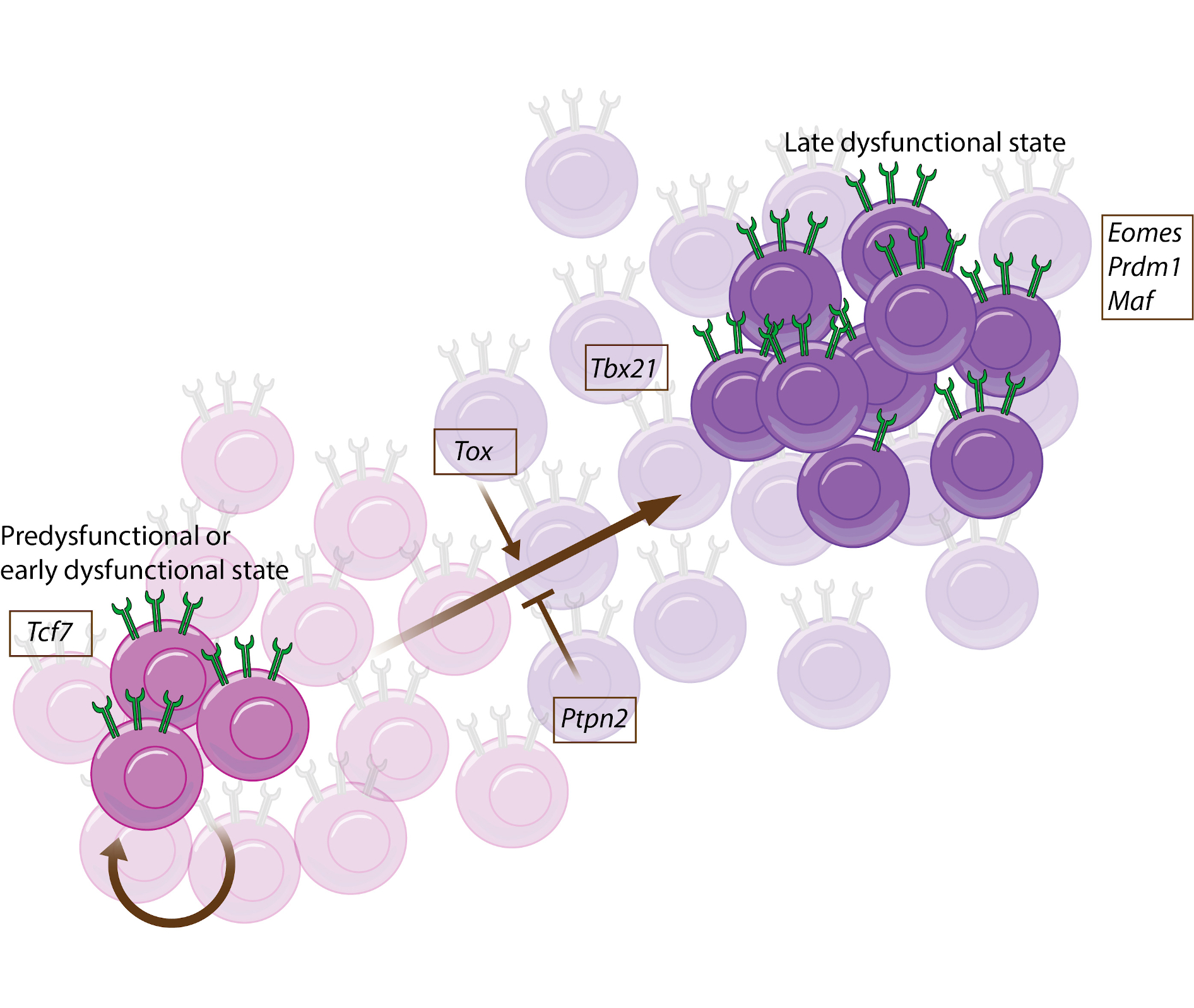
The T cell infiltrates that are formed in human cancers are a modifier of natural disease progression and also determine the probability of clinical response to cancer immunotherapies. Recent technological advances that allow the single-cell analysis of phenotypic and transcriptional states have revealed a vast heterogeneity of intratumoural T cell states, both within and between patients, and the observation of this heterogeneity makes it critical to understand the relationship between individual T cell states and therapy response. This Review covers our current knowledge of the T cell states that are present in human tumours and the role that different T cell populations have been hypothesized to play within the tumour microenvironment, with a particular focus on CD8+ T cells. The three key models that are discussed herein are as follows: (1) the dysfunction of T cells in human cancer is associated with a change in T cell functionality rather than inactivity; (2) antigen recognition in the tumour microenvironment is an important driver of T cell dysfunctionality and the presence of dysfunctional T cells can hence be used as a proxy for the presence of a tumour-reactive T cell compartment; (3) a less dysfunctional population of tumour-reactive T cells may be required to drive a durable response to T cell immune checkpoint blockade.


Conflict of interest statement

Competing interests

The authors declare no competing interests.

Figures

Figure 1
Figure 1. Model of intratumoral CD8+ T cell states
This schematic depicts a model describing the characteristics of, and possible connections between, the major CD8+ T cell states in human tumors, as based on data from–,–,,. Observations from the different studies that support this model are listed in Supplementary Table S1. In brief, the naïve-like cells described in non-small-cell lung cancer (NSCLC), hepatocellular carcinoma (HCC), colorectal cancer (CRC), basal cell cancer (BCC), and melanoma show a strong resemblance to the (central-)memory populations described by Sade-Feldman et al. and Clarke et al.,. Based on the expression of granzyme K (GZMK), intermediate expression of inhibitory molecules, and relatively low clonality, the T lymphocyte population described in Sade-Feldman et al. and the effector–memory population described in Zhang et al. were considered similar to the pre-dysfunctional cell states observed in melanoma, HCC, and NSCLC. While additional research is required to determine their extent of overlap, the tissue resident memory T (TRM) population described in triple-negative breast cancer (TNBC) and the HAVCR2+ TRM cells in NSCLC are here aligned with the dysfunctional state described in all other studies. The cell state definitions from Azizi et al. could not be integrated into the model presented here and the effector–memory subset reported by Savas et al. may be composed of a mixture of pre-dysfunctional and cytotoxic cells, based on the combined expression of GZMK and killer cell lectin-like receptor subfamily G member 1 (KLRG1). In this model, we propose that the development of (pre-)dysfunctional cell states is predominantly driven by tumor-specific cues such as tumor antigen recognition and/or tumor-specific environmental factors (here referred to as tumor microenvironment (TME)-induced differentiation). Cytotoxic cell states are also encountered in healthy tissues,,, indicating that the underlying differentiation process is not strictly tumor-specific (here referred to as TME-independent differentiation). Cytotoxic effector T cells are depicted as a population that is most likely developmentally distinct from the cells along the (pre-)dysfunctional axis, but additional research is required to clarify whether cytotoxic effector cells indeed originate from a distinct pool of cells, or whether they are connected to the (pre-)dysfunctional axis in some situations (as depicted by the dashed two-way arrow). Note that both trajectory and T cell receptor (TCR) sharing analyses indicate that the pre-dysfunctional and dysfunctional cells form a continuum of cell states, rather than well-demarcated populations. The line graph shows approximate levels of proliferation, CXC-chemokine ligand 13 (CXCL13) expression, and the expression of inhibitory receptors by pre-dysfunctional, early dysfunctional, and late dysfunctional CD8+ T cells. CCR7, CC-chemokine receptor 7; CX3CR1, CX3C chemokine receptor 1; CTLA4, cytotoxic lymphocyte-associated antigen 4; FCGR3A, Fcγ receptor IIIA; IL7R, interleukin 7 receptor; LAG3, lymphocyte activation gene 3; PDCD1, programmed cell death 1; PRF1, perforin 1; TCF7, transcription factor 7.
Figure 2
Figure 2. Model for the development of CD8+ T cell dysfunction and the effect of PD1 blockade
a) Potential drivers of dysfunction are the suboptimal priming of CD8+ T cells (I) and continuous T cell receptor (TCR) triggering (II) in combination with environmental factors, which may be cytokines such as tumor growth factor β (TGFβ) and interleukin 10 (IL-10), but also metabolic conditions such as hypoxia (III) at the tumor site. T cell priming is here depicted to occur in the lymph node, but could potentially also occur (in tertiary lymphoid structures (TLS)) at the tumor site. b) Proposed effects of anti-programmed cell death protein 1 (PD1) therapy on the CD8+ T cell states in and outside of the tumor microenvironment (TME). Mouse model studies have shown that anti-PD1 induces proliferation (denoted by the circular arrow) and conversion of pre- (and/or early) dysfunctional CD8+ T cells towards a more late dysfunctional phenotype (1) and that durable tumor control may require the proliferative capacity of a pre-dysfunctional or early dysfunctional cell population that, contrary to the late dysfunctional population, expresses transcription factor 7 (TCF7), which encodes TCF1,. Note that it is unclear whether this TCF1-positive population contains naïve-like T cells as well. PD1 blockade may also directly increase the effector function of pre- or early dysfunctional cells (2), as well as the effector function of (late) dysfunctional cells (3). It remains to be established whether tumor regression upon PD1 blockade primarily occurs through the activity of a reactivated pre-dysfunctional or early dysfunctional cell pool, or through the activity of late dysfunctional cells that may be formed as their progeny. A recent report in human basal cell carcinoma (BCC) has provided evidence for the possible replacement of the TCR repertoire of the dysfunctional T cell pool upon anti-PD1 treatment (4). This could be due to an influx of new cells (although the relevance of the systemic T cell compartment to the anti-tumor effects of anti-PD1 has been debated,), or expansion of lowly abundant pre-existing intratumoral clones. Finally, it remains a possibility that new T cell states (either of cells that newly infiltrated the tumor, or of pre-existing intratumoral cells) might develop upon anti-PD1 treatment; although no strong evidence in favor of such a model has thus far been obtained in mouse models or human samples (5). In this model, programmed cell death protein 1 (PD1) and PD1 ligand 1 (PDL1) are only depicted on cells in cases where the interaction is blocked by anti-PD1 therapy and are left out on other cells for clarity. Note that the model depicted here is based on the assumption that tumor-reactivity is enriched in the CD8+ T cells that reside along the (pre-)dysfunctional axis, while the cytotoxic effector T cell pool (shown in Figure 1) is enriched for bystander CD8+ T cells.
Figure 3
Figure 3. Hallmarks of intratumoral tumor-reactive CD8+ T cells
This schematic depicts protein markers and functional properties that are enriched in tumor-reactive CD8+ T cells (i.e. T cells that express a tumor-reactive T cell receptor (TCR), irrespective of their functional capacity) relative to bystander CD8+ T cells at the tumor site. Note that none of these characteristics by themselves identify tumor-reactive CD8+ T cells with absolute precision and that for some of these markers (4-1BB, GITR, and CXC-chemokine ligand 13 (CXCL13)), the evidence is less well established. LAG3, lymphocyte activation gene 3 protein; PD1, programmed cell death protein 1; TIM3, T-cell immunoglobulin mucin receptor 3.
None

References

    1. Clemente CG, et al. Prognostic value of tumor infiltrating lymphocytes in the vertical growth phase of primary cutaneous melanoma. Cancer. 1996;77:1303–1310. - PubMed
    1. Galon J, et al. Type, density, and location of immune cells within human colorectal tumors predict clinical outcome. Science (80-. ) 2006;313:1960–1964. - PubMed
    1. Zhang L, et al. Intratumoral T Cells, Recurrence, and Survival in Epithelial Ovarian Cancer. N Engl J Med. 2003;348:203–213. - PubMed
    1. Fridman WH, Pages F, Sauts-Fridman C, Galon J. The immune contexture in human tumours: Impact on clinical outcome. Nature Reviews Cancer. 2012;12:298–306. - PubMed
    1. Rosenberg SA, Restifo NP. Adoptive cell transfer as personalized immunotherapy for human cancer. Science (80-. ) 2015;348:62–68. - PMC - PubMed

Highlighted references

    1. Azizi et al., Cell (2018): Single cell transcriptome analysis of immune cells in human breast cancer demonstrating that intratumoral T cells reside along a continuum that is driven by activation and terminal differentiation. In addition, cell state diversity was shown both between and within T cell clones.

    1. Clarke et al., J. Exp. Med. (2019): Single cell transcriptome analysis of T cells in human lung cancer describing a PD1 and TIM3 expressing TRM cell subset that contains a highly proliferative subset and is enriched in lesions of patients responding to anti-PD1 therapy.

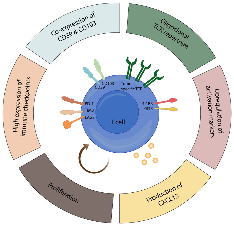
    1. Duhen et al., Nature Communications (2018): This study identifies CD103 and CD39 as markers of tumor-reactive T cells across multiple human cancer types.

    1. Guo et al., Nature Medicine (2018): Single cell transcriptome analysis of T cells in NSCLC, adjacent normal tissue, and blood showing the distribution of T cell states in these tissues and their relatedness based on TCR sharing between cell states in and outside the tumor. Moreover, this study provides evidence for dysfunctionality as a gradual state.

    1. Kurtulus et al., Immunity (2019): A mouse study that describes a PD1 low subset of T cells that respond to ICB and identifies Tcf7 expression to be required for response to anti-PD1 and anti-TIM3 combination therapy.

Publication types

MeSH terms