. 2021 Jun;18(6):1512-1527.
doi: 10.1038/s41423-020-0362-6. Epub 2020 Feb 5.

The deubiquitinase OTUB1 augments NF-κB-dependent immune responses in dendritic cells in infection and inflammation by stabilizing UBC13

Affiliations

The deubiquitinase OTUB1 augments NF-κB-dependent immune responses in dendritic cells in infection and inflammation by stabilizing UBC13

Floriana Mulas et al. Cell Mol Immunol. 2021 Jun.

Abstract

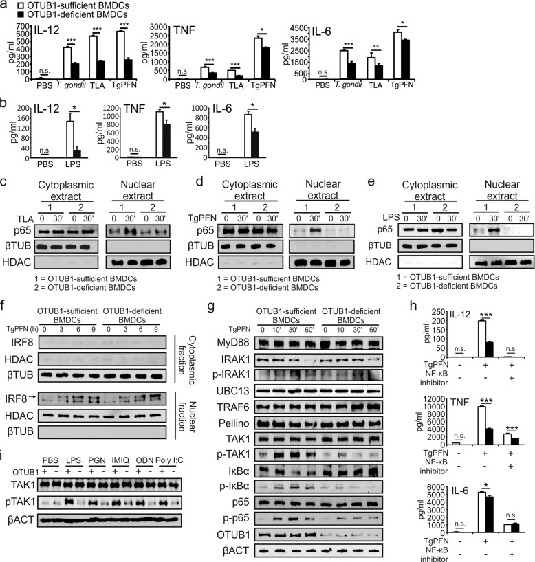
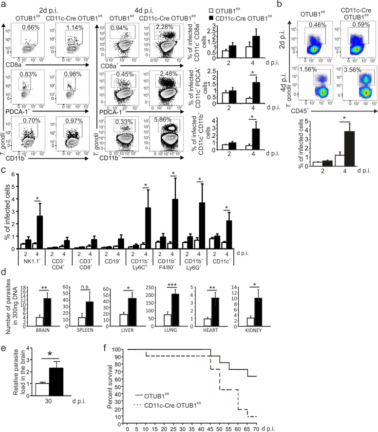
Dendritic cells (DCs) are indispensable for defense against pathogens but may also contribute to immunopathology. Activation of DCs upon the sensing of pathogens by Toll-like receptors (TLRs) is largely mediated by pattern recognition receptor/nuclear factor-κB (NF-κB) signaling and depends on the appropriate ubiquitination of the respective signaling molecules. However, the ubiquitinating and deubiquitinating enzymes involved and their interactions are only incompletely understood. Here, we reveal that the deubiquitinase OTU domain, ubiquitin aldehyde binding 1 (OTUB1) is upregulated in DCs upon murine Toxoplasma gondii infection and lipopolysaccharide challenge. Stimulation of DCs with the TLR11/12 ligand T. gondii profilin and the TLR4 ligand lipopolysaccharide induced an increase in NF-κB activation in OTUB1-competent cells, resulting in elevated interleukin-6 (IL-6), IL-12, and tumor necrosis factor (TNF) production, which was also observed upon the specific stimulation of TLR2, TLR3, TLR7, and TLR9. Mechanistically, OTUB1 promoted NF-κB activity in DCs by K48-linked deubiquitination and stabilization of the E2-conjugating enzyme UBC13, resulting in increased K63-linked ubiquitination of IRAK1 (IL-1 receptor-associated kinase 1) and TRAF6 (TNF receptor-associated factor 6). Consequently, DC-specific deletion of OTUB1 impaired the production of cytokines, in particular IL-12, by DCs over the first 2 days of T. gondii infection, resulting in the diminished production of protective interferon-γ (IFN-γ) by natural killer cells, impaired control of parasite replication, and, finally, death from chronic T. encephalitis, all of which could be prevented by low-dose IL-12 treatment in the first 3 days of infection. In contrast, impaired OTUB1-deficient DC activation and cytokine production by OTUB1-deficient DCs protected mice from lipopolysaccharide-induced immunopathology. Collectively, these findings identify OTUB1 as a potent novel regulator of DCs during infectious and inflammatory diseases.

Keywords: OTUB1; dendritic cell; innate immunity; signal transduction; ubiquitination.


Conflict of interest statement

The authors declare no competing interests.

Figures

Fig. 1
Fig. 1
OTUB1 expression is upregulated in DCs by T. gondii infection and LPS stimulation. a, b Mice were infected i.p. with five T. gondii cysts (a) or challenged with LPS (b) for the indicated time periods. CD11c+ cells were isolated from spleens by magnetic sorting, and OTUB1 expression was measured by WB analysis (upper panels). The lower panels show the relative expression of OTUB1 (n = 3 for each group). ch GM-CSF-expanded (c, e, and g) and FLT3L-expanded (d, f, and h) BMDCs generated from C57BL/6 mice were stimulated in vitro with tachyzoites at an MOI of 3 (c, d), 1 μg/ml TgPFN (e, f), or 500 ng/ml LPS (g, h) for the indicated time periods. OTUB1 expression in BMDCs was analyzed by WB analysis (upper panels). The lower panels show the relative expression of OTUB1 (n = 3 for each group). Data are displayed as the mean ± SD. *p < 0.05, **p < 0.01, and ***p < 0.001
Fig. 2
Fig. 2
OTUB1 deletion in DCs impairs the cytokine response during T. gondii infection and LPS stimulation by reducing NF-κB activation. a, b OTUB1-sufficient and OTUB1-deficient BMDCs were left untreated or stimulated with T. gondii tachyzoites (MOI = 3), different antigens (TLA and TgPFN) or LPS for 24 h. Concentrations of cytokines in the supernatant were analyzed by ELISA (n = 4). (ce) FLT3L-expanded BMDCs were left untreated or stimulated with TLA (c), TgPFN (d), or LPS (e) for 30 min. Levels of p65 in the cytoplasmic and nuclear fractions were analyzed by WB analysis. f FLT3L-expanded BMDCs were stimulated with TgPFN for the indicated time periods. IRF8 in the cytoplasmic and nuclear extracts was detected by WB analysis. g FLT3L-expanded BMDCs were stimulated with TgPFN for the indicated time periods. Whole-cell lysates were analyzed by WB analysis with the indicated antibodies. h FLT3L-expanded BMDCs were left untreated or stimulated with TgPFN in the presence or absence of NF-κB inhibitor for 24 h. Cytokine levels in the supernatants of cell cultures were measured by ELISA (n = 4). i FLT3L-expanded BMDCs were stimulated with LPS, PGN, IMIQ, ODN, and poly I:C for 30 min. Whole-cell lysates were analyzed by WB analysis with the indicated antibodies. Data are shown as the mean ± SD. *p < 0.05, **p < 0.01, and ***p < 0.001; n.s. not significant
Fig. 3
Fig. 3
OTUB1 stabilizes UBC13 by reducing its K48-linked polyubiquitination. ae Cytoplasmic proteins from unstimulated, TgPFN-stimulated (a, b, e) and LPS-stimulated (c, d) BMDCs were immunoprecipitated with the indicated antibodies. Immunoprecipitates and input were analyzed by WB analysis with the indicated antibodies. f OTUB1-sufficient and OTUB1-deficient FLT3L-expanded BMDCs were pretreated with TgPFN (1 μg/ml) for 1 h or left unstimulated. Then, cycloheximide (CHX, 100 μg/ml) was added for the indicated time period. Protein levels of UBC13 in whole-cell lysates were analyzed by WB analysis (upper panel). The lower panel shows the relative levels of UBC13 normalized to βACT levels (n = 3). g NIH 3T3 cells were transfected with OTUB1 siRNA for 36 h. Then, the cells were transfected with GFP, GFP-OTUB1, GFP-OTUB1 ΔN, or GFP-OTUB1 C91S plasmids. After 24 h, the cells were treated with CHX + LPS for 0, 3, and 6 h. Whole-cell lysates were then isolated and analyzed with the indicated antibodies. hk GM-CSF-expanded BMDCs were left unstimulated or stimulated with TgPFN in the presence of MG132. Cytoplasmic proteins were isolated and immunoprecipitated with anti-TRAF6 (h, i) and anti-IRAK1 (j, k) antibodies. Immunoprecipitates and input were analyzed with the indicated antibodies. l FLT3L-expanded BMDCs were left untreated or transduced with UBC13-expressing lentivirus or vector lentivirus for 72 h, followed by stimulation with TgPFN for 24 h. Cytokines in the supernatants of cell cultures were measured by flow cytometry (n = 4). Data are displayed as the mean ± SD (d) or mean ± SD (g). *p < 0.05, **p < 0.01, and ***p < 0.001; n.s. not significant
Fig. 4
Fig. 4
OTUB1 deletion in DC impairs the cytokine response during T. gondii infection. ac OTUB1fl/fl and CD11c-Cre OTUB1fl/fl mice were infected i.p. with 50,000 tachyzoites for 12 and 48 h, respectively. Peritoneal cells were collected by lavage of the peritoneal cavity and analyzed by flow cytometry to detect IL-12 production in CD11c+ CD8a+, CD11c+ PDCA-1+, and CD11c+ CD11b+ DCs (a); UBC13, MHC II, CD40, CD86 and CD80 expression in CD11c+ CD8a+, CD11c+ PDCA-1+, and CD11c+ CD11b+ DCs (b); and IFN-γ production in NK cells (c). Representative flow cytometry plots (left panels) and statistics (right panels) are shown (n = 8). Data are shown as the mean ± SD. *p < 0.05, **p < 0.01, and ***p < 0.001; n.s. not significant
Fig. 5
Fig. 5
OTUB1 deletion in DCs leads to impaired parasite control in toxoplasmosis. ac OTUB1fl/fl and CD11c-Cre OTUB1fl/fl mice were infected i.p. with 50,000 tachyzoites. Peritoneal cells were isolated from the peritoneal cavities and analyzed by flow cytometry at 2 and 4 days p.i. a Percentages of infected CD11c+ CD8a+, CD11c+ PDCA-1+, and CD11c+ CD11b+ DCs. b Percentages of infected peritoneal CD45+ cells in OTUB1fl/fl and CD11c-Cre OTUB1fl/fl mice. c Percentages of infected peritoneal leukocyte subpopulations in OTUB1fl/fl and CD11c-Cre OTUB1fl/fl mice. Representative FACS plots (upper panel) and statistics (lower panel) are shown (n = 10 for each group). d Mice were infected i.p. with 50,000 tachyzoites for 10 days. Parasite loads in different organs were determined by semiquantitative PCR of tissue DNA (n = 5 for each group). e, f Mice were infected i.p. with three cysts. Parasite load in the brain 30 days p.i. was determined by semiquantitative PCR of tissue DNA (n = 4 for each group) (e). Survival was monitored for 70 days p.i. (f) (n = 10 for each group). Data are expressed as the mean ± SD. *p < 0.05, **p < 0.01, and ***p < 0.001; n.s. not significant
Fig. 6
Fig. 6
IL-12 supplementation rescues the defect in parasite control in CD11c-Cre OTUB1fl/fl mice. a Experimental design scheme of IL-12 administration and analysis. Mice were i.p. injected with either PBS or 150 ng IL-12 daily from 0 to 3 days p.i. b The parasite load in peritoneal cells was determined by semiquantitative PCR of tissue DNA after infection with 50,000 tachyzoites for 4 days (n = 8 for each group). c Percentages of infected peritoneal CD45+ cells were analyzed by flow cytometry at day 4 after infection with 50,000 tachyzoites. Representative flow cytometry plots (left panel) and statistics (right panel) are shown (n = 8 for each group). d, e Percentages of infected peritoneal leukocyte subpopulations (d) and DC subsets (e) were analyzed by flow cytometry at day 4 after infection with 50,000 tachyzoites (n = 8 for each group). f Mice were i.p. infected with five cysts of the DX strain. At day 30 p.i., the parasite loads in the brains of surviving mice were determined by semiquantitative PCR. Two independent experiments with comparable results were performed. One representative experiment is shown (n = 4 for each group). g Intracerebral parasites in the brains of T. gondii-infected OTUB1fl/fl and CD11c-Cre OTUB1fl/fl mice at 30 days p.i. A PBS-treated OTUB1fl/fl mouse (top left) shows a single focus consisting of a few T. gondii cysts and some tachyzoites in the white matter of the frontal lobe. The brain of a PBS-treated CD11c-Cre OTUB1fl/fl mouse (top right) shows an increased number of parasitic foci (arrows) with T. gondii cysts and tachyzoites in the white matter of the frontal lobe. In both an OTUB1fl/fl and a CD11c-Cre OTUB1fl/fl mouse, IL-12 application reduced the intracerebral parasitic load, with only single cysts scattered throughout the frontal lobe (arrows). Immunohistochemistry with polyclonal rabbit anti-T. gondii (BioGenex, Fremont, CA, USA) and slight counterstaining with hemalum; original magnification ×200; scale bar (ad): 50 µm. The photomicrographs shown are representative of three mice per experimental group. Similar results were obtained in a second independent experiment. h The survival of the mice was monitored daily up to 30 days after infection with cysts of strain 5 DX (n = 8 for each group). Data are displayed as the mean ± SD. *p < 0.05, and ***p < 0.001; n.s. not significant
Fig. 7
Fig. 7
OTUB1 deficiency decreases LPS-induced mortality by reducing cytokine production. a OTUB1fl/fl and CD11c-Cre OTUB1fl/fl mice were i.p. administered 10 mg/kg LPS. After 3 h, blood was collected, and cytokines in the sera were analyzed by ELISA (n = 9). b OTUB1fl/fl and CD11c-Cre OTUB1fl/fl mice were i.p. administered a low dose of LPS (10 mg/kg), and survival was monitored daily for 6 days (n = 10). Data are displayed as the mean ± SD. *p < 0.05 and **p < 0.01; n.s. not significant

References

    1. Eisenbarth SC. Dendritic cell subsets in T cell programming: location dictates function. Nat. Rev. Immunol. 2019;19:89–103. - PMC - PubMed
    1. Rodrigues PF, et al. Distinct progenitor lineages contribute to the heterogeneity of plasmacytoid dendritic cells. Nat. Immunol. 2018;19:711–722. - PMC - PubMed
    1. Andrade WA, et al. Combined action of nucleic acid-sensing Toll-like receptors and TLR11/TLR12 heterodimers imparts resistance to Toxoplasma gondii in mice. Cell Host Microbe. 2013;13:42–53. - PMC - PubMed
    1. Yarovinsky F, et al. TLR11 activation of dendritic cells by a protozoan Profilin-like protein. Science (80-.). 2005;308:1626–1629. - PubMed
    1. Raetz M, et al. Cooperation of TLR12 and TLR11 in the IRF8-dependent IL-12 response to Toxoplasma gondii profilin. J. Immunol. 2013;191:4818–4827. - PMC - PubMed

Publication types

MeSH terms