. 2020 Mar;52(3):331-341.
doi: 10.1038/s41588-019-0576-7. Epub 2020 Feb 5.

Comprehensive analysis of chromothripsis in 2,658 human cancers using whole-genome sequencing

Collaborators, Affiliations

Comprehensive analysis of chromothripsis in 2,658 human cancers using whole-genome sequencing

Isidro Cortés-Ciriano et al. Nat Genet. 2020 Mar.

Erratum in

Abstract

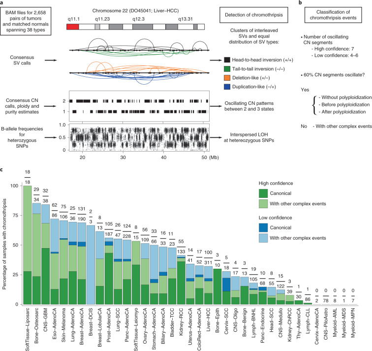
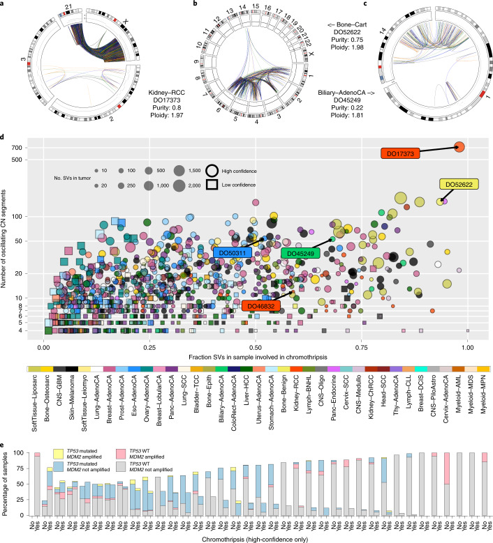
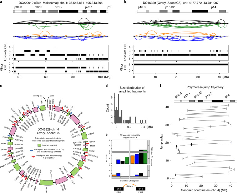
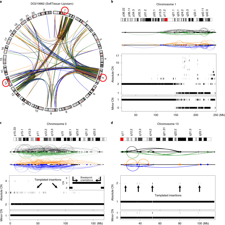
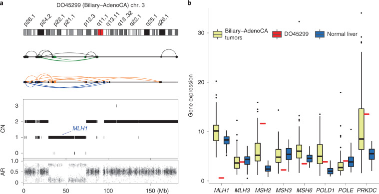
Chromothripsis is a mutational phenomenon characterized by massive, clustered genomic rearrangements that occurs in cancer and other diseases. Recent studies in selected cancer types have suggested that chromothripsis may be more common than initially inferred from low-resolution copy-number data. Here, as part of the Pan-Cancer Analysis of Whole Genomes (PCAWG) Consortium of the International Cancer Genome Consortium (ICGC) and The Cancer Genome Atlas (TCGA), we analyze patterns of chromothripsis across 2,658 tumors from 38 cancer types using whole-genome sequencing data. We find that chromothripsis events are pervasive across cancers, with a frequency of more than 50% in several cancer types. Whereas canonical chromothripsis profiles display oscillations between two copy-number states, a considerable fraction of events involve multiple chromosomes and additional structural alterations. In addition to non-homologous end joining, we detect signatures of replication-associated processes and templated insertions. Chromothripsis contributes to oncogene amplification and to inactivation of genes such as mismatch-repair-related genes. These findings show that chromothripsis is a major process that drives genome evolution in human cancer.


Conflict of interest statement

C.-Z.Z. is a co-founder and equity holder of Pillar Biosciences.

Figures

Fig. 1
Fig. 1. Overview of the chromothripsis-calling method and the frequency of events across 37 cancer types.
a, Example of a region displaying the characteristic features of chromothripsis: cluster of interleaved SVs with equal proportions of SV types (that is, fragment joins), a CN profile that oscillates between two states and interspersed LOH. Details of the criteria are described in the Methods. Both the color scheme and the abbreviations shown in this figure are used throughout the manuscript. b, Classification of chromothripsis events. In a canonical event, more than 60% of the segments oscillate between two CN states; a tumor is classified as canonical if it showed at least one canonical chromothripsis event. c, Percentage of patients with chromothripsis events across the entire cohort. The fractions at the top of the bars are the number of tumors that showed high-confidence chromothripsis out of the total number of tumors of that type. The cancer type abbreviations used across the manuscript are as follows: Biliary-AdenoCA, biliary adenocarcinoma; Bladder-TCC, bladder transitional cell carcinoma; Bone-Benign, bone cartilaginous neoplasm, osteoblastoma and bone osteofibrous dysplasia; Bone-Epith, bone neoplasm, epithelioid; Bone-Osteosarc, sarcoma, bone; Breast-AdenoCA, breast adenocarcinoma; Breast-DCIS, breast ductal carcinoma in situ; Breast-LobularCA, breast lobular carcinoma; Cervix-AdenoCA, cervix adenocarcinoma; Cervix-SCC, cervix squamous cell carcinoma; CNS-GBM, central nervous system glioblastoma; CNS-Oligo, CNS oligodenroglioma; CNS-Medullo, CNS medulloblastoma; CNS-PiloAstro, CNS pilocytic astrocytoma; ColoRect-AdenoCA, colorectal adenocarcinoma; Eso-AdenoCA, esophagus adenocarcinoma; Head-SCC, head-and-neck squamous cell carcinoma; Kidney-ChRCC, kidney chromophobe renal cell carcinoma; Kidney-RCC, kidney renal cell carcinoma; Liver-HCC, liver hepatocellular carcinoma; Lung-AdenoCA, lung adenocarcinoma; Lung-SCC, lung squamous cell carcinoma; Lymph-CLL, lymphoid chronic lymphocytic leukemia; Lymph-BNHL, lymphoid mature B-cell lymphoma; Lymph-NOS, lymphoid not otherwise specified; Myeloid-AML, myeloid acute myeloid leukemia; Myeloid-MDS, myeloid myelodysplastic syndrome; Myeloid-MPN, myeloid myeloproliferative neoplasm; Ovary-AdenoCA, ovary adenocarcinoma; Panc-AdenoCA, pancreatic adenocarcinoma; Panc-Endocrine, pancreatic neuroendocrine tumor; Prost-AdenoCA, prostate adenocarcinoma; Skin-Melanoma, skin melanoma; SoftTissue-Leiomyo, leiomyosarcoma, soft tissue; SoftTissue-Liposarc, liposarcoma, soft tissue; Stomach-AdenoCA, stomach adenocarcinoma; Thy-AdenoCA, thyroid low-grade adenocarcinoma; and Uterus-AdenoCA, uterus adenocarcinoma.
Fig. 2
Fig. 2. Heterogeneity of chromothripsis events.
ac, Examples of massive chromothripsis events on the background of quiescent genomes in samples from patients DO17373 (a), DO52622 (b) and DO45249 (c). d, The fraction of SVs involved in chromothripsis in each sample against the maximum number of contiguous oscillating CN segments for the high-confidence (circles) and low-confidence (squares) chromothripsis calls. e, Distribution of patients showing high-confidence chromothripsis, deleterious TP53 mutations and MDM2 amplification (CN ≥ 4). WT, wild-type allele.
Fig. 3
Fig. 3. Example of canonical chromothripsis events displaying templated insertions and evidence of MMBIR.
a, Evidence of chromothripsis in chromosome 1 in a skin-melanoma tumor with CN oscillations that span 3 CN levels and LOH. b, Example of a chromothripsis event in chromosome 4 involving low-level CN gains and absence of LOH in an ovarian adenocarcinoma. Segments at CN 3 correspond to templated insertions, as evidenced by their size, and breakpoint orientations at their edges. Breakpoints corresponding to interchromosomal SVs are depicted as colored dots in the SV profile, whereas intrachromosomal SVs are represented with black dots and colored arcs following the representation shown in Fig. 1. c, Reconstruction of the amplicon generated by the chromoanasynthesis event detected in chromosome 4 in tumor DO46329 (see b). Inverted segments are depicted in green. Red arrows highlight breakpoints with short microhomology tracts, whereas blue arrows indicate the presence of small insertions at the breakpoints. The CN for all segments is 3 unless otherwise indicated. d, Size distribution for the templated insertions forming the amplicon depicted in c. e, CN step plot for chromosome 4 indicating that most of the SVs mapped to chromosome 4 link genomic regions at CN 3. The x and y axes correspond to the CN level of the segments linked by a given SV. The color of the bars corresponds to the four types of SVs (that is, deletion-like, duplication-like, and head-to-head and tail-to-tail inversions) indicated in Fig. 1a and considered throughout the manuscript. f, Trajectory of the polymerase across chromosome 4 estimated from the template-switching events shown in c.
Fig. 4
Fig. 4. Example of a multichromosomal chromothripsis event in a soft-tissue liposarcoma co-localized with other complex events involving templated insertions.
a, Scaled circos plot of the entire genome for this tumor except for chromosome Y. bd, SV and CN profiles for chromosomes 1 (b), 5 (c) and 13 (d). Tens of CN oscillations and LOH in chromosome 1 are co-localized with additional rearrangements. The size, minor CN (from the allele with the lower number of copies) and orientation of the breakpoint junctions associated with the segments at CN 3 indicate that these are templated insertions. c, Inset: orientation of the breakpoint junctions at the edges of low-level CN gains originated from template switching (that is, − and + according to the annotation that we use in the manuscript).
Fig. 5
Fig. 5. Chromothripsis-mediated depletion of MLH1.
a, Chromothripsis event and expression levels of DNA MMR genes in the sample of patient DO45299 (biliary adenocarcinoma). b, Mean expression of DNA MMR genes in a panel of 16 biliary adenocarcinomas and 16 normal liver samples. Box plots in b show median, first and third quartiles (boxes), and the whiskers encompass observations within a distance of 1.5× the interquartile range from the first and third quartiles. AR, allelic ratio computed for heterozygous SNPs.

References

    1. Stephens PJ, et al. Massive genomic rearrangement acquired in a single catastrophic event during cancer development. Cell. 2011;144:27–40. doi: 10.1016/j.cell.2010.11.055. - DOI - PMC - PubMed
    1. Zhang C-Z, et al. Chromothripsis from DNA damage in micronuclei. Nature. 2015;522:179–184. doi: 10.1038/nature14493. - DOI - PMC - PubMed
    1. Korbel JO, Campbell PJ. Criteria for inference of chromothripsis in cancer genomes. Cell. 2013;152:1226–1236. doi: 10.1016/j.cell.2013.02.023. - DOI - PubMed
    1. Leibowitz ML, Zhang C-Z, Pellman D. Chromothripsis: a new mechanism for rapid karyotype evolution. Annu. Rev. Genet. 2015;49:183–211. doi: 10.1146/annurev-genet-120213-092228. - DOI - PubMed
    1. Notta F, et al. A renewed model of pancreatic cancer evolution based on genomic rearrangement patterns. Nature. 2016;538:378–382. doi: 10.1038/nature19823. - DOI - PMC - PubMed

Publication types