

Successful endovascular coil embolization of large pseudoaneurysm of ductus arteriosus diverticulum
- PMID: 32026144
- PMCID: PMC6966358
- DOI: 10.1186/s42155-019-0053-5
Successful endovascular coil embolization of large pseudoaneurysm of ductus arteriosus diverticulum
Abstract
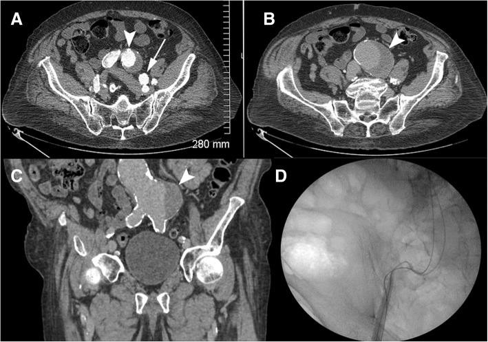
Background: Pseudoaneurysm of the ductus arteriosus diverticulum, although rare in adults, may have catastrophic consequences if left untreated due to erosion and rupture of the pseudoaneurysm into adjacent thoracic structures. Although thoracic endovascular aortic repair (TEVAR) is the standard treatment method for aneurysm closure of the ductus arteriosus diverticulum, it was not possible in our patient secondary to marked aortoiliac access vessel tortuosity, significant vascular calcific burden, and an abdominal aortic aneurysm. We describe the first reported case of endovascular coil embolization being successfully used as the definite repair of a ductus arteriosus diverticulum pseudoaneurysm.
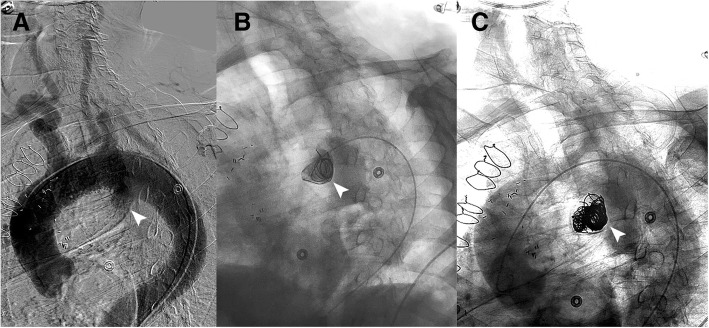
Case presentation: An 85-year-old man with history of severe coronary arterial disease presented with an enlarging pseudoaneurysm of a ductus arteriosus diverticulum. The diverticulum and thoracic aortic junction demonstrated the typical obtuse angles and wide neck, differentiating it from otherwise similar-appearing diagnostic considerations. Repair was attempted with conventional aortic stent graft but the patient's infrarenal abdominal aortic aneurysm and his heavily calcified, tortuous iliac vessels could not accommodate the 24Fr introducer sheath necessary for stent graft placement. Therefore, endovascular coil embolization was successfully completed through a 4Fr directional catheter. The patient tolerated the procedure well and was discharged from the hospital in good condition on post-embolization day six.
Conclusions: Endovascular coil embolization is an alternative treatment for ductus arteriosus diverticulum pseudoaneurysm closure in cases where the standard TEVAR method is unsuccessful. Instead of the wide entry point at the aorta we used the junction of the diverticulum and pseudoaneurysm as the "neck" for satisfactory and stable coil placement. Endovascular coil embolization alone may be a viable definitive therapy for occlusion of the ductus pseudoaneurysm component of the diverticulum in cases where complex anatomy or extensive vascular disease makes stent graft repair impractical if not impossible.
Keywords: Ductus arteriosus pseudoaneurysm; Endovascular coil embolization; TEVAR; Thoracic aneurysm; Thoracic aortic repair.
Conflict of interest statement
The authors declare that they have no conflict of interest.
Figures

References
-
- Addams-Williams JH, Collin N, Agrawal N, Armstrong S, Tierney PA (2005) Aneurysm of the diverticulum of the ductus arteriosus in the adult associated with left recurrent laryngeal nerve palsy: a case series and review of the literature. The Internet Journal of Otorhinolaryngology 4
-
- Hyung Ann J, Young Kim E, Mi Jeong Y, Ho Kim J, Sik Kim H, Choi H-Y. Morphologic evaluation of ductus diverticulum using multi - detector computed tomography: comparison with traumatic Pseudoaneurysm of the aortic isthmus. Iran J Radiol. 2016;13:e38016. doi: 10.5812/iranjradiol.38016. - DOI - PMC - PubMed
-
- Mitchell RS, Seifert FC, Miller DC, Jamieson SW, Shumway NE. Aneurysm of the diverticulum of the ductus arteriosus in the adult. Successful surgical treatment in five patients and review of the literature. J Thorac Cardiovasc Surg. 1983;86:400–408. - PubMed
LinkOut - more resources
Full Text Sources