Review
. 2020 May;38(5):411-433.
doi: 10.1007/s11604-020-00922-8. Epub 2020 Feb 5.

Computed tomographic evaluation of myocardial ischemia

Affiliations
Review

Computed tomographic evaluation of myocardial ischemia

Yuki Tanabe et al. Jpn J Radiol. 2020 May.

Abstract

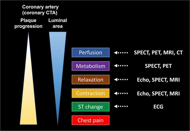
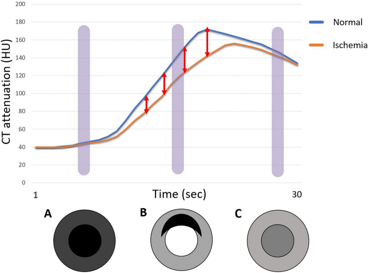
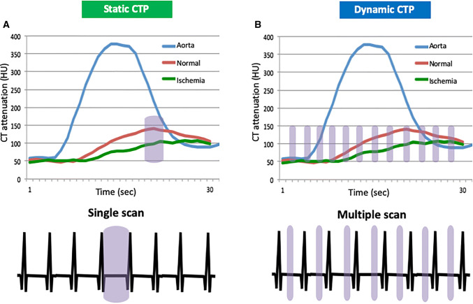
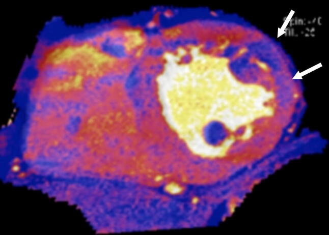
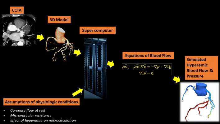
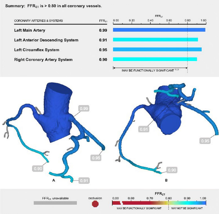
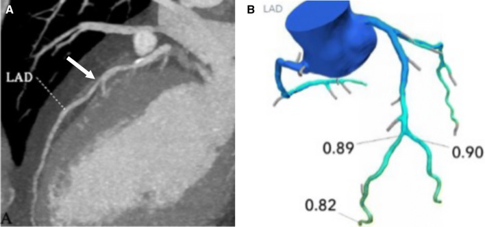
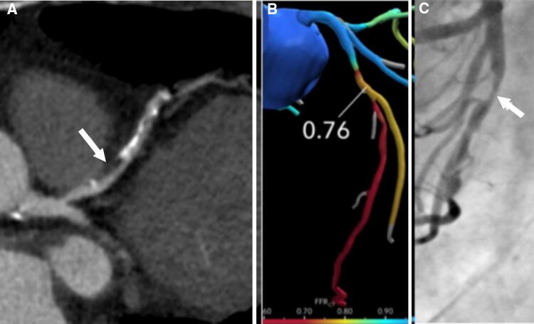
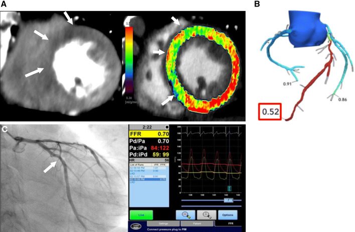
Myocardial ischemia is caused by a mismatch between myocardial oxygen consumption and oxygen delivery in coronary artery disease (CAD). Stratification and decision-making based on ischemia improves the prognosis in patients with CAD. Non-invasive tests used to evaluate myocardial ischemia include stress electrocardiography, echocardiography, single-photon emission computed tomography, and magnetic resonance imaging. Invasive fractional flow reserve is considered the reference standard for assessment of the hemodynamic significance of CAD. Computed tomography (CT) angiography has emerged as a first-line imaging modality for evaluation of CAD, particularly in the population at low to intermediate risk, because of its high negative predictive value; however, CT angiography does not provide information on the hemodynamic significance of stenosis, which lowers its specificity. Emerging techniques, e.g., CT perfusion and CT-fractional flow reserve, help to address this limitation of CT, by determining the hemodynamic significance of coronary artery stenosis. CT perfusion involves acquisition during the first pass of contrast medium through the myocardium following pharmacological stress. CT-fractional flow reserve uses computational fluid dynamics to model coronary flow, pressure, and resistance. In this article, we review these two functional CT techniques in the evaluation of myocardial ischemia, including their principles, technology, advantages, limitations, pitfalls, and the current evidence.

Keywords: Computed tomography; Coronary artery disease; Fractional flow reserve; Myocardial ischemia; Myocardial perfusion.


Conflict of interest statement

Yuki Tanabe declares that he has no conflict of interest. Akira Kurata declares that he has no conflict of interest. Takuya Matsuda declares that he has no conflict of interest. Kazuki Yoshida declares that he has no conflict of interest. Dhiraj Baruah is medical educator for Boehringer-Ingelheim pharmaceuticals, Inc. Teruhito Kido declares that he has no conflict of interest. Teruhito Mochizuki declares that he has no conflict of interest. Prabhakar Rajiah declares that he has no conflict of interest.

Figures

Fig. 1
Fig. 1
Illustration showing the progressive pathological conditions in the myocardial ischemic cascade. Coronary artery atherosclerosis progresses and leads to myocardial hypoperfusion because of plaque progression and luminal stenosis. Myocardial hypoperfusion is followed by metabolic abnormalities, diastolic dysfunction, systolic dysfunction, and ECG abnormalities, culminating in chest pain. The right column shows the modalities that can be used to detect abnormalities at each step of the cascade. CT computed tomography, CTA computed tomography angiography, ECG electrocardiogram, SPECT single-photon emission computed tomography, PET positron emission tomography, MRI magnetic resonance imaging
Fig. 2
Fig. 2
Time attenuation curves for normal myocardium (blue) and ischemic myocardium (orange). At the optimal scan time (B), stress CT perfusion demonstrates a clear distinction between a normal and ischemic myocardium because of large differences in attenuation between normal and abnormal myocardium. Stress CT perfusion cannot distinguish normal from ischemic myocardium if the timing is too early (A) or too late (C). CT computed tomography, HU Hounsfield unit
Fig. 3
Fig. 3
Static and dynamic CTP techniques. a Static CTP data are acquired during a single phase of first pass of contrast in the myocardium. b Dynamic CTP data are acquired at multiple phases of first pass of contrast in the myocardium. Blue box means scan duration. CTP computed tomography perfusion, HU Hounsfield unit
Fig. 4
Fig. 4
A dual-energy CT scan overlaid with iodine in the short-axis plane obtained from a static stress myocardial CTP scan showing a perfusion defect in the mid anterolateral septum (arrow). CT computed tomography, CTP computed tomography perfusion
Fig. 5
Fig. 5
Comprehensive CTP protocol. In the stress-first protocol, CTP is first acquired with pharmacological stress. The rest CTP is acquired 10–20 min later and also serves as coronary CTA. Nitroglycerin and β-blockers are administered during this phase to obtain high-quality coronary CTA images. LIE-CT scans can be obtained 5–10 min following the second CTP scan. In the rest-first protocol, rest images are acquired first and stress images are acquired later. CTA computed tomography angiography, CTP computed tomography perfusion, LIE-CT late iodine enhancement computed tomography
Fig. 6
Fig. 6
Static CTP scans for single-vessel disease in a 68-year-old woman with chest pain. She had hypertension and dyslipidemia. a A short-axis CTP image following stress shows a subendocardial perfusion defect in the mid anterolateral wall (arrows). b A rest CTP image in the same position as a shows no perfusion defect, consistent with myocardial ischemia. CTP computed tomography perfusion
Fig. 7
Fig. 7
Dynamic CTP scans for single-vessel disease in an 87-year-old man with chest pain. He had hypertension and a history of smoking. Short-axis (a) and two-chamber (b) grayscale stress dynamic CTP images showing a subendocardial perfusion defect in the anterolateral, anterior, and anteroseptal segments (arrows). c A CT-MBF color-coded image shows that the MBF in the ischemic myocardium is lower than in the remote myocardium. CTP computed tomography perfusion, CT-MBF computed tomography derived-myocardial blood flow
Fig. 8
Fig. 8
Dynamic CTP scans for triple-vessel disease in an 84-year-old woman with chest pain. a Short-axis view of a dynamic CTP (grayscale) shows a subendocardial perfusion defect in the entire circumference of the heart (white arrows). b A CT-MBF color-coded image shows low CT-MBF throughout the heart. SPECT during c stress and d at rest shows a reversible perfusion defect in the lateral wall but no marked perfusion defect in the other regions. Invasive coronary angiography of the e right and f left coronary arteries shows severe triple-vessel disease. In this case, single-photon emission tomography did not accurately detect the presence of triple-vessel disease, known as balanced ischemia. CTP computed tomography perfusion, CT-MBF computed tomography derived-myocardial blood flow, SPECT single-photon emission computed tomography
Fig. 9
Fig. 9
Illustration showing the CT-FFR technique. The CCTA data are segmented and a three-dimensional model is generated. This model is then processed by a supercomputer using assumptions of physiological conditions to solve the Navier–Stokes equation and generate a hyperemic model of coronary flow and pressure. CCTA coronary computed tomography angiography, CT computed tomography, FFR fractional flow reserve
Fig. 10
Fig. 10
Normal CT-FFR report for a 59-year-old man with atypical chest pain. The CT-FFR report shows the coronary arteries color-coded according to their CT-FFR values. In this patient, all the major coronary arteries show normal values (> 0.8). CT computed tomography, FFR fractional flow reserve
Fig. 11
Fig. 11
a A curved multiplanar reconstruction image of the LAD in a 52-year-old man with chest pain showing moderate stenosis of the mid LAD (arrow). b CT-FFR in the same patient shows a value of 0.89 in the mid LAD, which is within normal limits, indicating that there is no lesion-specific ischemia. The patient was referred for medical management. Therefore, CT-FFR helped to avoid ICA. CT computed tomography, FFR fractional flow reserve, ICA invasive coronary angiography, LAD left anterior descending artery
Fig. 12
Fig. 12
a A curved multiplanar reconstruction image of the LAD in a 66-year-old woman showing a non-calcified plaque in the proximal LAD causing moderate luminal stenosis (arrow). b CT-FFR shows a value of 0.76 in the proximal LAD, which is indicative of hemodynamically significant stenosis. c The patient was referred for ICA, which confirmed a significant stenosis with a decreased FFR of 0.65. CT computed tomography, FFR fractional flow reserve, ICA invasive coronary angiography, LAD left anterior descending artery
Fig. 13
Fig. 13
CTP versus CT-FFR. a CTP image and CT-MBF map showing a perfusion defect and lower CT-MBF in the anteroseptal wall (arrows). b A CT-FFR image in the same patient showing abnormal CT-FFR with a low value of 0.52 at the LAD. c ICA showing a stenotic lesion in the mid LAD with an abnormal FFR of 0.70. CTP computed tomography perfusion, CT-MBF computed tomography derived-myocardial blood flow, CT computed tomography, FFR fractional flow reserve, ICA invasive coronary angiography, LAD left anterior descending artery

References

    1. Mori H, Torii S, Kutyna M, Sakamoto A, Finn AV, Virmani R, et al. Coronary artery calcification and its progression: what does it really mean? JACC Cardiovasc Imaging. 2018;11:127–142. doi: 10.1016/j.jcmg.2017.10.012. - DOI - PubMed
    1. Nesto RW, Kowalchuk GJ. The ischemic cascade: temporal sequence of hemodynamic, electrocardiocardiographic, and symptomatic expressions of ischemia. Am J Cardiol. 1987;59:23–30. doi: 10.1016/0002-9149(87)90192-5. - DOI - PubMed
    1. Stillman AE, Oudkerk M, Bluemke DA, de Boer MJ, Bremerich J, Garcia EV, et al. Imaging the myocardial ischemic cascade. Int J Cardiovasc Imaging. 2018;34:1249–1263. doi: 10.1007/s10554-018-1330-4. - DOI - PubMed
    1. Renker M, Baumann S, Rier J, Ebersberger U, Fuller SR, Batalis NI, et al. Imaging coronary artery disease and the myocardial ischemic cascade: clinical principles and scope. Radiol Clin N Am. 2015;53:261–269. doi: 10.1016/j.rcl.2014.11.010. - DOI - PubMed
    1. Tonino PA, De Bruyne B, Pijls NH, Siebert U, Ikeno F, van’t Veer M, et al. Fractional flow reserve versus angiography for guiding percutaneous coronary intervention. N Engl J Med. 2009;360:213–224. doi: 10.1056/NEJMoa0807611. - DOI - PubMed

LinkOut - more resources