. 2020 Jul 1;168(1):15-22.
doi: 10.1093/jb/mvaa016.

Old but not obsolete: an enhanced high-speed immunoblot

Affiliations

Old but not obsolete: an enhanced high-speed immunoblot

Sayuri L Higashi et al. J Biochem. .

Erratum in

Abstract

The immunoblotting technique (also known as western blotting) is an essential tool used in biomedical research to determine the relative size and abundance of specific proteins and protein modifications. However, long incubation times severely limit its throughput. We have devised a system that improves antigen binding by cyclic draining and replenishing (CDR) of the antibody solution in conjunction with an immunoreaction enhancing agent. Biochemical analyses revealed that the CDR method reduced the incubation time of the antibodies, and the presence of a commercial immunoreaction enhancing agent altered the affinity of the antibody, respectively. Combination of the CDR method with the immunoreaction enhancing agent considerably enhanced the output signal and further reduced the incubation time of the antibodies. The resulting high-speed immunoblot can be completed in 20 min without any loss in sensitivity. Further, the antibodies are fully reusable. This method is effective for both chemiluminescence and fluorescence detection. Widespread adoption of this technique could dramatically boost efficiency and productivity across the life sciences.

Keywords: chemiluminescence; fluorescence; immunoblot; immunoreaction enhancing agent; western blot.


Figures

Fig. 1.
Fig. 1.
(A) Effect of CGS on the binding of anti-6X His tag antibody. Purified (His)6-tagged EGFP protein (40 µg/ml) was immobilized and the binding of different concentrations of anti-6X His tag antibody under static conditions was measured. Inset, scatchard plot. Red, CGS; blue, BSA. (B) Enhanced and accelerated binding of anti-6X His tag antibody achieved by the combination of CGS and CDR. Purified (His)6-tagged EGFP protein (1 mg/ml) was immobilized and the binding of anti-6X His tag antibody (3.3 nM) was monitored over time in the presence or absence of CGS. Red line, CGS; blue lines, BSA; solid lines, CDR method; dashed lines, static method.
Fig. 2.
Fig. 2.
Optimization and validation of immunoblot solutions with chemiluminescence detection. (A) Different amounts of cell lysates (1:2 serial dilutions from 8.8 µg/lane) were separated by SDS-PAGE, followed by immunoblot with mouse anti-ß-actin antibody (1:3,000 dilution). (B) Conditioned media containing the secreted (His)6 tagged AP (8 × 10⁻¹⁴ mol/lane) was separated, followed by transfer to PVDF membranes. Each membrane was subjected to immunoblot with different concentrations of anti-6X His tag antibody (1:2 serial dilutions from 400 ng/ml). The membranes were imaged as a single image and dotted lines indicate the border of individual membranes.
Fig. 3.
Fig. 3.
Application of enhanced chemiluminescence immunoblot to other common antibodies. (A) Detection of induced phosphorylation of Erk1/2 by PMA treatment. Cell lysates (20 µg/lane) from HeLa cells treated with either DMSO or PMA were subjected to immunoblot with rabbit anti-phospho Erk1/2 antibody. The membranes were imaged as a single image and dotted line indicates the border of individual membranes. After stripping the bound antibody, the membranes were re-probed with rabbit anti-Erk1/2 antibody. (B) Detection of exogenous expression of GFP. Cell lysates (20 µg/lane) from 293 cells transfected with pEGFP were subjected to immunoblot with chicken anti-GFP antibody. The membranes were imaged as a single image and dotted line indicates the border of individual membranes. After stripping the bound antibody, the membranes were re-probed with mouse anti-ß-actin antibody. Source data for the re-probed blots are available in Supplementary Fig. S7.
Fig. 4.
Fig. 4.
Other applications of enhanced and accelerated chemiluminescence immunoblot. (A) Detection of tyrosine phosphorylation induced by overexpression of chicken v-src. Cell lysates (20 µg/lane) from 293 cells transfected with pLNCX-v-src were separated by SDS-PAGE, followed by immunoblot with anti-phospho tyrosine antibody. The blots were imaged as a single image and dotted line indicates the border of individual membranes. After stripping the bound antibody, the membranes were re-probed with mouse anti-ß-actin antibody. Source data for the re-probed blot are available in Supplementary Fig. S7. (B) Detection of GFAP. Cell lysates (10 µg/lane) from primary cultured mouse astrocytes were subjected to immunoblot with anti-GFAP antibody. The blots were imaged as a single image and dotted line indicates the border of individual membranes. M, marker; L, lysates.
Fig. 5.
Fig. 5.
CDR in conjunction with CGS in fluorescence immunoblot exhibits great dynamic range with reduced incubation time. (A) Different amounts of the secreted (His)6 tagged AP (1:2 serial dilutions from 16 × 10−14 mol/slot in duplicate) were loaded and the membranes were blocked at 4°C for 60 min with ODS. The membranes were incubated for indicated times with 200 ng/ml of anti-6X His tag antibody diluted either in 10% CGS-1 under CDR condition (top) or ODS-T under static condition (bottom). After rapid washing, the membranes were incubated for 15 min with goat anti-rabbit IgG-IRDye 680RD (1:5,000 dilution) either in 10% CGS-2 under CDR condition (top) or ODS-T under static condition (bottom). (B) Quantitation of raw images was implemented and the average of fluorescent intensity at each condition was plotted. (C) Left: different amounts of the secreted (His)6 tagged AP (1:2 serial dilutions from 8 x 10⁻¹⁴ mol/lane) were subjected to immunoblot. The membranes were incubated with anti-6X His tag antibody (200 ng/ml) diluted either in 10% CGS-1 under CDR condition for 1 h (top) or ODS-T under static condition for 16 h (bottom). After rapid washing, the membranes were incubated for 15 min with goat anti-rabbit IgG-IRDye 680RD (1:5,000 dilution) either in 10% CGS-2 under CDR condition (top) or ODS-T under static condition (bottom). Source data are available in Supplementary Fig. S7. Right: quantitation of raw images was implemented and the fluorescent intensity was plotted.
Fig. 6.
Fig. 6.
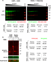
Simultaneous detection of multiple targets in fluorescence immunoblot using CDR in conjunction with CGS. (A) Different amounts of lysates (1:2 serial dilutions from 10 µg/lane) from 293 cells transfected with pAPTAG5 were separated and transferred to PVDF membrane. The blot was probed with anti-6X His tag and anti-ß-actin antibodies, followed by IRDye 800CW goat anti-rabbit IgG and IRDye 680RD goat anti-mouse IgG. Left: antibodies diluted with CGS under CDR condition; right: antibodies diluted with ODS-T under static condition. (B) Cell lysates (20 µg/lane) from HeLa cells treated with either DMSO or PMA were subjected to fluorescence immunoblot with rabbit anti-phospho Erk1/2 antibody and mouse anti-Erk1/2 antibody. Source data are available in Supplementary Fig. S7.
Fig. 7.
Fig. 7.
Schematic illustration of an enhanced and accelerated CDR immunoblot compared to a traditional static method. An immunoblot that utilizes the static method requires at least 150 min to obtain result. Whereas, the equivalent image can be achieved with the CDR method in as early as in 20 min.

References

    1. Schuck P., Zhao H. (2010) The role of mass transport limitation and surface heterogeneity in the biophysical characterization of macromolecular binding processes by SPR biosensing. Methods Mol. Biol. 627, 15–54 - PMC - PubMed
    1. Svitel J., Boukari H., Van Ryk D., Willson R.C., Schuck P. (2007) Probing the functional heterogeneity of surface binding sites by analysis of experimental binding traces and the effect of mass transport limitation. Biophys. J. 92, 1742–1758 - PMC - PubMed
    1. Li J., Zrazhevskiy P., Gao X. (2016) Eliminating size-associated diffusion constraints for rapid on-surface bioassays with nanoparticle probes. Small 12, 1035–1043 - PMC - PubMed
    1. Wang H., Katagiri Y., McCann T.E., Unsworth E., Goldsmith P., Yu Z.X., Tan F., Santiago L., Mills E.M., Wang Y., Symes A.J., Geller H.M. (2008) Chondroitin-4-sulfation negatively regulates axonal guidance and growth. J Cell Sci. 121, 3083–3091 - PMC - PubMed
    1. Laemmli U.K. (1970) Cleavage of structural proteins during the assembly of the head of bacteriophage T4. Nature. 227, 680–685 - PubMed