. 2020 Apr;295(1):210-217.
doi: 10.1148/radiol.2020200274. Epub 2020 Feb 6.

Emerging 2019 Novel Coronavirus (2019-nCoV) Pneumonia

Affiliations

Emerging 2019 Novel Coronavirus (2019-nCoV) Pneumonia

Fengxiang Song et al. Radiology. 2020 Apr.

Erratum in

Abstract

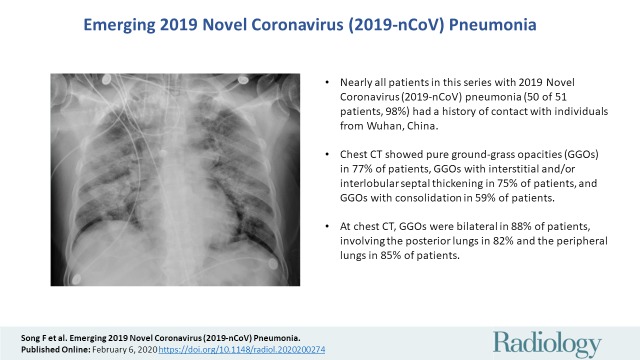
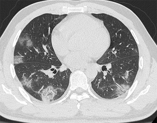
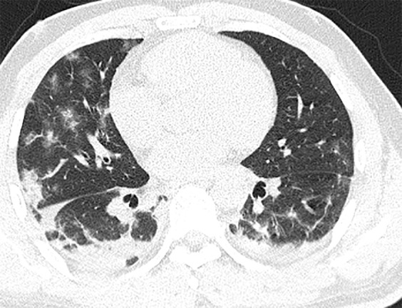
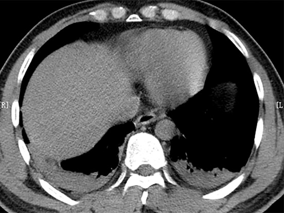
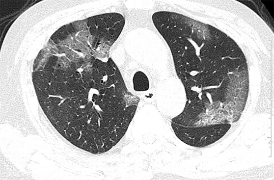
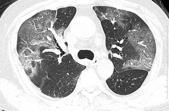
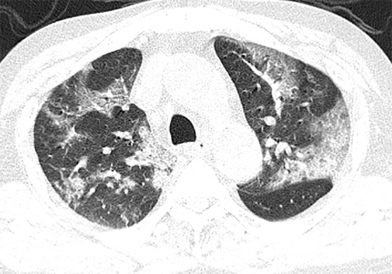
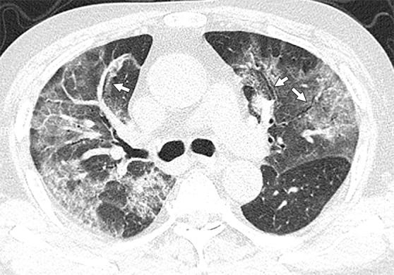
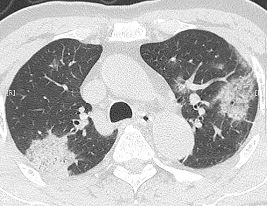
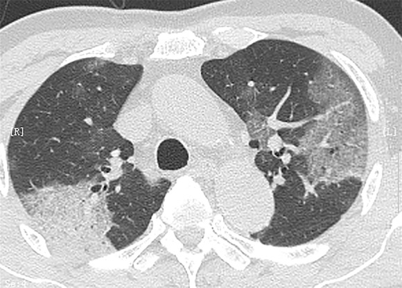
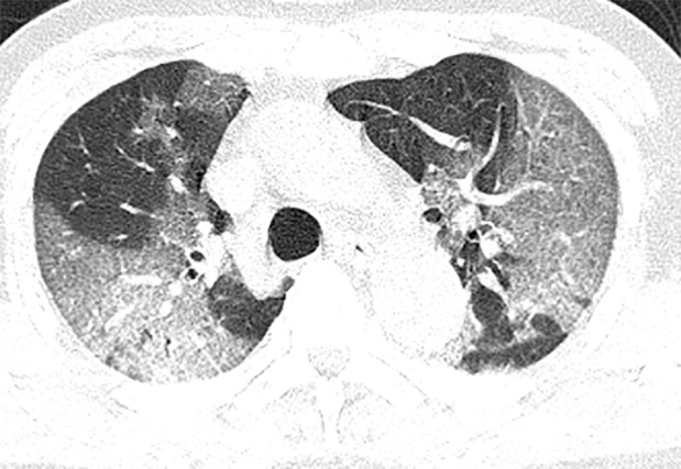
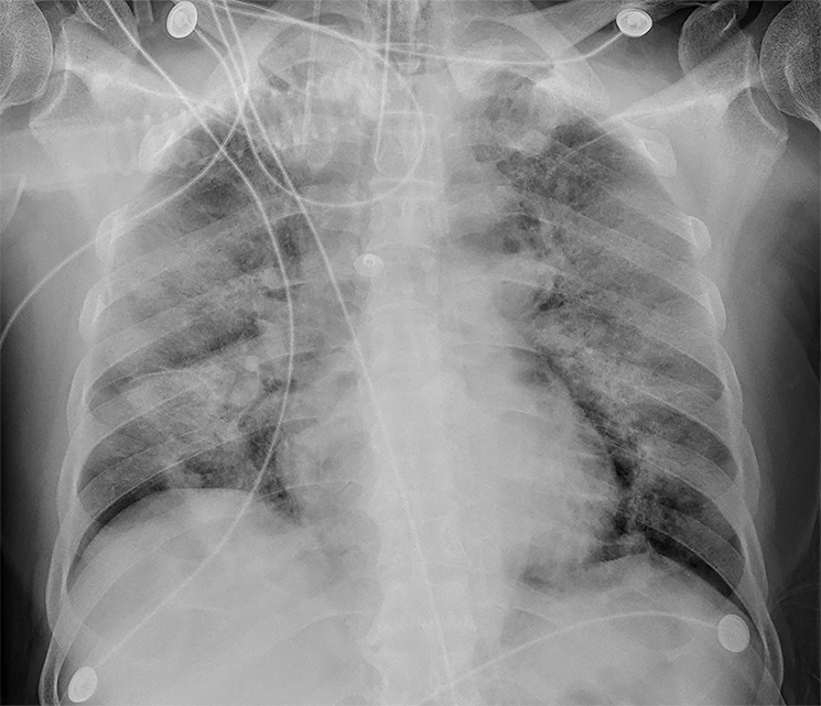
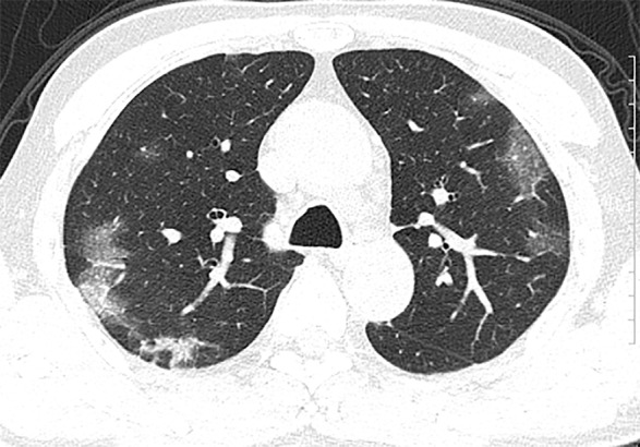
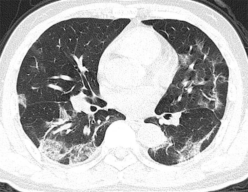
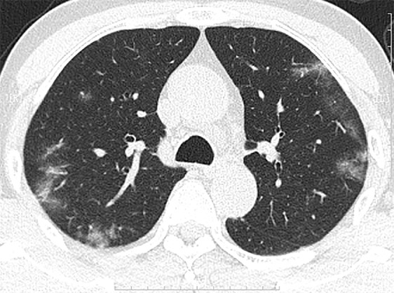
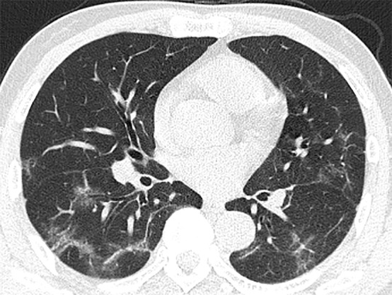
BackgroundThe chest CT findings of patients with 2019 Novel Coronavirus (2019-nCoV) pneumonia have not previously been described in detail.PurposeTo investigate the clinical, laboratory, and imaging findings of emerging 2019-nCoV pneumonia in humans.Materials and MethodsFifty-one patients (25 men and 26 women; age range 16-76 years) with laboratory-confirmed 2019-nCoV infection by using real-time reverse transcription polymerase chain reaction underwent thin-section CT. The imaging findings, clinical data, and laboratory data were evaluated.ResultsFifty of 51 patients (98%) had a history of contact with individuals from the endemic center in Wuhan, China. Fever (49 of 51, 96%) and cough (24 of 51, 47%) were the most common symptoms. Most patients had a normal white blood cell count (37 of 51, 73%), neutrophil count (44 of 51, 86%), and either normal (17 of 51, 35%) or reduced (33 of 51, 65%) lymphocyte count. CT images showed pure ground-glass opacity (GGO) in 39 of 51 (77%) patients and GGO with reticular and/or interlobular septal thickening in 38 of 51 (75%) patients. GGO with consolidation was present in 30 of 51 (59%) patients, and pure consolidation was present in 28 of 51 (55%) patients. Forty-four of 51 (86%) patients had bilateral lung involvement, while 41 of 51 (80%) involved the posterior part of the lungs and 44 of 51 (86%) were peripheral. There were more consolidated lung lesions in patients 5 days or more from disease onset to CT scan versus 4 days or fewer (431 of 712 lesions vs 129 of 612 lesions; P < .001). Patients older than 50 years had more consolidated lung lesions than did those aged 50 years or younger (212 of 470 vs 198 of 854; P < .001). Follow-up CT in 13 patients showed improvement in seven (54%) patients and progression in four (31%) patients.ConclusionPatients with fever and/or cough and with conspicuous ground-glass opacity lesions in the peripheral and posterior lungs on CT images, combined with normal or decreased white blood cells and a history of epidemic exposure, are highly suspected of having 2019 Novel Coronavirus (2019-nCoV) pneumonia.© RSNA, 2020.


Figures

None
Graphical abstract
Figure 1a:
Figure 1a:
(a) Baseline CT images at admission of a 35-year-old man show multiple patchy areas of pure ground-glass opacity (GGO); GGO with reticular and/or interlobular septal thickening; and patchy, focal, often rounded peribronchovascular and subpleural opacities associated with reticulation and architectural distortion. Lesions are mostly distributed in peripheral and posterior part of lungs. (b, c) Follow-up CT images on day 5 after admission show prominent progression with increased size and density of lesions, greater consolidation, and with interval new bilateral pleural effusions and likely bibasilar compression atelectasis (c).
Figure 1b:
Figure 1b:
(a) Baseline CT images at admission of a 35-year-old man show multiple patchy areas of pure ground-glass opacity (GGO); GGO with reticular and/or interlobular septal thickening; and patchy, focal, often rounded peribronchovascular and subpleural opacities associated with reticulation and architectural distortion. Lesions are mostly distributed in peripheral and posterior part of lungs. (b, c) Follow-up CT images on day 5 after admission show prominent progression with increased size and density of lesions, greater consolidation, and with interval new bilateral pleural effusions and likely bibasilar compression atelectasis (c).
Figure 1c:
Figure 1c:
(a) Baseline CT images at admission of a 35-year-old man show multiple patchy areas of pure ground-glass opacity (GGO); GGO with reticular and/or interlobular septal thickening; and patchy, focal, often rounded peribronchovascular and subpleural opacities associated with reticulation and architectural distortion. Lesions are mostly distributed in peripheral and posterior part of lungs. (b, c) Follow-up CT images on day 5 after admission show prominent progression with increased size and density of lesions, greater consolidation, and with interval new bilateral pleural effusions and likely bibasilar compression atelectasis (c).
Figure 2a:
Figure 2a:
(a, b) Baseline CT images at admission of a 75-year-old man show multiple patchy areas of pure ground-glass opacity (GGO) and GGO with reticular and/or interlobular septal thickening. (c, d) Follow-up CT images on day 3 after admission show overlap of organizing pneumonia with diffuse alveolar damage in that it is more diffuse (not rounded) and associated with underlying reticulation, prominent progression with increased size and density of the lesions, and with more consolidations. Air bronchogram is also shown in d (arrows). Interlobular septal thickening does not seem to be major component. There is reticulation in many of cases of both suspected organizing pneumonia and diffuse alveolar damage.
Figure 2b:
Figure 2b:
(a, b) Baseline CT images at admission of a 75-year-old man show multiple patchy areas of pure ground-glass opacity (GGO) and GGO with reticular and/or interlobular septal thickening. (c, d) Follow-up CT images on day 3 after admission show overlap of organizing pneumonia with diffuse alveolar damage in that it is more diffuse (not rounded) and associated with underlying reticulation, prominent progression with increased size and density of the lesions, and with more consolidations. Air bronchogram is also shown in d (arrows). Interlobular septal thickening does not seem to be major component. There is reticulation in many of cases of both suspected organizing pneumonia and diffuse alveolar damage.
Figure 2c:
Figure 2c:
(a, b) Baseline CT images at admission of a 75-year-old man show multiple patchy areas of pure ground-glass opacity (GGO) and GGO with reticular and/or interlobular septal thickening. (c, d) Follow-up CT images on day 3 after admission show overlap of organizing pneumonia with diffuse alveolar damage in that it is more diffuse (not rounded) and associated with underlying reticulation, prominent progression with increased size and density of the lesions, and with more consolidations. Air bronchogram is also shown in d (arrows). Interlobular septal thickening does not seem to be major component. There is reticulation in many of cases of both suspected organizing pneumonia and diffuse alveolar damage.
Figure 2d:
Figure 2d:
(a, b) Baseline CT images at admission of a 75-year-old man show multiple patchy areas of pure ground-glass opacity (GGO) and GGO with reticular and/or interlobular septal thickening. (c, d) Follow-up CT images on day 3 after admission show overlap of organizing pneumonia with diffuse alveolar damage in that it is more diffuse (not rounded) and associated with underlying reticulation, prominent progression with increased size and density of the lesions, and with more consolidations. Air bronchogram is also shown in d (arrows). Interlobular septal thickening does not seem to be major component. There is reticulation in many of cases of both suspected organizing pneumonia and diffuse alveolar damage.
Figure 3a:
Figure 3a:
Serial imaging after admission of a 71-year-old man. (a) Baseline CT images on January 21, 2020, show consolidation of right upper lobe and ground-glass opacity (GGO) with consolidation and reticular and/or interlobular septal thickening of left upper lobe of left upper lobe; and patchy, focal, often rounded peribronchovascular and subpleural opacities associated with reticulation and architectural distortion. (b) Two days later, CT images show increased size of lesions in both lungs and decreased density in GGO lesions. (c) However, GGO on both lungs were larger on day 4 after admission. (d) Bedside portable chest radiograph on day 6 following admission shows diffusely increased opacities in both lungs, with relative bibasilar sparing.
Figure 3b:
Figure 3b:
Serial imaging after admission of a 71-year-old man. (a) Baseline CT images on January 21, 2020, show consolidation of right upper lobe and ground-glass opacity (GGO) with consolidation and reticular and/or interlobular septal thickening of left upper lobe of left upper lobe; and patchy, focal, often rounded peribronchovascular and subpleural opacities associated with reticulation and architectural distortion. (b) Two days later, CT images show increased size of lesions in both lungs and decreased density in GGO lesions. (c) However, GGO on both lungs were larger on day 4 after admission. (d) Bedside portable chest radiograph on day 6 following admission shows diffusely increased opacities in both lungs, with relative bibasilar sparing.
Figure 3c:
Figure 3c:
Serial imaging after admission of a 71-year-old man. (a) Baseline CT images on January 21, 2020, show consolidation of right upper lobe and ground-glass opacity (GGO) with consolidation and reticular and/or interlobular septal thickening of left upper lobe of left upper lobe; and patchy, focal, often rounded peribronchovascular and subpleural opacities associated with reticulation and architectural distortion. (b) Two days later, CT images show increased size of lesions in both lungs and decreased density in GGO lesions. (c) However, GGO on both lungs were larger on day 4 after admission. (d) Bedside portable chest radiograph on day 6 following admission shows diffusely increased opacities in both lungs, with relative bibasilar sparing.
Figure 3d:
Figure 3d:
Serial imaging after admission of a 71-year-old man. (a) Baseline CT images on January 21, 2020, show consolidation of right upper lobe and ground-glass opacity (GGO) with consolidation and reticular and/or interlobular septal thickening of left upper lobe of left upper lobe; and patchy, focal, often rounded peribronchovascular and subpleural opacities associated with reticulation and architectural distortion. (b) Two days later, CT images show increased size of lesions in both lungs and decreased density in GGO lesions. (c) However, GGO on both lungs were larger on day 4 after admission. (d) Bedside portable chest radiograph on day 6 following admission shows diffusely increased opacities in both lungs, with relative bibasilar sparing.
Figure 4a:
Figure 4a:
(a, b) Baseline CT images at admission of a 56-year-old man show multiple patchy areas of organizing pneumonia with some areas of interstitial and/or interlobular septal thickening and “strip shaped” consolidation (patchy, focal, often rounded, peribronchovascular and subpleural opacities associated with reticulation and architectural distortion). These abnormalities are mostly distributed in peripheral and posterior parts of lungs. (c, d) Follow-up CT images on day 5 after admission show interval improvement and absorption with fewer lesions and decreased lesion density.
Figure 4b:
Figure 4b:
(a, b) Baseline CT images at admission of a 56-year-old man show multiple patchy areas of organizing pneumonia with some areas of interstitial and/or interlobular septal thickening and “strip shaped” consolidation (patchy, focal, often rounded, peribronchovascular and subpleural opacities associated with reticulation and architectural distortion). These abnormalities are mostly distributed in peripheral and posterior parts of lungs. (c, d) Follow-up CT images on day 5 after admission show interval improvement and absorption with fewer lesions and decreased lesion density.
Figure 4c:
Figure 4c:
(a, b) Baseline CT images at admission of a 56-year-old man show multiple patchy areas of organizing pneumonia with some areas of interstitial and/or interlobular septal thickening and “strip shaped” consolidation (patchy, focal, often rounded, peribronchovascular and subpleural opacities associated with reticulation and architectural distortion). These abnormalities are mostly distributed in peripheral and posterior parts of lungs. (c, d) Follow-up CT images on day 5 after admission show interval improvement and absorption with fewer lesions and decreased lesion density.
Figure 4d:
Figure 4d:
(a, b) Baseline CT images at admission of a 56-year-old man show multiple patchy areas of organizing pneumonia with some areas of interstitial and/or interlobular septal thickening and “strip shaped” consolidation (patchy, focal, often rounded, peribronchovascular and subpleural opacities associated with reticulation and architectural distortion). These abnormalities are mostly distributed in peripheral and posterior parts of lungs. (c, d) Follow-up CT images on day 5 after admission show interval improvement and absorption with fewer lesions and decreased lesion density.

Comment in

References

    1. ProMED-mail. https://promedmail.org/promed-post/?id=6864153. Accessed January 7, 2020.
    1. Huang C, Wang Y, Li X, et al. . Clinical features of patients infected with 2019 novel coronavirus in Wuhan, China. Lancet 2020 Jan 24 [Epub ahead of print] [Published correction appears in Lancet 2020 Jan 30.]. - PMC - PubMed
    1. World Health Organization . Novel coronavirus – Thailand (ex-China). http://www.who.int/csr/don/14-january-2020-novel-coronavirus-thailand/en/. Published January 14, 2020. Accessed January 19, 2020.
    1. World Health Organization . Novel coronavirus – Japan (ex-China). http://www.who.int/csr/don/17-january-2020-novel-coronavirus-japan-ex-ch.... Published January 17, 2020. Accessed January 19, 2020.
    1. World Health Organization . Novel coronavirus – Republic of Korea (ex-China). http://www.who.int/csr/don/21-january-2020-novel-coronavirus-republic-of.... Published January 21, 2020. Accessed January 23, 2020.

MeSH terms