. 2020 May 1;130(5):2657-2672.
doi: 10.1172/JCI128867.

Exosomes mediate sensory hair cell protection in the inner ear

Affiliations

Exosomes mediate sensory hair cell protection in the inner ear

Andrew M Breglio et al. J Clin Invest. .

Abstract

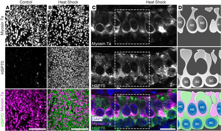
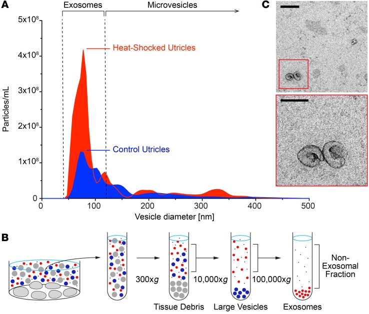
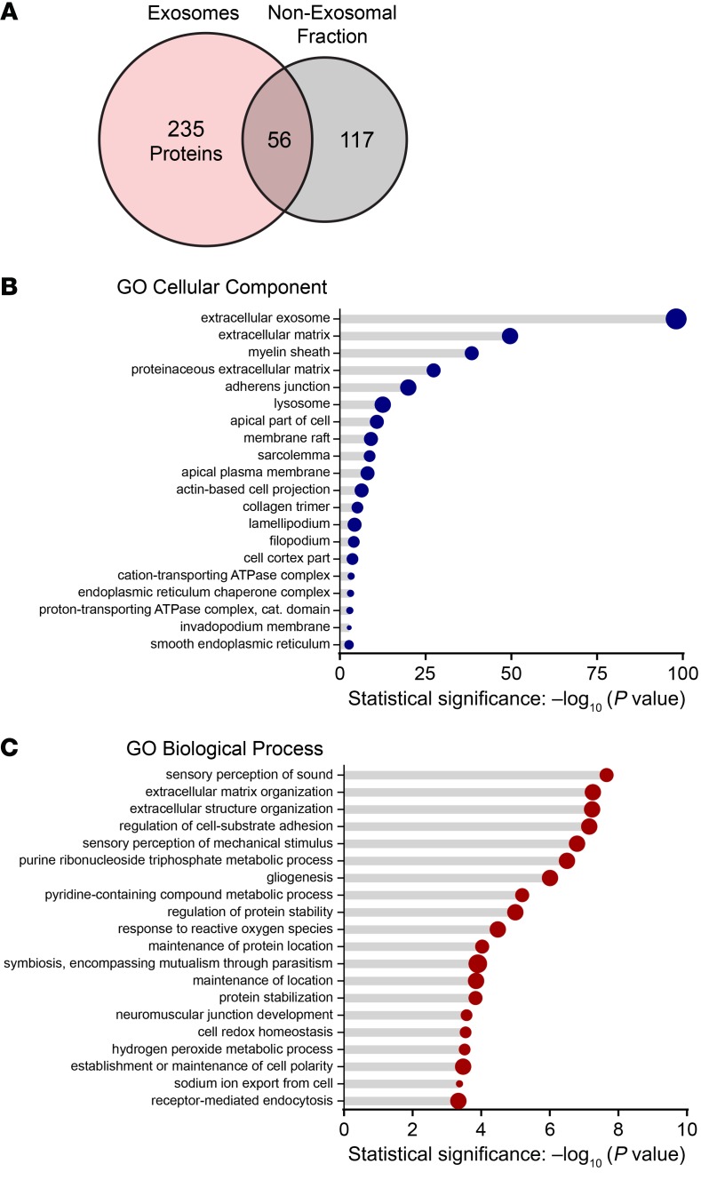
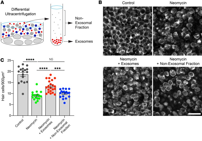
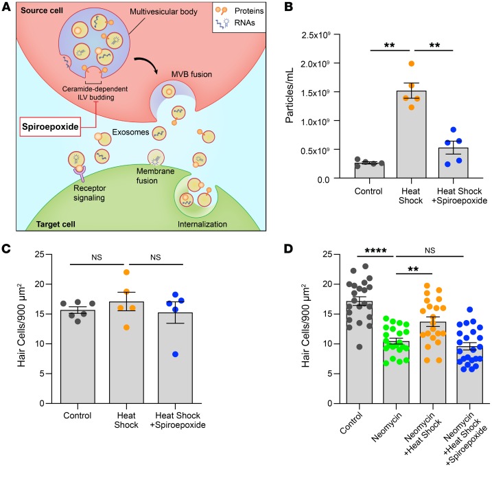
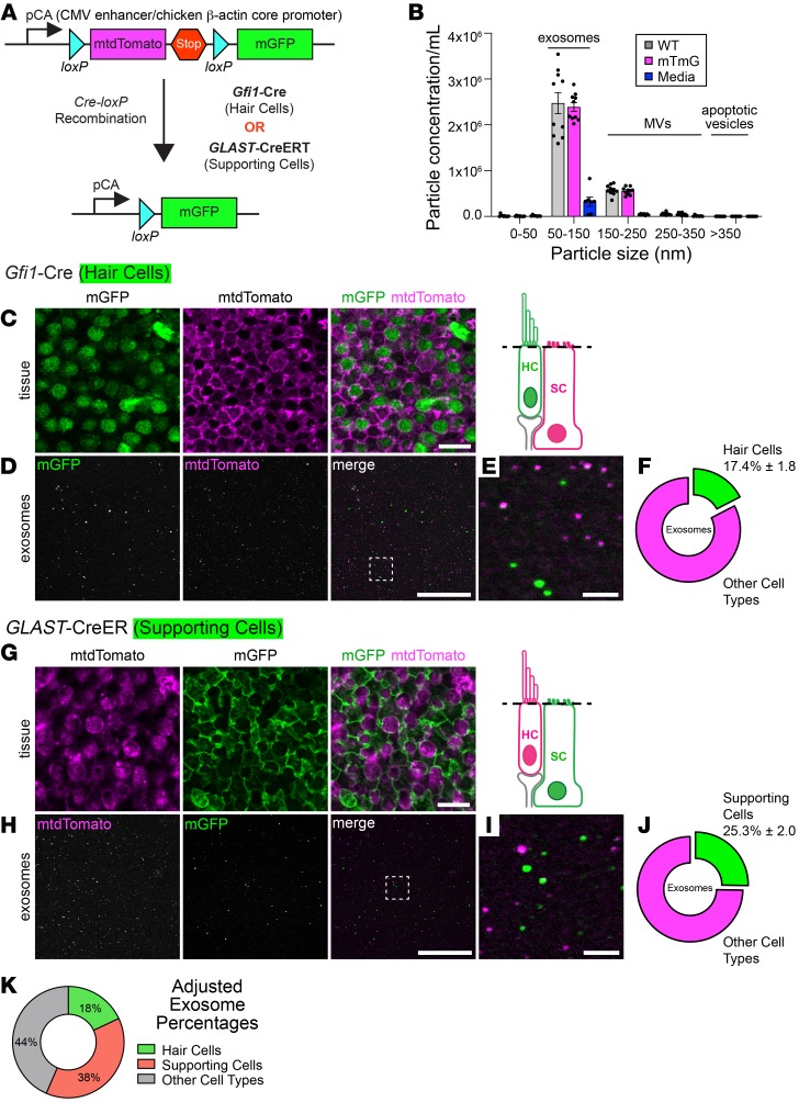
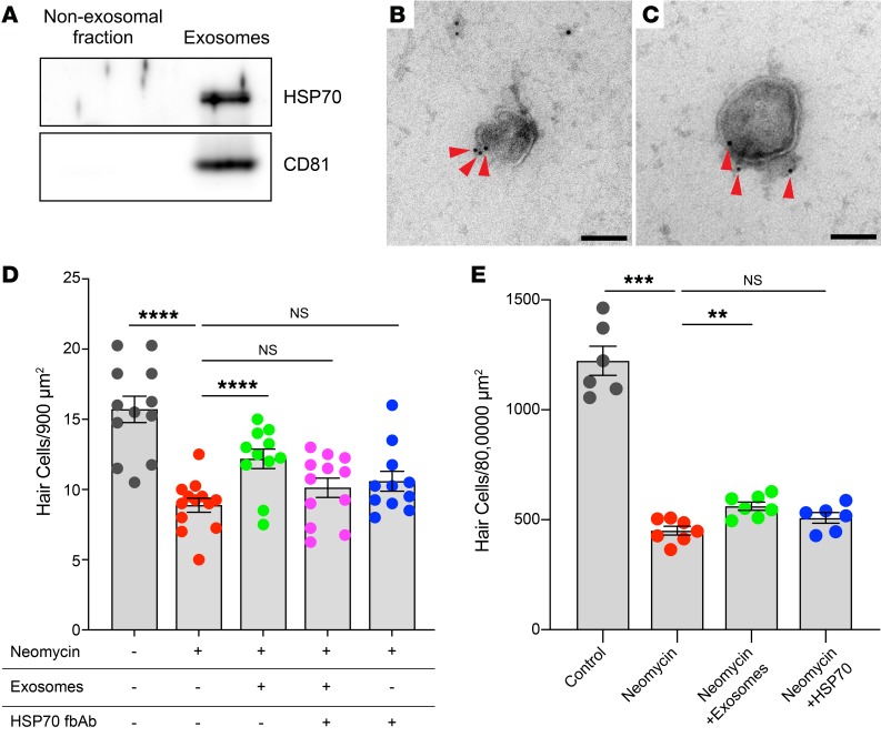
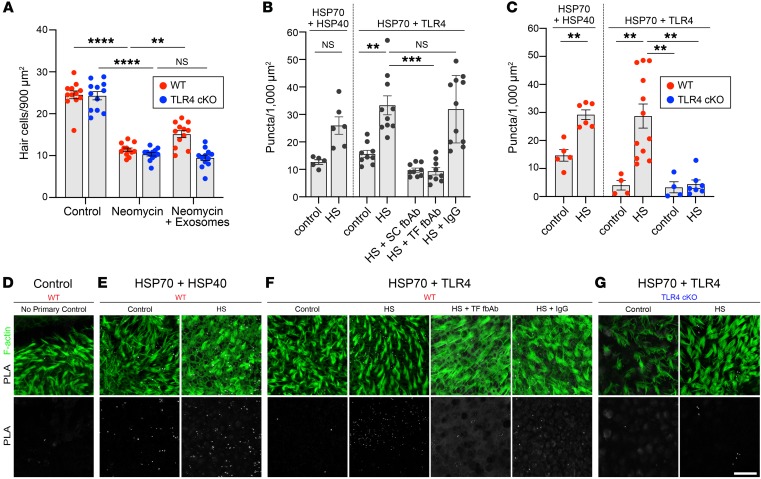
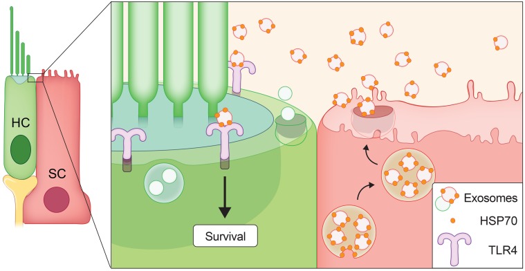
Hair cells, the mechanosensory receptors of the inner ear, are responsible for hearing and balance. Hair cell death and consequent hearing loss are common results of treatment with ototoxic drugs, including the widely used aminoglycoside antibiotics. Induction of heat shock proteins (HSPs) confers protection against aminoglycoside-induced hair cell death via paracrine signaling that requires extracellular heat shock 70-kDa protein (HSP70). We investigated the mechanisms underlying this non-cell-autonomous protective signaling in the inner ear. In response to heat stress, inner ear tissue releases exosomes that carry HSP70 in addition to canonical exosome markers and other proteins. Isolated exosomes from heat-shocked utricles were sufficient to improve survival of hair cells exposed to the aminoglycoside antibiotic neomycin, whereas inhibition or depletion of exosomes from the extracellular environment abolished the protective effect of heat shock. Hair cell-specific expression of the known HSP70 receptor TLR4 was required for the protective effect of exosomes, and exosomal HSP70 interacted with TLR4 on hair cells. Our results indicate that exosomes are a previously undescribed mechanism of intercellular communication in the inner ear that can mediate nonautonomous hair cell survival. Exosomes may hold potential as nanocarriers for delivery of therapeutics against hearing loss.

Keywords: Apoptosis survival pathways; Cell Biology; Neurodegeneration; Neuroscience.


Conflict of interest statement

Conflict of interest: Matthew Wood is a founder and director of Evox Therapeutics.

Figures

Figure 1
Figure 1. HSP70 is upregulated in supporting cells upon heat shock.
Mouse utricles were cultured under control or heat shock conditions, fixed 6 hours later, and labeled with Abs against myosin 7a (hair cell marker, magenta) and inducible HSP70 (green). (A and B) Confocal images of whole-mount utricles show sensory hair cells surrounded by glia-like supporting cells. (A) Inducible HSP70 was not detected under control culture conditions. (B) Heat shock induced HSP70 specifically in supporting cells, with very little induction in hair cells. (C) Cryosection of a heat-shocked utricle confirmed negligible induction of HSP70 (green) in hair cells (labeled with myosin 7a, magenta), whereas HSP70 was substantially upregulated in supporting cells. Nuclei in AC were stained with DAPI (blue). (D) Schematics showing locations of hair cells (HC, magenta) and supporting cells (SC, green) in boxes outlined in C, with nuclei indicated in blue in the lower right panel. Scale bars: 50 μm (A and B) and 10 μm (C).
Figure 2
Figure 2. Heat stress stimulates exosome release from inner ear tissue.
(A) Nanoparticle tracking analysis of conditioned media from utricles cultured under control conditions shows release of exosome-sized (~50–150 nm diameter) particles from control utricles. Heat shock resulted in a 2.4-fold increase in exosome release. Horizontal bars denote typical size ranges of exosomes and microvesicles. (B) Schematic of differential ultracentrifugation procedure used to isolate exosomes from utricle-conditioned culture medium. This process sequentially sediments extracellular components of decreasing size (tissue debris, gray; large vesicles, blue), with exosomes (red) isolated in the final pellet. (C) Isolated exosomes from utricle-conditioned media visualized by TEM were approximately 90 nm in diameter and displayed canonical cup-shaped morphology. Scale bars: 200 nm (top); 100 nm (enlarged inset).
Figure 3
Figure 3. Proteins identified in utricle exosomes.
Proteins associated with exosomes and non-exosomal proteins secreted from heat-shocked utricles were identified using tandem mass spectrometry. Exosomes were isolated from conditioned media via size-exclusion chromatography and analyzed in parallel with the exosome-depleted media (non-exosomal fraction). (A) A total of 291 unique protein families were identified in exosomes, 56 of which were also found in the non-exosomal fraction. (B) The 20 most significantly enriched GO cellular component terms for proteins identified in utricle-derived exosomes. (C) The 20 most significantly enriched biological process GO terms for proteins identified in exosomes from utricles.
Figure 4
Figure 4. Isolated exosomes protect against neomycin-induced hair cell death.
Utricles were cultured for 24 hours in neomycin, with or without the addition of exosomes isolated from heat-shocked utricles. (A) Exosomes and the non-exosomal fraction (supernatant) were purified from utricle-conditioned media using differential ultracentrifugation (see Figure 2B) and applied to neomycin-treated utricles. (B) Fixed utricles were labeled with the hair cell marker myosin 7a, and images were acquired using laser scanning confocal microscopy. Representative z sections from surface preparations of utricle whole mounts are shown. Scale bar: 20 μm. (C) Neomycin caused hair cell death. Application of isolated exosomes significantly improved hair cell survival. In contrast, no protective effect was observed when the non-exosomal fraction (i.e., exosome-depleted conditioned media) was added. Each data point represents the average hair cell density of an individual utricle. n = 16–20 utricles per condition from 4 independent experiments. Data indicate the mean ± SEM. ***P < 0.001 and ****P < 0.0001, by Brown-Forsythe and Welsh ANOVA followed by Dunnett’s T3 multiple comparisons test.
Figure 5
Figure 5. Exosomes are required for the protective effect of heat shock.
(A) Exosomes containing proteins, nucleic acids, and lipids are released from source cells (red) and can modify the biological state of target cells (green) via a variety of interactions. Within the source cell, exosome biogenesis occurs via budding of intraluminal vesicles (ILV) into the lumen of a multivesicular body (MVB) (purple), a process that requires the sphingolipid ceramide. The N-SMase inhibitor spiroepoxide blocks ceramide production and inhibits exosome biogenesis. (B) Inhibition of exosome biogenesis reduced the number of exosome-sized particles in conditioned media from heat-shocked utricles. Data indicate the mean ± SEM for 5 NTA captures. (C) Quantification of surviving hair cells in utricles demonstrated that reduced exosome release in the presence of spiroepoxide was not caused by cytotoxicity. n = 5–6 utricles per condition. (D) Utricles were cultured for 24 hours in neomycin, with or without heat shock and with or without spiroepoxide. Neomycin caused hair cell death, whereas heat shock improved survival of neomycin-exposed hair cells. Inhibition of exosome biogenesis using spiroepoxide abolished the protective effect of heat shock. n = 21–23 utricles (shown as individual data points) per condition. Data indicate the mean ± SEM. **P < 0.01 and ****P < 0.0001, by Brown-Forsythe and Welsh ANOVA followed by Dunnett’s T3 multiple comparisons test (B and D) or 1-way ANOVA followed by Holm-Šídák multiple comparisons test (C).
Figure 6
Figure 6. Supporting cells release more exosomes than hair cells under heat stress.
(A) mTmG double-fluorescent reporter mice constitutively express myristoylated tdTomato. When crossed with Cre recombinase–expressing mice, the loxP-flanked mtdTomato cassette is deleted in all Cre-expressing cells, resulting in tissue-specific mGFP fluorescence. (B) NTA of conditioned media from heat-shocked utricles of mTmG mice (magenta) or age-matched WT mice (gray) shows that lipidation of fluorophores in mTmG mice did not affect exosome release. The culture media (blue) contributed 10% of particles to each size category. MVs, microvesicles. Data indicate the mean ± SEM and are from 2 independent experiments (n = 5 NTA captures from 22 utricles for each condition). (C) Utricles from mTmG mice crossed with Gfi1-Cre mice displayed mGFP-expressing hair cells (green), whereas supporting cells retained mtdTomato expression (magenta). Schematic depicts the focal plane. (D) Fluorescence emitted from utricle-derived exosomes from mTmG mice crossed with Gfi1-Cre mice. Box indicates the region magnified in E. (F) 17.4% of utricle-derived exosomes in D were mGFP positive. (G) Supporting cells in mTmG mice crossed with GLAST-CreER mice were mGFP positive (green). All other cells retained mtdTomato expression (magenta). (H) Fluorescence emitted from utricle-derived exosomes from mTmG mice crossed with GLAST-CreER mice. Box indicates the region magnified in I. (J) 25.3% of exosomes visualized in H were mGFP positive. (K) Contributions of hair cells and supporting cells to the total utricle-derived exosome population after taking into account Cre recombinase efficiency in hair cells (Gfi1-Cre = 96.5%) and supporting cells (GLAST-CreER = 64.5%). Results showed that 44% of exosomes were probably contributed by other cell types. Data in F and J are presented as the mean ± SEM and are from 3 experiments (n = 9–11 utricles per condition). Scale bars: 10 μm (C and G), 50 μm (D and H), and 5 μm (E and I).
Figure 7
Figure 7. Exosomes contain HSP70, and exosome-associated HSP70 is required for the protective effect of exosomes.
(A) Western blot shows the association of HSP70 with exosomes but not the non-exosomal fraction. Similarly, CD81 (exosome marker) was detected exclusively in exosomes. Exosomes and the non-exosomal fraction were isolated from heat-shocked utricles by differential ultracentrifugation (Figure 2B). Equivalent amounts of total protein were loaded for each lane. See complete unedited blots in the supplemental material. (B and C) Two representative Immunogold TEM micrographs of utricle-derived exosomes revealed HSP70 immunoreactivity (red arrowheads) near the exosomal membrane. Scale bars: 100 nm. (D) HSP70 was required for the protective effect of exosomes. Utricles were cultured for 24 hours in the presence of neomycin, with or without the addition of exosomes isolated from heat-shocked utricles. Exosomes significantly protected hair cells against neomycin-induced ototoxicity. Addition of an HSP70 fbAb abolished the protective effect of exosomes. Each data point represents the average hair cell density of an individual utricle from 4 independent experiments (n = 11–13 utricles per condition). (E) Non-exosomal HSP70 was not protective against neomycin-induced hair cell death. Utricles were cultured for 24 hours in the presence of neomycin, with the addition of either isolated exosomes or soluble (recombinant, non-exosomal) HSP70. Exogenous HSP70 failed to protect hair cells, whereas exosomes were protective. Data indicate the mean ± SEM. **P < 0.01, ***P < 0.001, ****P < 0.0001, by Brown-Forsythe and Welsh ANOVA followed by Dunnett’s T3 multiple comparisons test.
Figure 8
Figure 8. The protective effect of exosomes requires interaction of exosomal HSP70 with TLR4 on hair cells.
(A) Exosomes improved hair cell survival in neomycin-exposed utricles from control (WT) littermates (red) but not from hair cell–specific TLR4-cKO mice (blue). Data indicate the mean ± SEM (n = 12 utricles per condition). **P < 0.01 and ****P < 0.0001, by 2-way ANOVA with Holm-Šídák multiple comparisons test. (B and C) A PLA was performed to detect interaction between exosomal HSP70 and HSP40 or HSP70 and TLR4. (B) Two different fbAbs against HSP70 (HS) abolished the interaction between HSP70 and TLR4, whereas IgG had no effect. SC, Santa Cruz Biotechnology; TF, Thermo Fisher Scientific. (C) Heat shock increased the interaction between HSP70 and HSP40 and between HSP70 and TLR4 in WT utricles. Hair cell–specific deletion of TLR4 abolished the PLA signal in heat-shocked utricles from TLR4-cKO mice. Data in B and C indicate the mean ± SEM and are shown as the average number of puncta per 1000 μm2 (n = 4–12 utricles per condition). **P < 0.01 and ***P < 0.001, by Brown-Forsythe and Welsh ANOVA followed by Dunnett’s T3 multiple comparisons test. (DG) Confocal images of PLA signals in utricles from WT (DF) or TLR4-cKO (G) mice under control or heat shock conditions. Top row, F-actin (green) and PLA signal (white); bottom row, PLA signal only (white). (D) Negative control (no primary Ab). (E) Heat shock induced HSP70 interaction with HSP40 in WT utricles. (F) Heat shock increased HSP70 interaction with TLR4 in WT utricles. HSP70 fbAbs inhibited interaction between HSP70 and TLR4 in WT utricles, independently of heat shock, whereas control IgG had no effect. (G) Hair cell–specific deletion of TLR4 abolished the HSP70-TLR4 interaction in TLR4-cKO mice. Scale bar: 20 μm.
Figure 9
Figure 9. Model for the role of exosomes as mediators of protection against hair cell death caused by ototoxic drugs.
Our data are consistent with a model in which heat stress induces HSP70 expression, predominately in supporting cells (Figure 1). Some of this HSP70 associates with exosomes (Table 1 and Figure 7, A and B) that are released into the extracellular environment (Figure 2A and Figure 5B). HSP70-carrying exosomes interact with TLR4 on the hair cell’s surface (Figure 8) to stimulate a pro-survival response (Figure 4, B and C, and Figure 5D). The significance of hair cell–derived exosomes (Figure 6, C–K) remains under investigation.

Comment in

References

    1. World Health Organization. WHO Global Estimates on Prevalence of Hearing Loss. http://www.who.int/deafness/estimates/en Accessed March 6, 2020.
    1. Francis SP, Kramarenko, Brandon CS, Lee FS, Baker TG, Cunningham LL. Celastrol inhibits aminoglycoside-induced ototoxicity via heat shock protein 32. Cell Death Dis. 2011;2:e195. - PMC - PubMed
    1. Francis SP, et al. A novel role of cytosolic protein synthesis inhibition in aminoglycoside ototoxicity. J Neurosci. 2013;33(7):3079–3093. doi: 10.1523/JNEUROSCI.3430-12.2013. - DOI - PMC - PubMed
    1. Hailey DW, Esterberg R, Linbo TH, Rubel EW, Raible DW. Fluorescent aminoglycosides reveal intracellular trafficking routes in mechanosensory hair cells. J Clin Invest. 2017;127(2):472–486. doi: 10.1172/JCI85052. - DOI - PMC - PubMed
    1. Huth ME, Ricci AJ, Cheng AG. Mechanisms of aminoglycoside ototoxicity and targets of hair cell protection. Int J Otolaryngol. 2011;2011:937861. - PMC - PubMed

Publication types

MeSH terms