Review
. 2020 Jan;22(1):29-46.
doi: 10.5853/jos.2019.02236. Epub 2020 Jan 31.

Neuroinflammation after Intracerebral Hemorrhage and Potential Therapeutic Targets

Affiliations
Review

Neuroinflammation after Intracerebral Hemorrhage and Potential Therapeutic Targets

Christine Tschoe et al. J Stroke. 2020 Jan.

Abstract

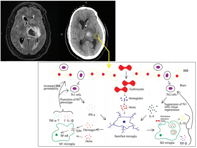
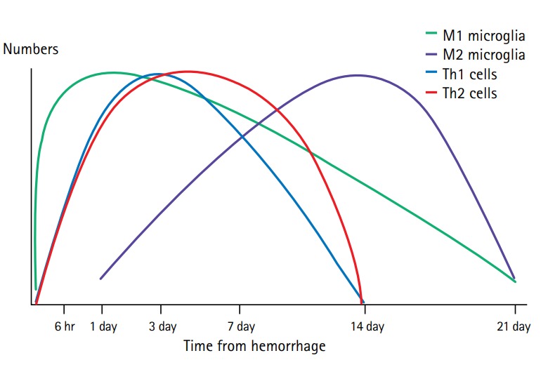
Spontaneous intracerebral hemorrhage (ICH) is a catastrophic illness causing significant morbidity and mortality. Despite advances in surgical technique addressing primary brain injury caused by ICH, little progress has been made treating the subsequent inflammatory cascade. Pre-clinical studies have made advancements identifying components of neuroinflammation, including microglia, astrocytes, and T lymphocytes. After cerebral insult, inflammation is initially driven by the M1 microglia, secreting cytokines (e.g., interleukin-1β [IL-1β] and tumor necrosis factor-α) that are involved in the breakdown of the extracellular matrix, cellular integrity, and the blood brain barrier. Additionally, inflammatory factors recruit and induce differentiation of A1 reactive astrocytes and T helper 1 (Th1) cells, which contribute to the secretion of inflammatory cytokines, augmenting M1 polarization and potentiating inflammation. Within 7 days of ICH ictus, the M1 phenotype coverts to a M2 phenotype, key for hematoma removal, tissue healing, and overall resolution of inflammation. The secretion of anti-inflammatory cytokines (e.g., IL-4, IL-10) can drive Th2 cell differentiation. M2 polarization is maintained by the secretion of additional anti-inflammatory cytokines by the Th2 cells, suppressing M1 and Th1 phenotypes. Elucidating the timing and trigger of the anti-inflammatory phenotype may be integral in improving clinical outcomes. A challenge in current translational research is the absence of an equivalent disease animal model mirroring the patient population and comorbid pathophysiologic state. We review existing data and describe potential therapeutic targets around which we are creating a bench to bedside translational research model that better reflects the pathophysiology of ICH patients.

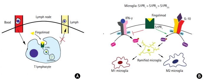
Keywords: Cerebral hemorrhage; Fingolimod hydrochloride; Immunomodulation; Neuroinflammation; Programmed death-1; Stroke.


Figures

Figure 1.
Figure 1.
Magnetic resonance imaging fluid attenuated inversion recovery (left) and computed tomography (right) illustrating cerebral edema surrounding the primary intracerebral hemorrhage. Cerebral edema is the result of the ongoing inflammatory response occurring in the presence of the breakdown products of the hematoma. The initial pro-inflammatory phase involves the activation of M1 microglia, upregulating the production of interleukin-1β (IL-1β), tumor necrosis factor-α (TNF-α), and other inflammatory products that are involved in potentiating cellular damage and the breakdown of the blood brain barrier (BBB). Once the M2 microglia are activated, there is release of anti-inflammatory cytokines IL-4, IL-10, and transforming growth factor-β (TGF-β), all involved in the regeneration of tissue and overall resolution of neuroinflammation. IFN-γ, interferon-γ; Th, T helper.
Figure 2.
Figure 2.
Timeline of inflammatory cells after intracerebral hemorrhage. These timelines are based on young, healthy male animal models with data based on normal immune responses. When accounting for age and comorbidities, the timing and activation of different immune cells may differ in the target population of patients with intracerebral hemorrhage. Th, T helper.
Figure 3.
Figure 3.
Programmed death-1 (PD-1)/programmed death-ligand 1 (PD-L1) driven polarization of microglia. In the setting of intracerebral hemorrhage, upregulation of PD-1 has been noted in the perihematomal tissue. After the administration of PD-L1, the overall number of T cells infiltrating the central nervous system was decreased, with decreased total T helper 1 (Th1) and Th17 cells and increased Th2 and T regulatory cells. There is also increased polarization to the M2 phenotype in the setting of signal transducer and activator of transcription 1 (STAT1) inhibition. IFN-γ, interferon-γ; JAK, Janus kinase; IL-4, interleukin-4.
Figure 4.
Figure 4.
Fingolimod’s proposed anti-inflammatory functions. (A) The sphingosine 1-phosphate (S1P) and sphingosine 1-phosphate receptor 1 (S1PR1) interactions on lymphocytes are key for exiting the lymph nodes. In the inactivated state, S1PR1 undergoes cyclical expression on circulating T cells. In the blood and lymph, S1PRs are typically downregulated in the presence of a high concentration of S1P. When the T cells circulate within the lymph nodes, S1PR expression is upregulated in the setting of low S1P concentrations. If an appropriate antigen is not encountered, T cells will exit, following the gradient of S1P. In the setting of fingolimod, the receptors internalize and are degraded. This prevents the sensing of the S1P gradient, thereby preventing the egress of the T cells. (B) In the central nervous system, S1PR1 is noted to be expressed in high concentrations on microglia. It is proposed that fingolimod increases the expression and phosphorylation of signal transducer and activator of transcription 3 (STAT3) and inhibiting STAT1, potentiating the polarization the M2 phenotype. IFN-γ, interferon-γ; JAK, Janus kinase; IL-10, interleukin 10.

References

    1. Qureshi AI, Mendelow AD, Hanley DF. Intracerebral haemorrhage. Lancet. 2009;373:1632–1644. - PMC - PubMed
    1. van Asch CJ, Luitse MJ, Rinkel GJ, van der Tweel I, Algra A, Klijn CJ. Incidence, case fatality, and functional outcome of intracerebral haemorrhage over time, according to age, sex, and ethnic origin: a systematic review and meta-analysis. Lancet Neurol. 2010;9:167–176. - PubMed
    1. Fogelholm R, Murros K, Rissanen A, Avikainen S. Long term survival after primary intracerebral haemorrhage: a retrospective population based study. J Neurol Neurosurg Psychiatry. 2005;76:1534–1538. - PMC - PubMed
    1. Writing Group Members, Lloyd-Jones D, Adams RJ, Brown TM, Carnethon M, Dai S, et al. Heart disease and stroke statistics-2010 update: a report from the American Heart Association. Circulation. 2010;121:e46–e215. - PubMed
    1. Qureshi AI, Suri MF, Nasar A, Kirmani JF, Ezzeddine MA, Divani AA, et al. Changes in cost and outcome among US patients with stroke hospitalized in 1990 to 1991 and those hospitalized in 2000 to 2001. Stroke. 2007;38:2180–2184. - PubMed

LinkOut - more resources