. 2020 May 5;118(9):2280-2296.
doi: 10.1016/j.bpj.2019.12.038. Epub 2020 Jan 16.

Nucleosomal DNA Dynamics Mediate Oct4 Pioneer Factor Binding

Affiliations

Nucleosomal DNA Dynamics Mediate Oct4 Pioneer Factor Binding

Jan Huertas et al. Biophys J. .

Abstract

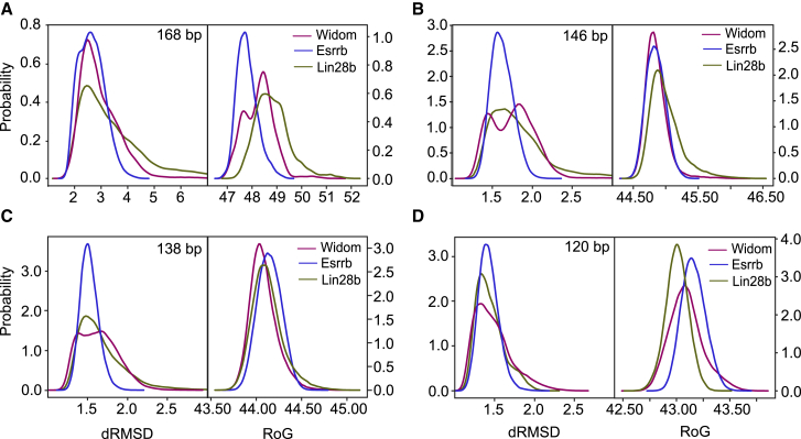
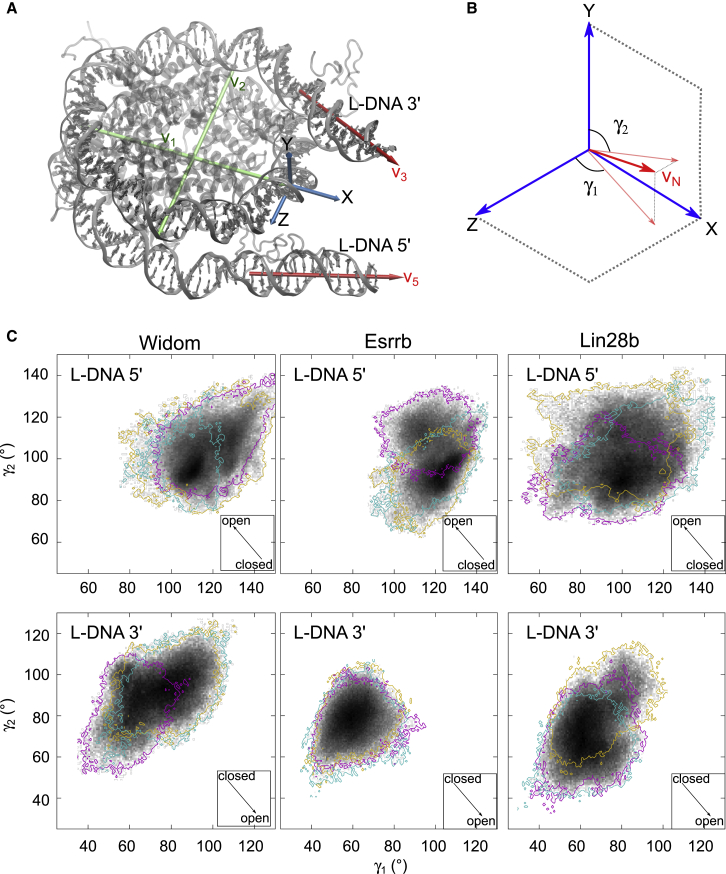
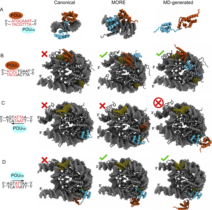
Transcription factor (TF) proteins bind to DNA to regulate gene expression. Normally, accessibility to DNA is required for their function. However, in the nucleus, the DNA is often inaccessible, wrapped around histone proteins in nucleosomes forming the chromatin. Pioneer TFs are thought to induce chromatin opening by recognizing their DNA binding sites on nucleosomes. For example, Oct4, a master regulator and inducer of stem cell pluripotency, binds to DNA in nucleosomes in a sequence-specific manner. Here, we reveal the structural dynamics of nucleosomes that mediate Oct4 binding from molecular dynamics simulations. Nucleosome flexibility and the amplitude of nucleosome motions such as breathing and twisting are enhanced in nucleosomes with multiple TF binding sites. Moreover, the regions around the binding sites display higher local structural flexibility. Probing different structures of Oct4-nucleosome complexes, we show that alternative configurations in which Oct4 recognizes partial binding sites display stable TF-DNA interactions similar to those observed in complexes with free DNA and compatible with the DNA curvature and DNA-histone interactions. Therefore, we propose a structural basis for nucleosome recognition by a pioneer TF that is essential for understanding how chromatin is unraveled during cell fate conversions.


Figures

Figure 1
Figure 1
Asymmetric nucleosome flexibility. (AC) Normalized, relative atomic fluctuations of the nonhydrogen DNA atoms calculated from the simulation ensembles of the three nucleosomes are shown: (A) Widom, (B) Esrrb, and (C) Lin28b. First, the absolute atomic fluctuations were calculated, and the average (mass weighted) per residue was then squared and weighted by 8/3π2 to obtain factor β analogs to crystallographic B-factors. The nucleosomes were colored by ββˆ, where βˆ is the mean β of the core nucleosomal DNA (146 bp). The residues with fluctuations smaller and larger than the average fluctuations of the core DNA are in blue and red, respectively, with darkest red corresponding to ββˆ = 2 to avoid the masking of fluctuations by the very high values found in the L-DNA. (DE) Phase space sampling of the core histone tails during the simulation ensembles is shown. The positions of all Cα atoms of the H3 (D) and H2B (E) tails are shown at every 20 ns. The colors indicate the three nucleosomes—Widom (magenta), Esrrb (blue), and Lin28b (green)—and are maintained throughout this article. To see this figure in color, go online.
Figure 2
Figure 2
Sequence-dependent nucleosome flexibility. Distributions of the RMSD of interatomic distances in the DNA sugar-phosphate backbone (dRMSD, left) and radius of gyration (RoG, right) from the simulation ensembles of the three nucleosomes. The distributions were calculated for different regions of DNA: (A) the entire simulated DNA (168 bp), (B) the core nucleosomal DNA (146 bp) obtained by excluding 11 bp of L-DNA at each end, (C) nucleosomal DNA obtained by excluding 15 bp of DNA at each end (138 bp), and (D) nucleosomal DNA corresponding to the complete inner DNA turn and half of the outer DNA turn obtained by excluding 23 bp of DNA at each end (120 bp). To see this figure in color, go online.
Figure 3
Figure 3
Motions of the linker DNA arms. (A) The coordinate system definition is given. The XYZ reference system was defined as follows (see Materials and Methods for details): X was defined along the dyad axis, Y as the cross products between a vector v1 defined along the dyad axis and a vector v2 defined to be approximately orthogonal to v1 and to intersect v1 as close as possible to the center of the nucleosome, and Z as the cross product between X and Y. (B) Definition of the angles, γ1 and γ2, ισ γιϖεν. γ1 is the angle between the projection of the vector vN defined along the helical axis of the L-DNA on the XZ plane and the Z axis. γ2 is the angle formed between the projection of vN on the XY plane and the Y axis. An increase of γ1 indicates opening at the 3′ L-DNA but closing at the 5′ L-DNA, whereas an increase of γ2 indicates closing at the 3′ L-DNA and opening at the 5′ L-DNA. (C) Two-dimensional histograms showing the sampling of the γ1/γ2 conformational space for both 5′ L-DNA and 3′ L-DNA. The colors (cyan, magenta, and gold) indicate the sampling covered by each of the three 1 μs simulations. The arrows in the square inserts indicate the direction of the nucleosome opening. To see this figure in color, go online.
Figure 4
Figure 4
Correlations of nucleosome opening-closing motions. Evolution of the angles γ1 and γ2 in the two first PCs. After PCA of the ensemble of simulations for each nucleosome, pseudotrajectories along the two first eigenvectors were generated. Then, the motion of the L-DNA arms was analyzed as described in Fig. 3. The motions corresponding to the first and second eigenvectors are in red and blue, respectively. Light and dark colors indicate the low and high amplitudes of the motions, respectively. The arrows in the square inserts indicate the direction of the nucleosome opening. To see this figure in color, go online.
Figure 5
Figure 5
Twisting motions in the nucleosomes. (A) The coordinate system definition is given. The XYZ reference system was defined using the following approach (see also Materials and Methods): X is along a vector defined to connect the N1 atom of the pyrimidine with the N9 atom of the purine. Z is along the cross product between X and a vector defined from atom positions to be approximately orthogonal to X. The intersection of this vector with X is the origin of the coordinate system and is near the bp center. Y is the cross product between X and Z (see also Fig. S7A). (B) A schematic representation of the motion of a bp during nucleosome twisting is shown. In each snapshot along the PC trajectory, the coordinate system is redefined. Then, the displacement d of the current position of the origin from the position at the minimum amplitude and the angle α between the current Y axis and the Y axis at minimum amplitude (Y0) were calculated. (C) The range of α is plotted for each bp along the first five PC trajectories. Each point is colored by the value of d. Twisting motions are characterized by high values of α and low displacement values and are indicated with black arrows. To see this figure in color, go online.
Figure 6
Figure 6
Local flexibility in the nucleosomes. Distributions of dRMSD for different DNA segments are shown. The first segment was the 8 bp region centered on the dyad. The other segments were 8 bp long, extending from the dyad segment to the ends of the 146 bp nucleosome core particle. (A) The position in the nucleosome of the dyad and the regions containing the Oct4 binding sites (in lime) are shown. (B) dRMSD distributions for all segments of each nucleosome are shown. The dyad and the Oct4 binding sites are in lime. Lines indicate the median (solid) and the first and third quartiles (dashed). To see this figure in color, go online.
Figure 7
Figure 7
Structural basis for Oct4-nucleosome recognition. (A) Structures of Oct4 used to build Oct4-nucleosome complexes are shown. From left to right, a schematic representation of the canonical configuration found in the crystal structure of an Oct4 dimer bound to palindromic DNA (6) is followed by structural views of the canonical configuration, MORE homodimer configuration (originally found in the crystal structure of Oct1-Oct1-DNA complex and modeled in our previous work (10)), and an example of a configuration obtained from MD simulations of free Oct4. The recognized bases are highlighted in red. (BD) Oct4-nucleosome complexes are shown. From left to right, a schematic representation indicating which domain is bound in a sequence-specific manner is followed by structural models of Oct4-nucleosome complexes built using the canonical, MORE, and MD-generated configurations. (B) POUS is bound sequence specifically. (C and D) POUHD is bound sequence specifically either in an orientation as observed in structures of Oct4-DNA complexes (C) or in an orientation in which an AT bp step on the opposite strand is recognized (D). The black arrows indicate the orientation of the POUHD binding. The POUS and POUHD are in orange and cyan cartoons, and the nucleosome core is in gray cartoons. To see this figure in color, go online.

References

    1. Luger K., Mäder A.W., Richmond T.J. Crystal structure of the nucleosome core particle at 2.8 A resolution. Nature. 1997;389:251–260. - PubMed
    1. Zaret K.S., Mango S.E. Pioneer transcription factors, chromatin dynamics, and cell fate control. Curr. Opin. Genet. Dev. 2016;37:76–81. - PMC - PubMed
    1. Takahashi K., Yamanaka S. Induction of pluripotent stem cells from mouse embryonic and adult fibroblast cultures by defined factors. Cell. 2006;126:663–676. - PubMed
    1. Soufi A., Donahue G., Zaret K.S. Facilitators and impediments of the pluripotency reprogramming factors’ initial engagement with the genome. Cell. 2012;151:994–1004. - PMC - PubMed
    1. Soufi A., Garcia M.F., Zaret K.S. Pioneer transcription factors target partial DNA motifs on nucleosomes to initiate reprogramming. Cell. 2015;161:555–568. - PMC - PubMed

Publication types