. 2020 May:134:115260.
doi: 10.1016/j.bone.2020.115260. Epub 2020 Feb 3.

Regulation of mesenchymal stem cell differentiation on microstructured titanium surfaces by semaphorin 3A

Affiliations

Regulation of mesenchymal stem cell differentiation on microstructured titanium surfaces by semaphorin 3A

Ethan M Lotz et al. Bone. 2020 May.

Abstract

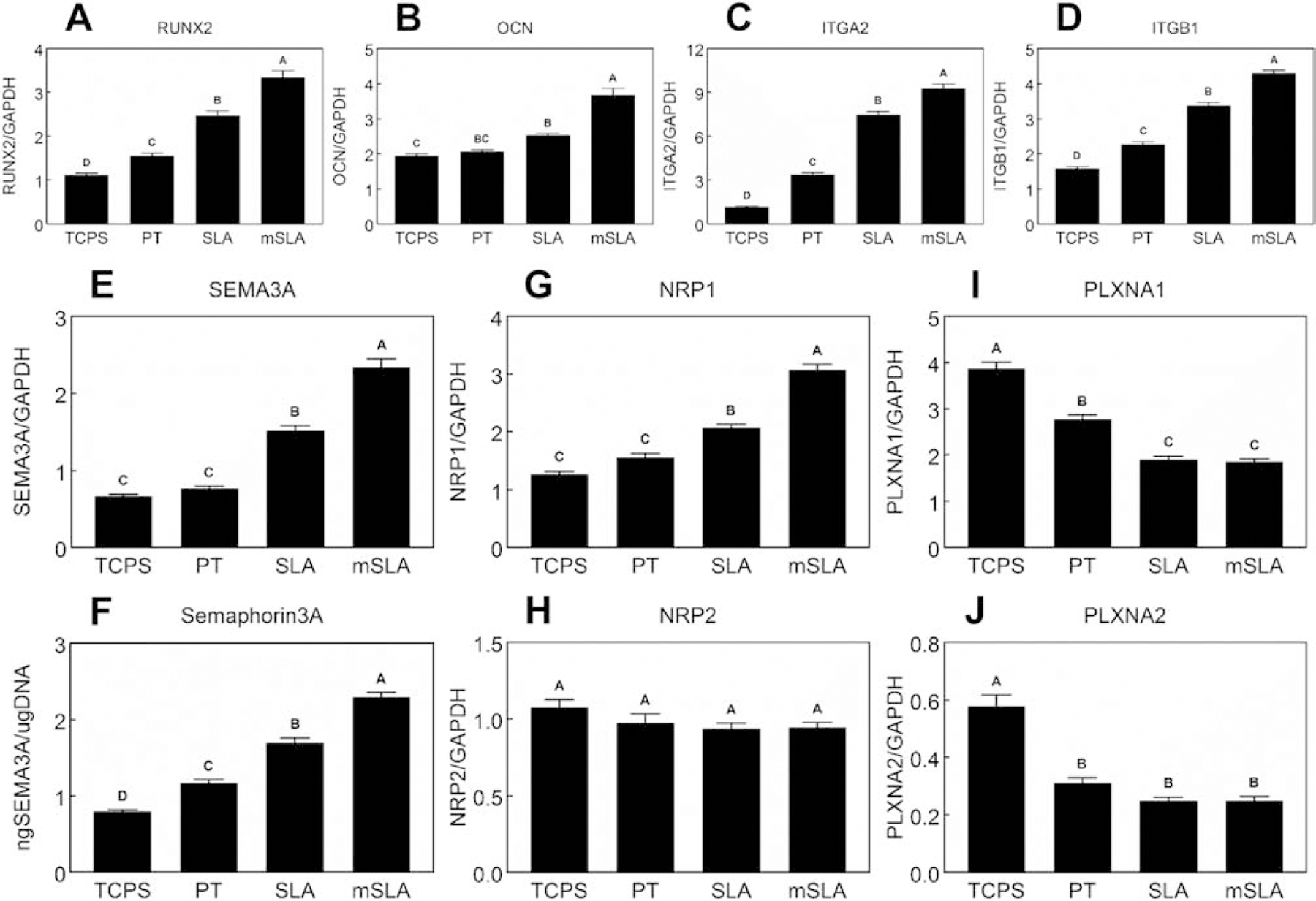
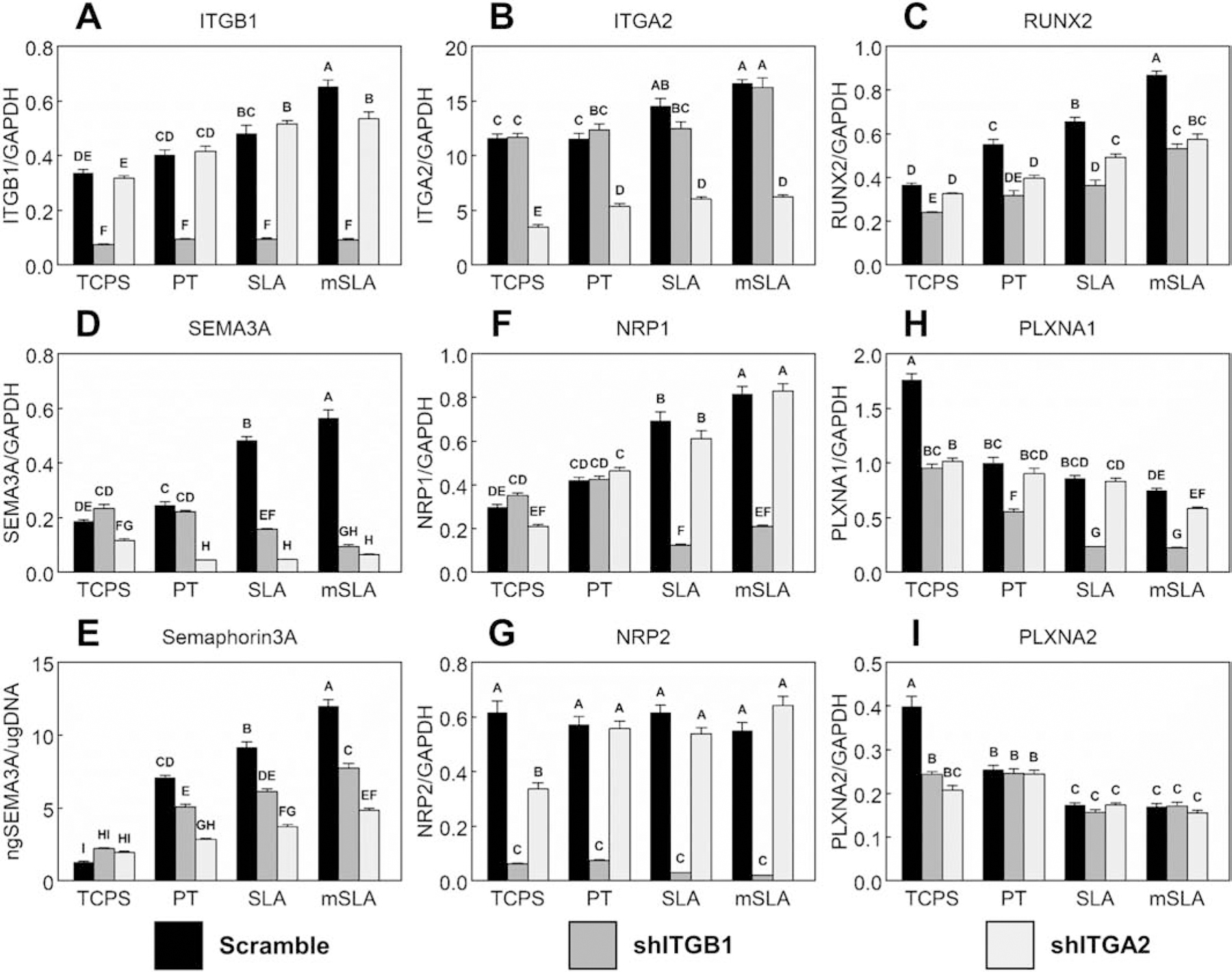
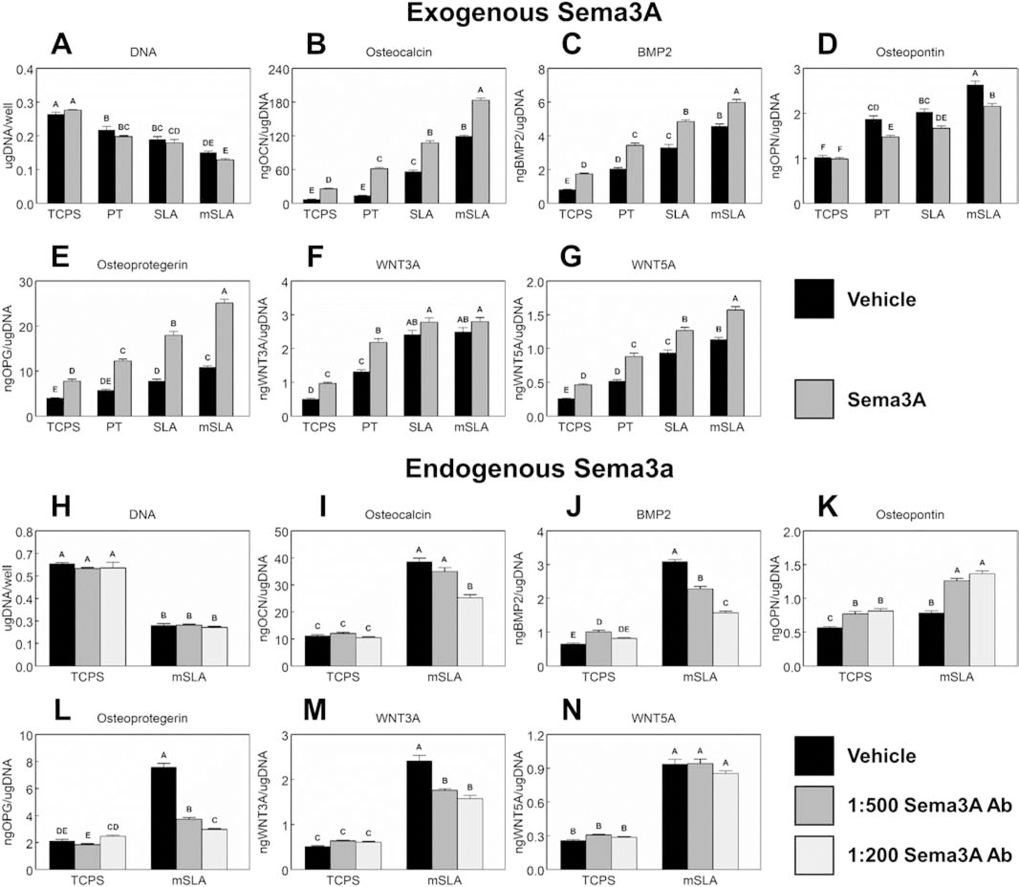
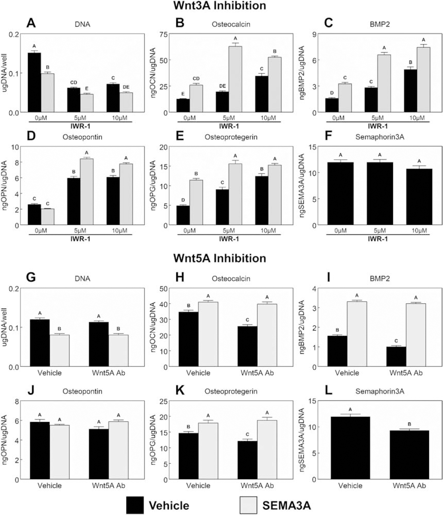
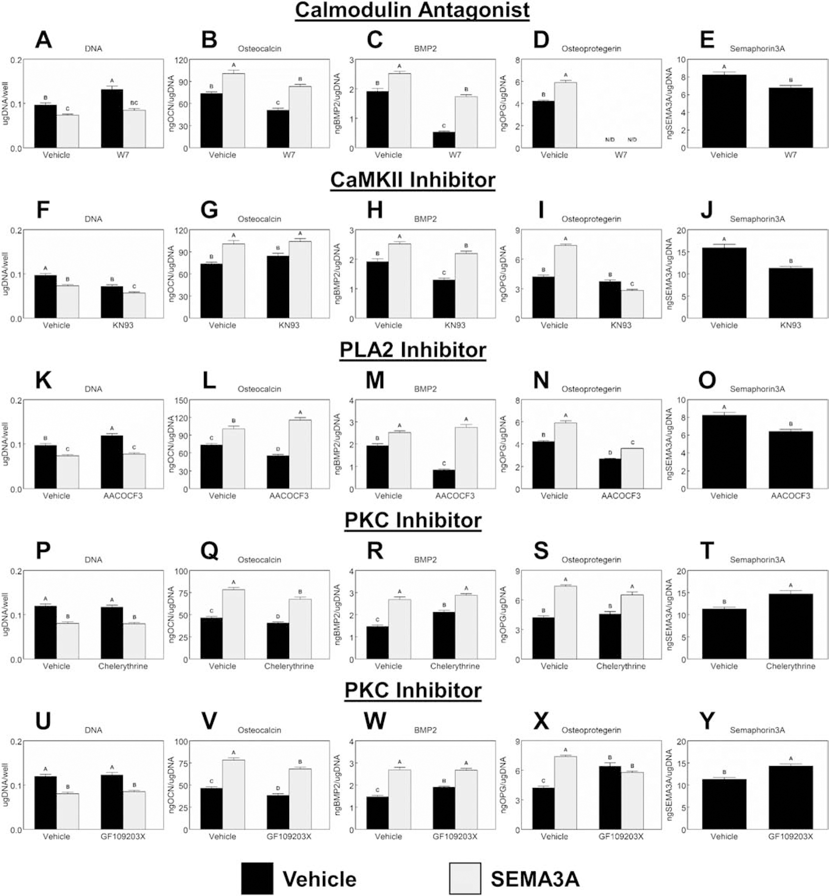
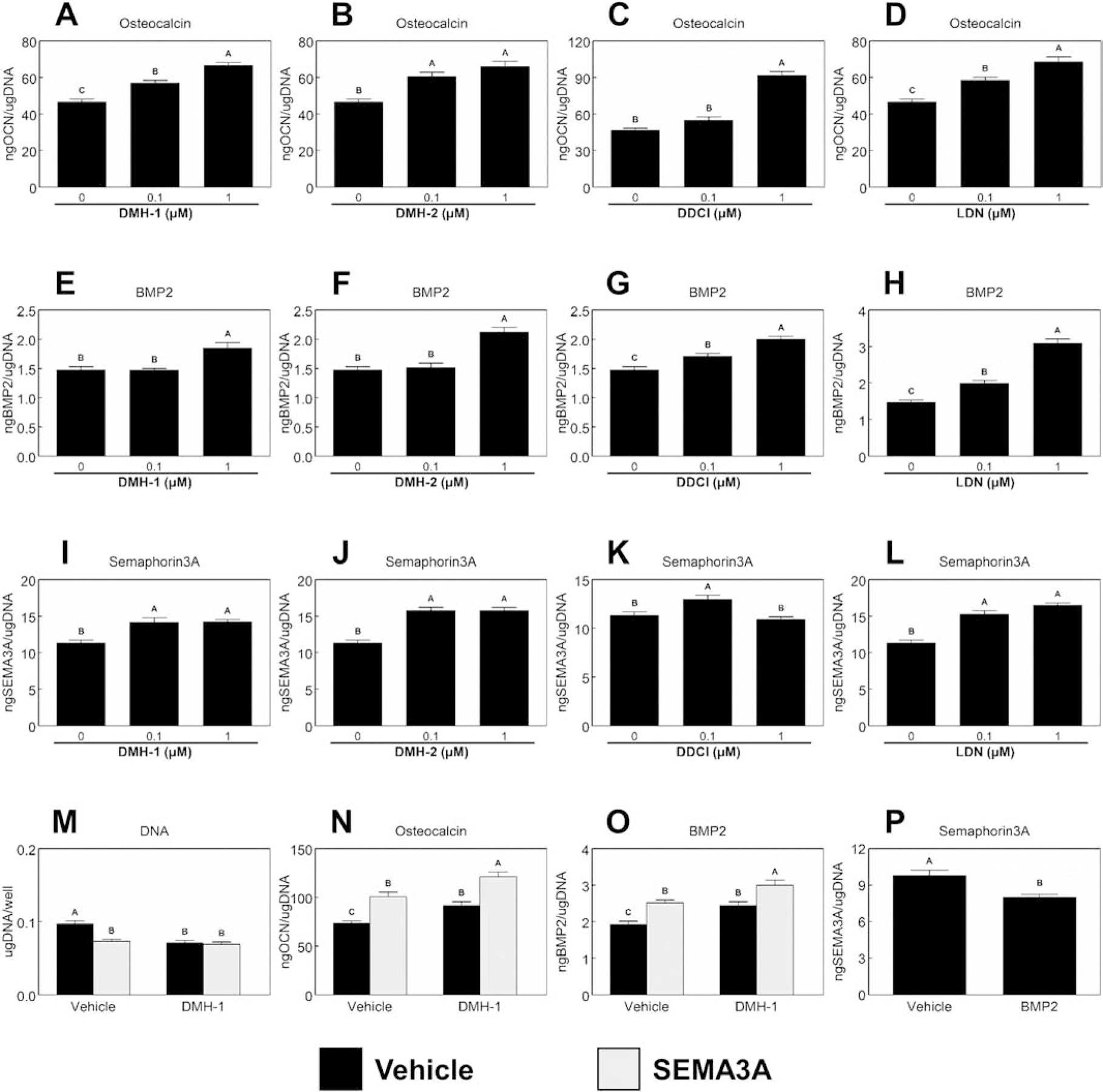
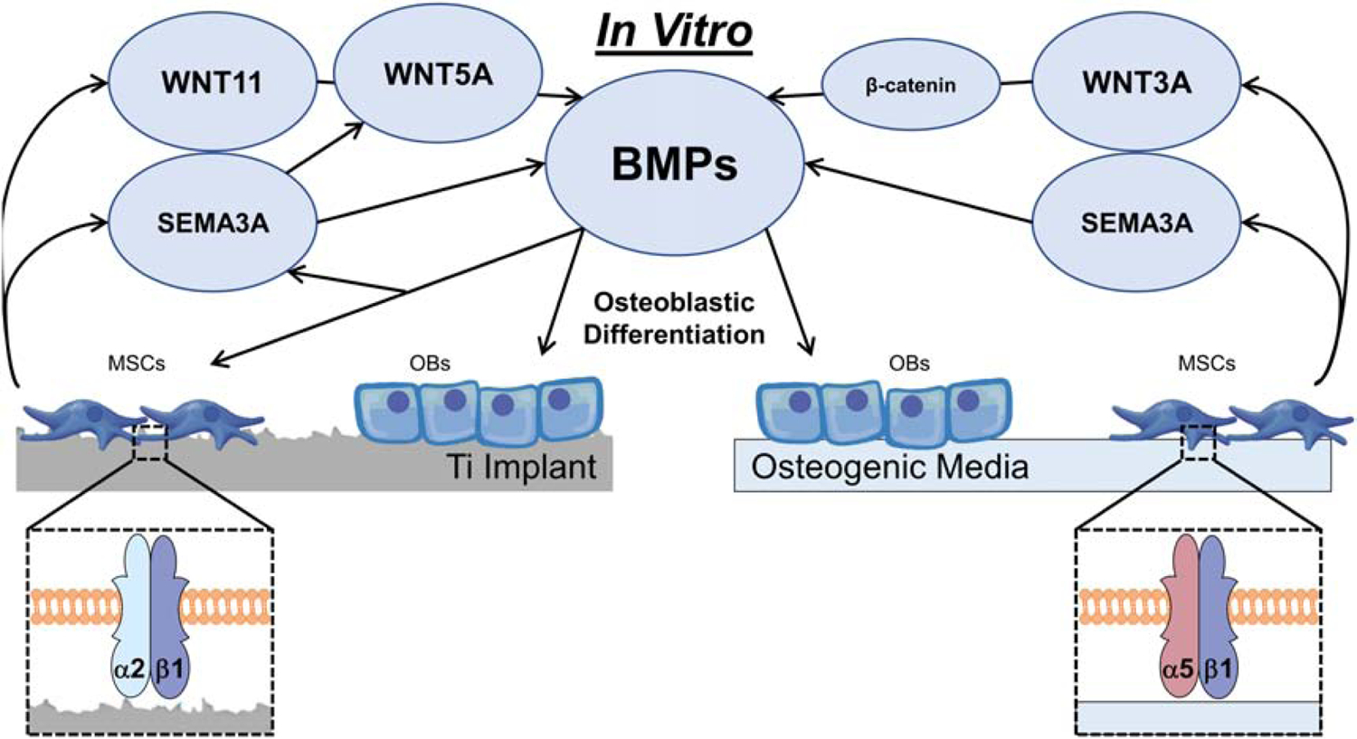
Peri-implant bone formation depends on the ability of mesenchymal stem cells (MSCs) to colonize implant surfaces and differentiate into osteoblasts, but the precise mechanisms controlling this process remain unclear. In vitro, MSCs undergo osteoblastic differentiation on microstructured titanium (Ti) surfaces in the absence of exogenous media supplements and produce factors that promote osteogenesis while regulating osteoclast activity, including semaphorins. The goal of this study was to evaluate the role of semaphorin 3A (Sema3A) on surface-mediated osteoblastic differentiation and determine the hierarchy of this signaling cascade. Human MSCs were cultured on 15 mm grade 2 smooth (pretreatment, PT), hydrophobic-microrough (sand blasted/acid etched, SLA), hydrophilic-microrough Ti (mSLA) (Institut Straumann AG, Basel, Switzerland), or tissue culture polystyrene (TCPS). Expression of SEMA3A family proteins increased after 7 days of culture, and the increased expression in response to microstructured Ti was dependent on recognition of the surface by integrin α2β1. Exogenous Sema3A increased differentiation whereas differentiation was decreased in cells treated with a Sema3A antibody. Furthermore, Sema3A influenced the production of osteoprotegerin and osteopontin suggesting it as an important local regulator of bone remodeling. Inhibition of Wnt3A and Wnt5A revealed that activation of Sema3A occurs downstream of Wnt5A and may facilitate the translocation of β-catenin bypassing the canonical Wnt3A initiating signal associated with osteoblastic differentiation. Furthermore, chemical inhibition of calmodulin (CaM), Ca2+/calmodulin-dependent protein kinase (CaMKII), phospholipase A2 (PLA2), protein kinase C (PKC), and BMP receptors suggest that Sema3A could serve as a feedback mechanism for both Wnt5A and BMP2. Here, we show novel roles for Sema3A family proteins in the surface-dependent modulation of MSCs as well as important interactions with pathways known to be associated with osteoblastic differentiation. Moreover, their effects on bone remodeling markers have significant implications for peri-implant bone remodeling and downstream modulation of osteoclastic activity. These results suggest that Sema3A aids in peri-implant bone formation through regulation on multiple stages of osseointegration, making it a potential target to promote osseointegration in patients with compromised bone remodeling.

Keywords: Implant; Mesenchymal stem cell; Osteoblast; Semaphorin3A; Surface topography; Titanium.


Figures

Figure 1.
Figure 1.. Effect of surface microstructured and hydrophilicity on MSC semaphorin expression and production.
MSCs were cultured on TCPS or Ti substrates. After 7d, mRNA levels of RUNX2 (A), OCN (B), ITGA2 (C), ITGB1 (D), SEMA3A (E) as well as Semaphorin3A protein levels (F), NRP1 (G), NRP2 (H), PLXNA1 (I), and PLXNA2 (J) were measured. Data shown are the mean ± standard error (SE) of six independent samples. Groups not sharing a letter are statistically significant at α=0.05.
Figure 2.
Figure 2.. Effect of integrin signaling on MSC semaphorin expression and production.
MSCs (WT scramble), MSCs with silenced ITGB1 (shITGB1), and MSCs with silenced ITGA2 (shITGA2) were plated on TCPS or Ti substrates. After 7d, mRNA levels of RUNX2 (A), OCN (B), ITGA2 (C), ITGB1 (D), SEMA3A (E) as well as Semaphorin3A protein levels (F), NRP1 (G), NRP2 (H), PLXNA1 (I), and PLXNA2 (J) were measured. Data shown are the mean ± standard error (SE) of six independent samples. Groups not sharing a letter are statistically significant at α=0.05.
Figure 3.
Figure 3.. Autocrine and paracrine effects of Sema3A on MSC response to surface microstructure and hydrophilicity.
MSCs plated on TCPS, PT, SLA, or mSLA surfaces were cultured with 1μg/ml Sema3A (A – G) or 1:500 or 1:200 AbSema3A (H – N) for 7d. Cells were then treated with fresh media for 24h. After 24h, media were collected, and cell lysates were assayed for DNA content (A, H). Media were assayed for osteocalcin (B, I), BMP2 (C, J), osteopontin (D, K), osteoprotegerin (E, L), WNT3A (F, M), and WNT5A (G, N). Data shown are the mean ± standard error (SE) of six independent samples. Groups not sharing a letter are statistically significant at α=0.05.
Figure 4.
Figure 4.. Effect of Wnt signaling and Sema3A on MSC response to microstructured and hydrophilic surfaces.
MSCs were cultured on TCPS or Ti substrates. Cultures supplemented with or without 1μg/mL Sema3A were treated with either 5μM or 10μM IWR-1 (A – F) to inhibit Wnt3A signaling or a 1:200 dilution of a polyclonal anti-Wnt5A antibody (G – L) to inhibit Wnt5A signaling for 7d. Cells were then treated with fresh media for 24h. After 24h, media were collected, and cell lysates were assayed for DNA content (A, G). Media were assayed for osteocalcin (B, H), BMP2 (C, I), osteopontin (D, J), osteoprotegerin (E, K) and Semaphorin3A (F, L). Data shown are the mean ± standard error (SE) of six independent samples. Groups not sharing a letter are statistically significant at α=0.05.
Figure 5.
Figure 5.. Effect of CaM, CaMKII, PLA2, and PKC inhibition in addition to Sema3A addition on MSC response to microstructured and hydrophilic surfaces.
Downstream activation of calmodulin (CaM), Ca2+/calmodulin-dependent protein kinase (CaMKII), and phospholipase A2 (PLA2) were inhibited by treating cultures with 10μM W7 (A – E), 10μM KN93 (F – J), or 10μM AACOCF3 (K – O) respectively with or without the addition of 1μg/mL Sema3A. Protein kinase C (PKC) was inhibited using either 1μM chelerythrine chloride (P – T) or 1μM GF109203X (U – Y) with or without the addition of 1μg/mL Sema3A for 7d. Cells were then treated with fresh media for 24h. After 24h, media were collected, and cell lysates were assayed for DNA content. Media were assayed for osteocalcin, BMP2, osteoprotegerin, and Semaphorin3A. Data shown are the mean ± standard error (SE) of six independent samples. Groups not sharing a letter are statistically significant at α=0.05.
Figure 6.
Figure 6.. Effect of BMP2 signaling and Sema3A on MSC response to microstructured and hydrophilic surfaces.
0.1μM or 1μM of two chemicals known to inhibit ALK2, ALK3, and ALK6 [DMH-2 and dorsomorphin dihydrochloride (DDCl)]; LDN 193189 dihydrochloride (ALK2 and ALK3 inhibitor) or DMH-1 (ALK2 inhibitor) were used to treat surface cultured MSCs. After 7d, cells were treated with fresh media for 24h. After 24h, media were collected and assayed for osteocalcin (A – D), BMP2 (E – H), and Semaphorin3A (I – L). MSCs cultured on mSLA were also treated with 10μM DMH-1 with or without the addition of 1μg/mL Sema3A for 7d. Cultures were then treated for 24h with fresh media. After 24h, media were collected, and cell lysates were assayed for DNA content (M). Media were assayed for osteocalcin (N) and BMP2 (O). The production of Sema3A after the addition of 40ng/mL recombinant human BMP2 was also measured (P). Data shown are the mean ± standard error (SE) of six independent samples. Groups not sharing a letter are statistically significant at α=0.05.
Figure 7.
Figure 7.
Proposed role of Sema3A on the osteoblastic differentiation of mesenchymal stem cells cultured on microstructured Ti surfaces or in osteogenic media.

References

    1. Mellis DJ, Itzstein C, Helfrich MH, Crockett JC, The skeleton: A multi-functional complex organ. The role of key signalling pathways in osteoclast differentiation and in bone resorption, J. Endocrinol (2011). 10.1530/JOE-11-0212. - DOI - PubMed
    1. Cochran DL, A comparison of endosseous dental implant surfaces, J. Periodontol 70 (1999) 1523–1539. 10.1902/jop.1999.70.12.1523. - DOI - PubMed
    1. Gittens RA, Olivares-Navarrete R, Schwartz Z, Boyan BD, Implant osseointegration and the role of microroughness and nanostructures: Lessons for spine implants, Acta Biomater 10 (2014) 3363–3371. 10.1016/j.actbio.2014.03.037. - DOI - PMC - PubMed
    1. Gittens RA, Scheideler L, Rupp F, Hyzy SL, Geis-Gerstorfer J, Schwartz Z, Boyan BD, A review on the wettability of dental implant surfaces II: Biological and clinical aspects, Acta Biomater 10 (2014) 2907–2918. 10.1016/j.actbio.2014.03.032. - DOI - PMC - PubMed
    1. Boyan BD, Lotz EM, Schwartz Z, Roughness and hydrophilicity as osteogenic biomimetic surface properties, Tissue Eng. Part A 23 (2017) 1479–1489. 10.1089/ten.TEA.2017.0048. - DOI - PMC - PubMed

Publication types