. 2020 Apr:133:115262.
doi: 10.1016/j.bone.2020.115262. Epub 2020 Feb 4.

Tissue-nonspecific alkaline phosphatase is an anti-inflammatory nucleotidase

Affiliations

Tissue-nonspecific alkaline phosphatase is an anti-inflammatory nucleotidase

L Bessueille et al. Bone. 2020 Apr.

Abstract

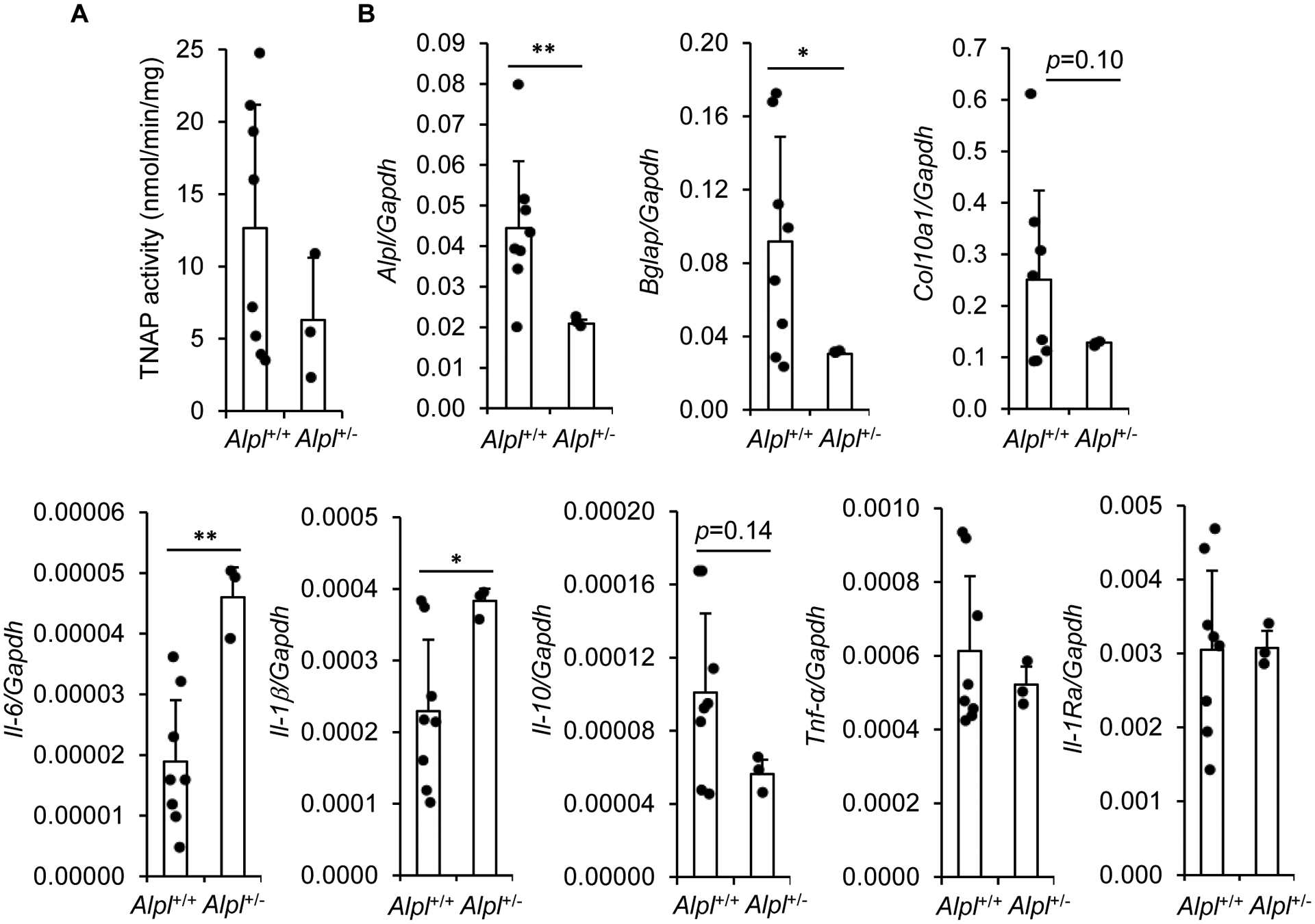
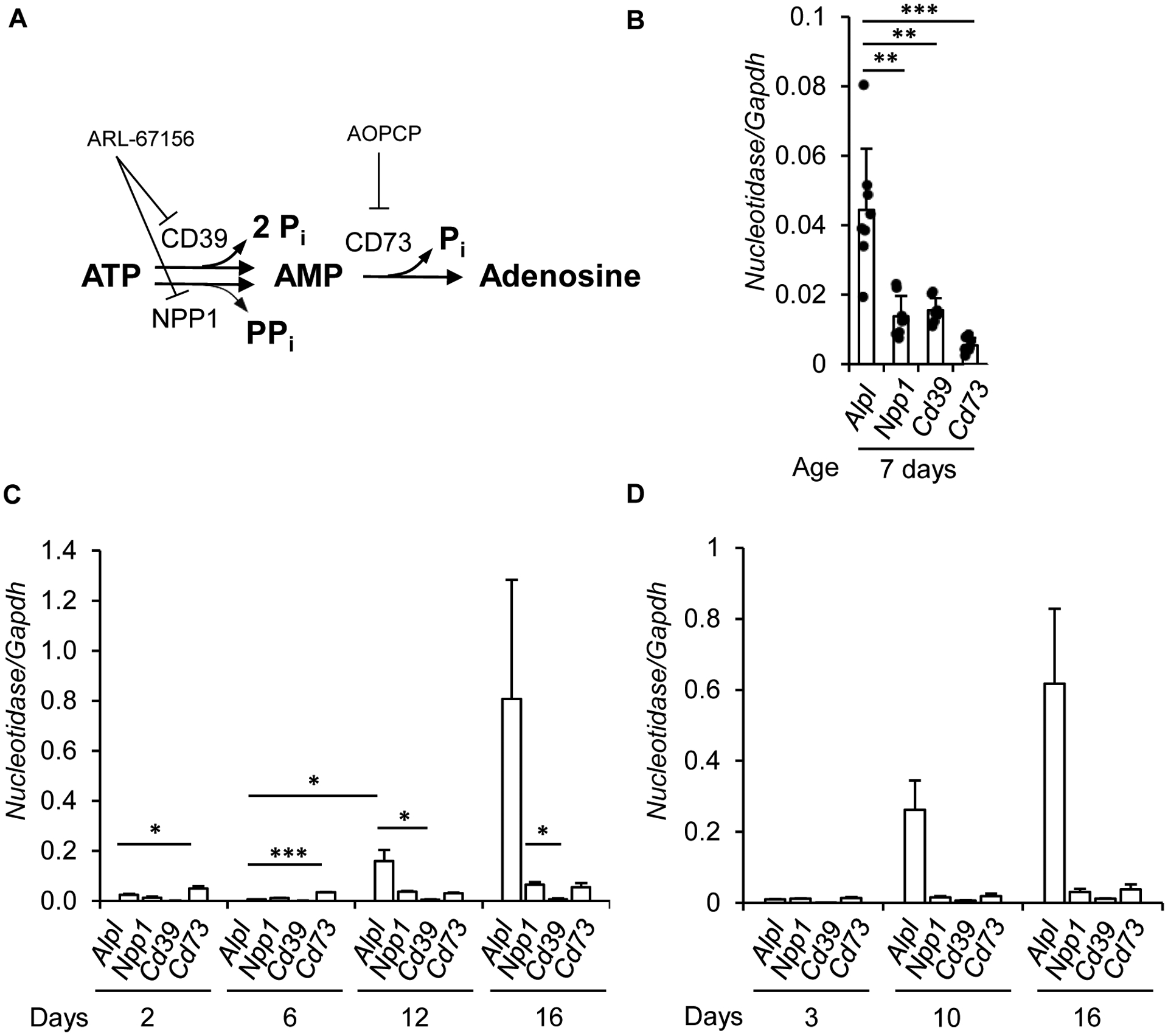
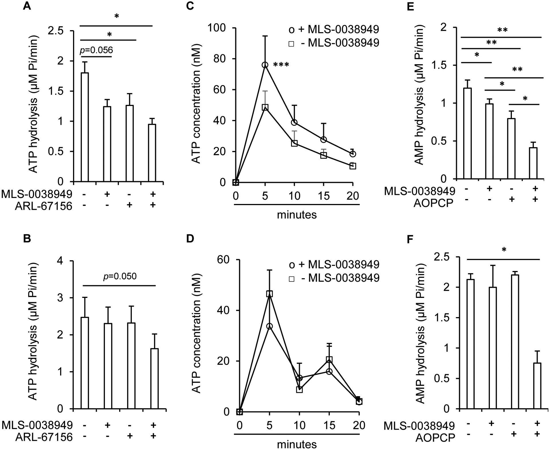
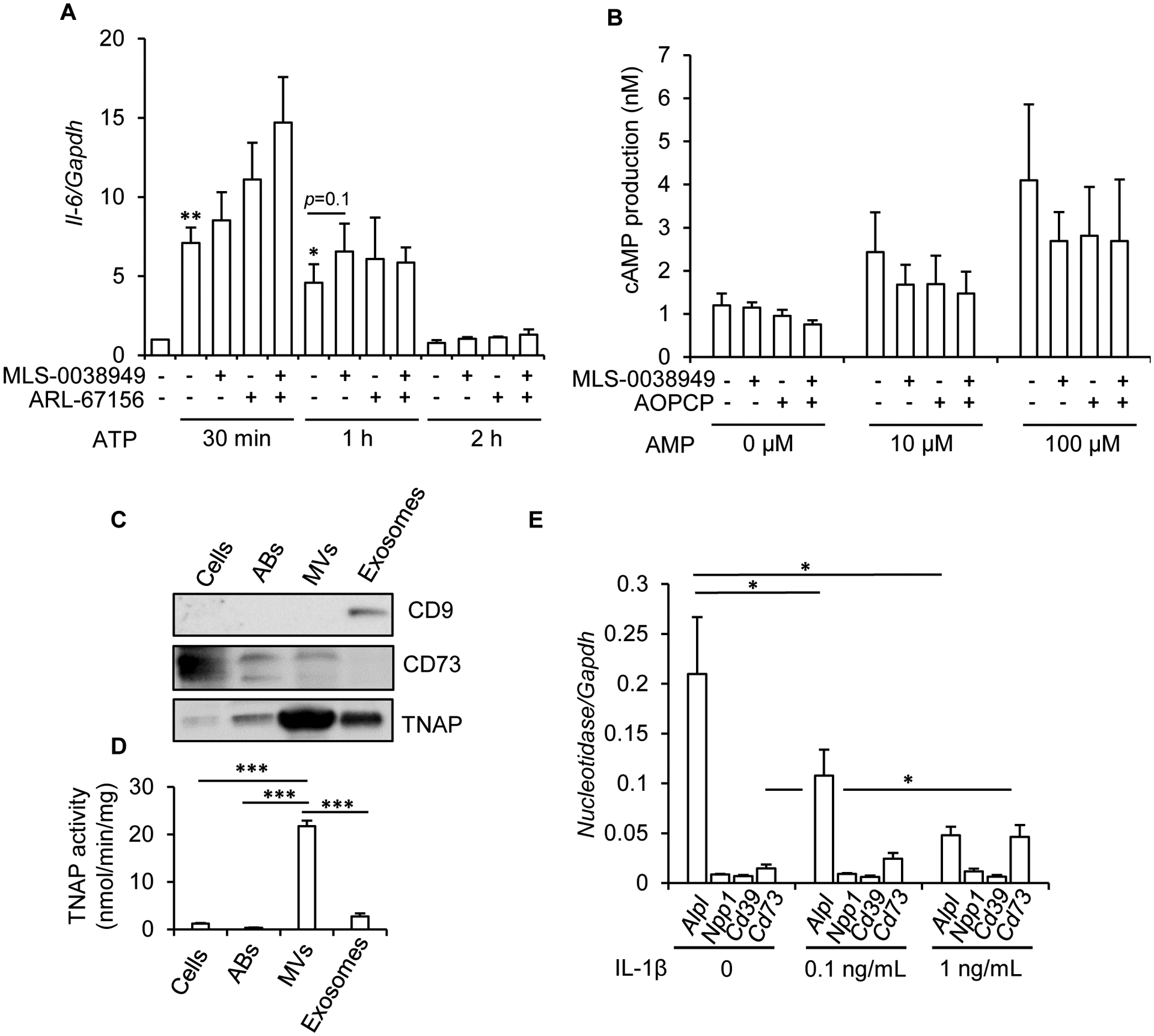
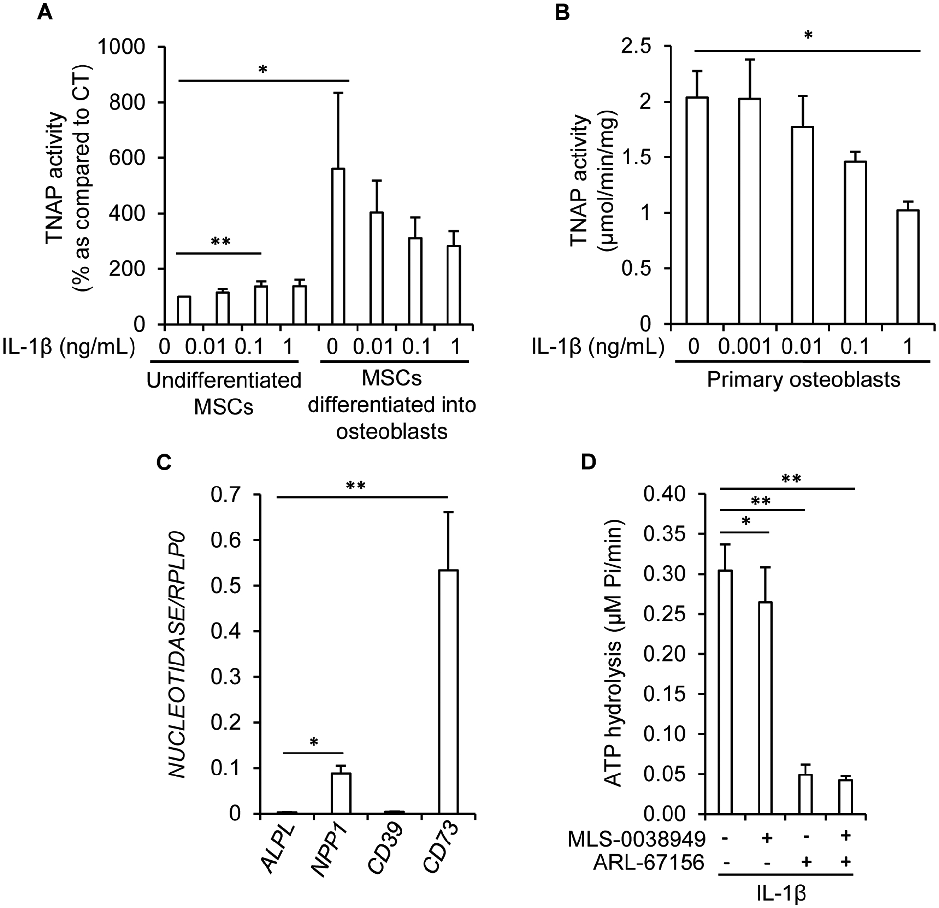
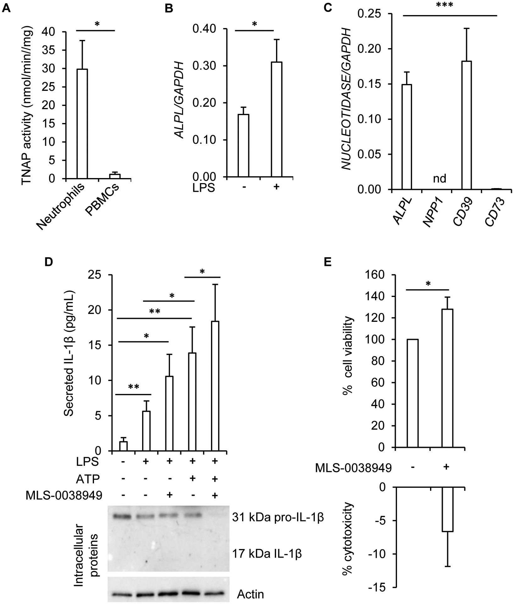
Tissue-nonspecific alkaline phosphatase (TNAP) is necessary for skeletal mineralization by its ability to hydrolyze the mineralization inhibitor inorganic pyrophosphate (PPi), which is mainly generated from extracellular ATP by ectonucleotide pyrophosphatase phosphodiesterase 1 (NPP1). Since children with TNAP deficiency develop bone metaphyseal auto-inflammations in addition to rickets, we hypothesized that TNAP also exerts anti-inflammatory effects relying on the hydrolysis of pro-inflammatory adenosine nucleotides into the anti-inflammatory adenosine. We explored this hypothesis in bone metaphyses of 7-day-old Alpl+/- mice (encoding TNAP), in mineralizing hypertrophic chondrocytes and osteoblasts, and non-mineralizing mesenchymal stem cells (MSCs) and neutrophils, which express TNAP and are present, or can be recruited in the metaphysis. Bone metaphyses of 7-day-old Alpl+/- mice had significantly increased levels of Il-1β and Il-6 and decreased levels of the anti-inflammatory Il-10 cytokine as compared with Alpl+/+ mice. In bone metaphyses, murine hypertrophic chondrocytes and osteoblasts, Alpl mRNA levels were much higher than those of the adenosine nucleotidases Npp1, Cd39 and Cd73. In hypertrophic chondrocytes, inhibition of TNAP with 25 μM of MLS-0038949 decreased the hydrolysis of AMP and ATP. However, TNAP inhibition did not significantly modulate ATP- and adenosine-associated effects in these cells. We observed that part of TNAP proteins in hypertrophic chondrocytes was sent from the cell membrane to matrix vesicles, which may explain why TNAP participated in the hydrolysis of ATP but did not significantly modulate its autocrine pro-inflammatory effects. In MSCs, TNAP did not participate in ATP hydrolysis nor in secretion of inflammatory mediators. In contrast, in neutrophils, TNAP inhibition with MLS-0038949 significantly exacerbated ATP-associated activation and secretion of IL-1β, and extended cell survival. Collectively, these results demonstrate that TNAP is a nucleotidase in both hypertrophic chondrocytes and neutrophils, and that this nucleotidase function is associated with autocrine effects on inflammation only in neutrophils.

Keywords: ATP; Hypophosphatasia; Inflammation; Nucleotidase; Tissue-nonspecific alkaline phosphatase.


Conflict of interest statement

Declaration of competing interest None.

Figures

Figure 1:
Figure 1:. Disturbed levels of hypertrophic chondrocyte markers and inflammatory cytokines in the metaphysis-containing bone fragments of Alpl+/− mice.
A: TNAP activity in the metaphysis-containing femurs and tibias of 7-day-old Alpl+/+ and Alpl+/− mice. B: RT-qPCR analysis of mRNA levels in the metaphysis-containing femurs and tibias of 7-day-old Alpl+/+ and Alpl+/− mice. * indicates a statistical difference with p<0.05; ** a difference with p<0.01.
Figure 2:
Figure 2:. expression of TNAP, and ectonucleotidases in metaphysis-containing bone fragments, hypertrophic chondrocytes and osteoblasts.
A: enzymes known to be involved in ATP hydrolysis into adenosine, and their inhibitor. B: RT-qPCR analysis of nucleotidase-encoding mRNA levels in the metaphysis-containing femurs and tibias of 7-day-old wildtype mice. Mouse primary chondrocytes (C, n=4 independent experiments) and primary osteoblasts (D, n=3) were cultured as detailed in the Materials and methods section and mRNA levels were quantified by RT-qPCR. * indicates a statistical difference with p<0.05; ** a difference with p<0.01; *** a difference with p<0.001.
Figure 3:
Figure 3:. ectonucleotidase activity of TNAP in hypertrophic chondrocytes and osteoblasts.
Mouse primary chondrocytes (A, C and E) and primary osteoblasts (B, D and F) were cultured as detailed in the Materials and methods section for 16 days. Hydrolysis of the substrate (10 μM ATP or 25 μM AMP) into Pi was measured by the Malachite green assay in the presence or absence of 25 μM of MLS-0038949 and 100 μM of ARL-67156 (A, n=5 and B, n=4), or in the presence or absence of 25 μM of MLS-0038949 and 100 μM of AOPCP (E, n=5 and F, n=4). Endogenously released ATP was quantified with the bioluminescent Luciferin-Luciferase reaction in presence (ο) or absence (□) of MLS-0038949 (C, n=5 and D, n=3). * indicates a statistical difference with p<0.05; ** a difference with p<0.01; *** a difference with p<0.001.
Figure 4:
Figure 4:. absence of anti-inflammatory effects of TNAP in hypertrophic chondrocytes.
Mouse primary chondrocytes were differentiated into hypertrophic chondrocytes for 16 days as detailed in the Materials and methods section. A: effect of MLS-0038949 and/or ARL-67156 on ATP-stimulated Il-6 expression (n=8). To eliminate the inter-experimental variations in Il-6 levels, Il-6/Gapdh ratio in untreated cells was set to 1. B: effect of MLS-0038949 and/or AOPCP on AMP-stimulated intracellular cAMP production (n=4). C: western-blot analysis of TNAP, CD73, and CD9 in hypertrophic chondrocyte cells, and in apoptotic bodies (ABs), MVs and exosomes released by these cells (n=3, a representative result is shown; the same quantity of protein was used in all conditions). D: TNAP activity in hypertrophic chondrocyte cells, and in ABs, MVs and exosomes released by these cells (n=4). E: effect of a 24-h treatment with IL-1β on nucleotidase levels, as determined by RT-qPCR (n=4). * indicates a statistical difference with p<0.05; ** a difference with p<0.01, and *** a difference with p<0.001.
Figure 5:
Figure 5:. absence of anti-inflammatory effects of TNAP in MSCs.
A: effect of a 7-day treatment with IL-1β on TNAP activity in MSCs left undifferentiated or differentiated into osteoblasts (n=6). To eliminate the inter-experimental variations, TNAP activity was set to 100% in untreated cells. B: effect of a 7-day treatment with IL-1β on TNAP activity in murine primary osteoblasts (n=3). C: quantification of nucleotidase mRNA levels in undifferentiated MSCs by RT-qPCR (n=8 independent experiments with MSCs from 4 different donors). D: in undifferentiated MSCs pre-treated for 4 days with 1 ng/mL of IL-1β, the hydrolysis of 10 μM of ATP into Pi was measured by the Malachite green assay in the presence or absence of 25 μM of MLS-0038949 and 100 μM of ARL-67156 (n=8 with MSCs from 4 different donors). * indicates a statistical difference with p<0.05; ** a difference with p<0.01.
Figure 6:
Figure 6:. anti-inflammatory ectonucleotidase function of TNAP in neutrophils.
A: TNAP activity measured in freshly isolated human neutrophils (n=5 for neutrophils and n=3 for PBMCs). B: effect of 0.5 μg/mL of LPS on ALPL mRNA levels as determined by RT-qPCR (n=6 independent experiments from 4 different donors). C: RT-qPCR analysis of nucleotidase transcripts in freshly isolated human neutrophils (n=5; nd: not detected). D: quantification by ELISA of extracellular levels of IL-1β in neutrophils primed with 0.5 μg/mL of LPS for 3 h and further stimulated with 2 mM of ATP during 45 min (n=9 independent experiments from 4 different donors). A representative western-blot experiment of intracellular IL-1β is shown. E: quantification of cell viability and cell death with MTT and LDH assays respectively in cells treated with 25 μM of MLS-0038949 for 24 h (n=7 independent experiments from 4 different donors). * indicates a statistical difference with p<0.05; ** a difference with p<0.01; *** a difference with p<0.001.

References

    1. Aghajanian P, Mohan S, The art of building bone: emerging role of chondrocyte-to-osteoblast transdifferentiation in endochondral ossification, Bone Res 6 (2018) 19. - PMC - PubMed
    1. Murshed M, Harmey D, Millán JL, McKee MD, Karsenty G, Unique coexpression in osteoblasts of broadly expressed genes accounts for the spatial restriction of ECM mineralization to bone, Genes Dev 19(9) (2005) 1093–104. - PMC - PubMed
    1. Deng H, Huang X, Yuan L, Molecular genetics of the COL2A1-related disorders, Mutat Res Rev Mutat Res 768 (2016) 1–13. - PubMed
    1. Bishop N, Bone Material Properties in Osteogenesis Imperfecta, J Bone Miner Res 31(4) (2016) 699–708. - PubMed
    1. Girschick HJ, Mornet E, Beer M, Warmuth-Metz M, Schneider P, Chronic multifocal non-bacterial osteomyelitis in hypophosphatasia mimicking malignancy, BMC Pediatr 7 (2007) 3. - PMC - PubMed

Publication types