. 2020 Feb 6;21(1):129.
doi: 10.1186/s12864-020-6459-6.

A transcriptomic and proteomic atlas of expression in the Nezara viridula (Heteroptera: Pentatomidae) midgut suggests the compartmentalization of xenobiotic metabolism and nutrient digestion

Affiliations

A transcriptomic and proteomic atlas of expression in the Nezara viridula (Heteroptera: Pentatomidae) midgut suggests the compartmentalization of xenobiotic metabolism and nutrient digestion

Shane Denecke et al. BMC Genomics. .

Abstract

Background: Stink bugs are an emerging threat to crop security in many parts of the globe, but there are few genetic resources available to study their physiology at a molecular level. This is especially true for tissues such as the midgut, which forms the barrier between ingested material and the inside of the body.

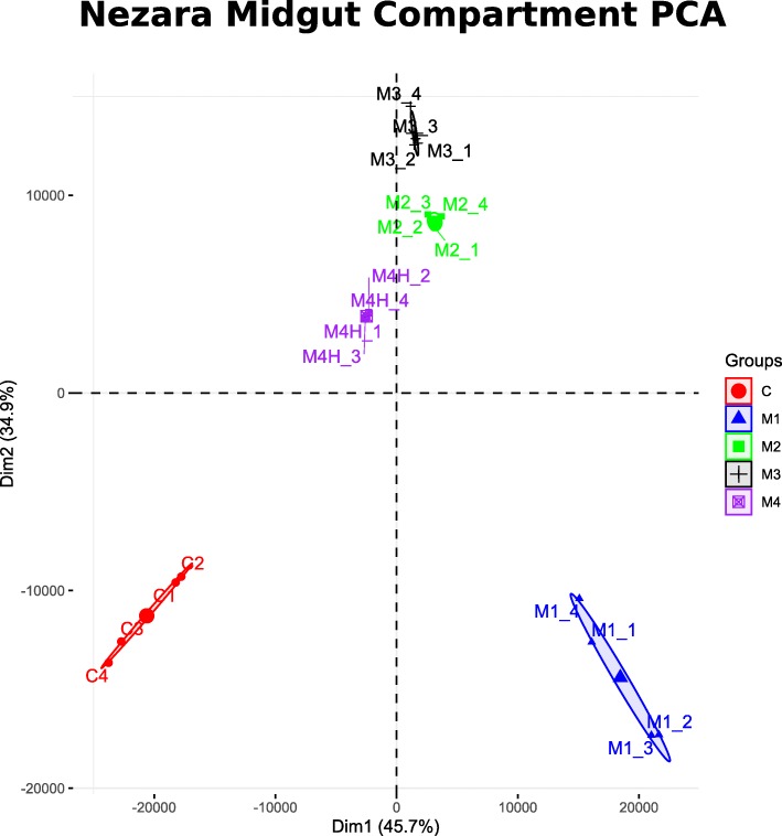
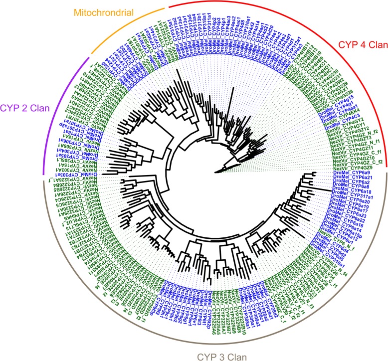
Results: Here, we focus on the midgut of the southern green stink bug Nezara viridula and use both transcriptomic and proteomic approaches to create an atlas of expression along the four compartments of the anterior-posterior axis. Estimates of the transcriptome completeness were high, which led us to compare our predicted gene set to other related stink bugs and Hemiptera, finding a high number of species-specific genes in N. viridula. To understand midgut function, gene ontology and gene family enrichment analyses were performed for the most highly expressed and specific genes in each midgut compartment. These data suggested a role for the anterior midgut (regions M1-M3) in digestion and xenobiotic metabolism, while the most posterior compartment (M4) was enriched in transmembrane proteins. A more detailed characterization of these findings was undertaken by identifying individual members of the cytochrome P450 superfamily and nutrient transporters thought to absorb amino acids or sugars.

Conclusions: These findings represent an initial step to understand the compartmentalization and physiology of the N. viridula midgut at a genetic level. Future studies will be able to build on this work and explore the molecular physiology of the stink bug midgut.

Keywords: Midgut; Nezara viridula; P450; Proteomics; Southern green stink bug; Transcriptomics; Transporter.


Conflict of interest statement

Authors SD, PI, and AI are funded as a part of a joint collaboration with Bayer Crop Sciences. Authors SG and RN are employed by Bayer Crop Sciences.

Figures

Fig. 1
Fig. 1
Comparative gene sets among insects. a A phylogeny is shown constructed from 221 single-copy genes present in all species included in this analysis. The Pentatomidae (red) form a cluster within the Hemiptera (yellow) order which forms a sister clade to Holometabola (blue). The tree is rooted with the crustacean Daphnia pulex (not shown). Black dots indicate nodes with bootstrap support > 75%, whereas gray dots indicate nodes with bootstrap support between 50 and 75%. The scale bar is in substitutions per site. b Orthology profile of stinkbugs (names shown in red), compared to other insects. Note the large fraction of species-specific genes in N. viridula (Nviri) which is very similar to what has been previously documented for the pea aphid A. pisum (Apisu). Species names prefixed with “[T]” indicate that the unigene set was obtained from a transcriptome assembly; for the remaining insect species the data were obtained from a genome assembly. Species names abbreviations: Nviri – N. viridula; Ahila – A. hilare; Pstal – P. stali; Hhaly – H. halys; Cruti – C. rutilans; Ofasc – Oncopeltus fasciatus; Rprol – Rhodnius prolixus; Clect – Cimex lectularius; Dcitr – Diaphorina citri; Apisu – A. pisum; Tcast – Tribolium castaneum; Dmela – Drosophila melanogaster; Dplex – Danaus plexippus; Amell – Apis mellifera
Fig. 2
Fig. 2
Shared transcript and protein expression. Venn diagrams are shown for both detected transcripts (a) and proteins (b), showing expression > 1 transcript per million (TPM) in each tissue. In each case, a sizable portion of the detected features are found across all midgut compartments, indicating a that many genes are expressed across the anterior-posterior axis. In both cases the M1 and M4 region display the most distinctiveness. The relatively lower number of proteins detected in the proteome compared to transcripts in the transcriptome is reflective of the sensitivities of these two technologies
Fig. 3
Fig. 3
Principle Component Analysis. The results of a principle component analysis of the expression of all unigenes is shown. The first two principle components explain a total of 78% of the total variation detected within the RNA-seq data. Each shape and color represent a distinct sample (M1: Blue triangle, M2: Green square, M3: Black cross, M4: purple crossed square, carcass: red circle). The variation in each sample is shown with an ellipse which encompass all replicates in that sample
Fig. 4
Fig. 4
Gene expression patterns along the midgut. The results of the fuzzy-C means clustering is shown. Transcripts were grouped into eight categories based on their relative expression pattern, and all members with membership values > 0.6 were plotted. The darker shading on the plot indicates a larger number of individual transcripts which show that expression pattern. The top four clusters are composed of more complicated patterns, whereas the bottom four clusters display transcripts enriched specifically in one compartment
Fig. 5
Fig. 5
Phylogeny of identified P450s in N. viridula. A phylogenetic tree constructed of N. viridula P450s is shown along with other reference species. The names of each of the four CYP clans is shown at the outer rim. Individual P450s used in the tree were those N. viridula P450s identified in the current study (black), or P450s from D. melanogaster (dark red)
Fig. 6
Fig. 6
Enrichment of AAAP and APC transporters in the M4 midgut region. A boxplot is shown comparing the expression of all AAAP and APC transporters showing along the midgut. Only genes showing expression above >1TPM in either the M4 region or the anterior region (average of the M1-M3 region) were plotted. The M4 region of the gut displays an enrichment in both APC and AAAP expression compared to the anterior section of the midgut

References

    1. Catarino R, Ceddia G, Areal FJ, Park J. The impact of secondary pests on Bacillus thuringiensis (Bt) crops. Plant Biotechnol J. 2015;13:601–612. doi: 10.1111/pbi.12363. - DOI - PubMed
    1. Esquivel J, Dmitry LM, Walker AJ, Wolfgang R, Jeremy KG, Michael DT, et al. Nezara viridula (L.) In: McPherson JE, et al., editors. Invasive stink bugs and related species (Pentatomoidea) 1. Boca Raton: CRC Press; 2018. pp. 351–425.
    1. Billingsley PF, Lehane MJ. Biology of the insect Midgut. Dordrecht: Springer Netherlands; 1996. Structure and ultrastructure of the insect midgut; pp. 3–30.
    1. Huang J-H, Jing X, Douglas AE. The multi-tasking gut epithelium of insects. Insect Biochem Mol Biol. 2015;67:15–20. doi: 10.1016/j.ibmb.2015.05.004. - DOI - PMC - PubMed
    1. Engel P, Moran NA. The gut microbiota of insects – diversity in structure and function. FEMS Microbiol Rev. 2013;37:699–735. doi: 10.1111/1574-6976.12025. - DOI - PubMed

LinkOut - more resources