. 2020 Feb 7;21(1):10.
doi: 10.1186/s10194-020-1079-8.

The distribution of oxytocin and the oxytocin receptor in rat brain: relation to regions active in migraine

Affiliations

The distribution of oxytocin and the oxytocin receptor in rat brain: relation to regions active in migraine

Karin Warfvinge et al. J Headache Pain. .

Abstract

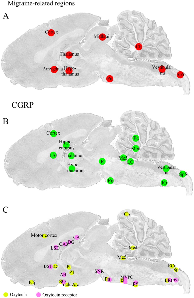
Background: Recent work, both clinical and experimental, suggests that the hypothalamic hormone oxytocin (OT) and its receptor (OTR) may be involved in migraine pathophysiology. In order to better understand possible central actions of OT in migraine/headache pathogenesis, we mapped the distribution of OT and OTR in nerve cells and fibers in rat brain with a focus on areas related to migraine attacks and/or shown previously to contain calcitonin gene related peptide (CGRP), another neuropeptide involved in migraine.

Methods: Distribution of OT and OTR in the adult, rat brain was qualitatively examined with immunohistochemistry using a series of well characterized specific antibodies.

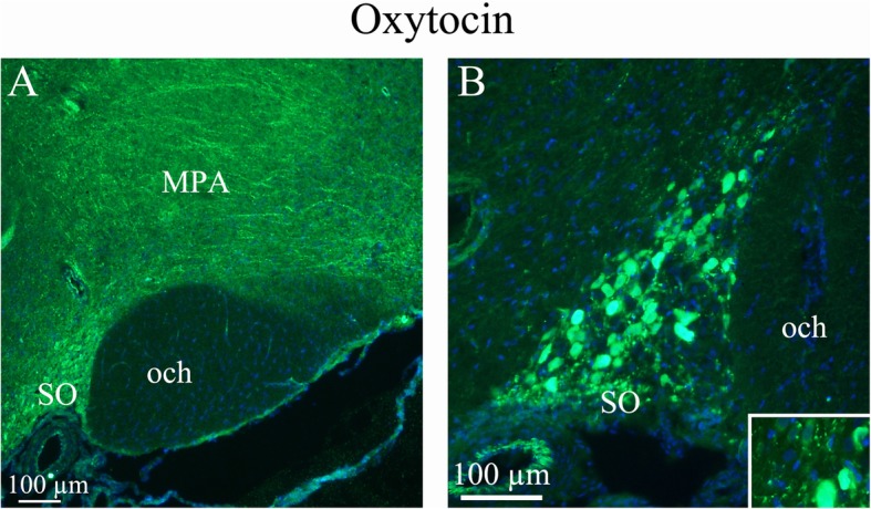
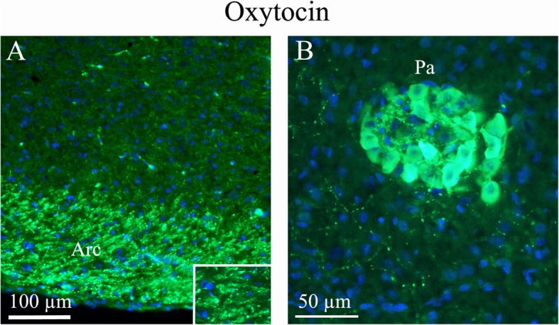
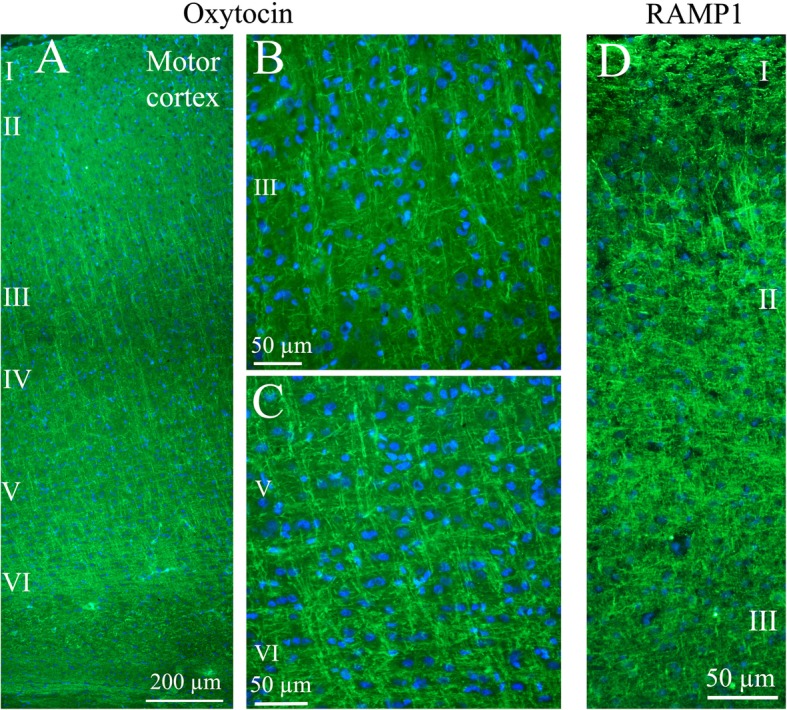
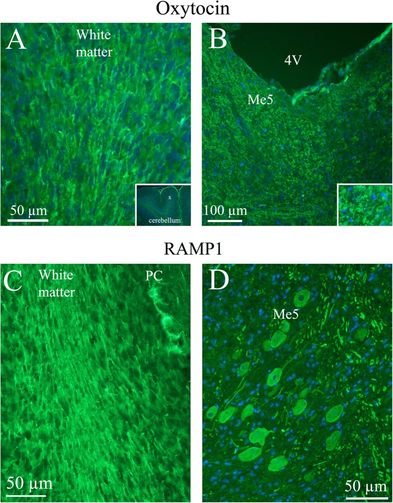
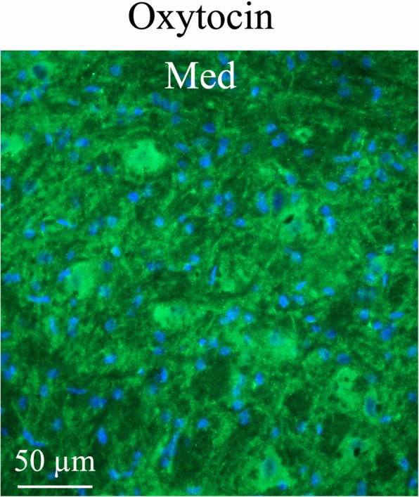
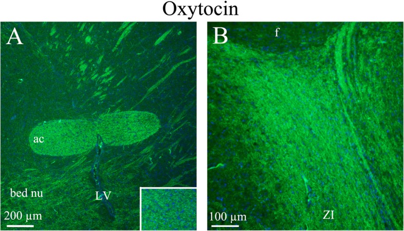
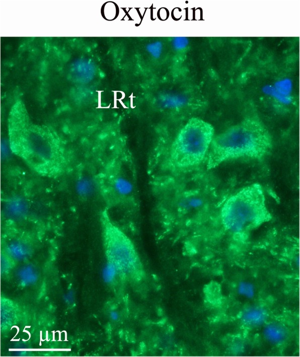
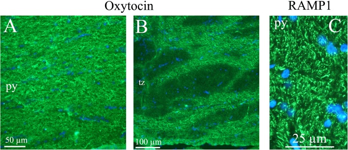
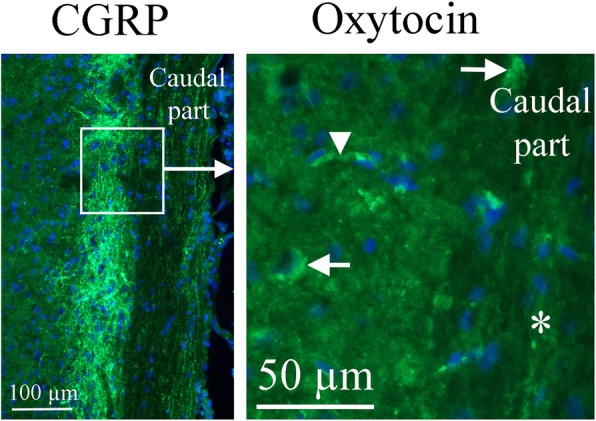
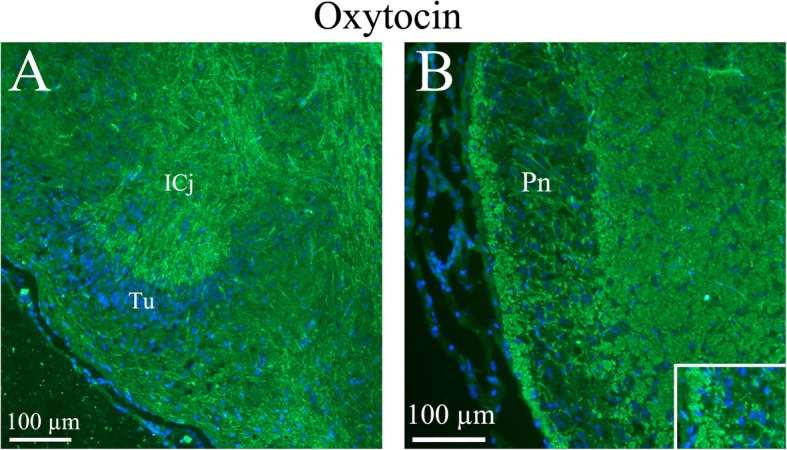
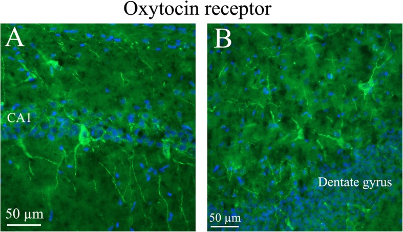
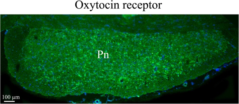
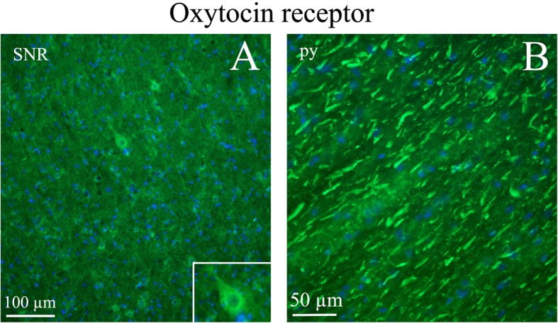
Results: As expected, OT was extensively localized in the cell somas of two hypothalamic nuclei, the supraoptic (SO or SON) and paraventricular nuclei (Pa or PVN). OT also was found in many other regions of the brain where it was localized mainly in nerve fibers. In contrast, OTR staining in the brain was mainly observed in cell somas with very little expression in fibers. The most distinct OTR expression was found in the hippocampus, the pons and the substantia nigra. In some regions of the brain (e.g. the amygdala and the hypothalamus), both OT and OTR were expressed (match). Mismatch between the peptide and its receptor was primarily observed in the cerebral and cerebellar cortex (OT expression) and hippocampus (OTR expression).

Conclusions: We compared OT/OTR distribution in the CNS with that of CGRP and identified regions related to migraine. In particular, regions suggested as "migraine generators", showed correspondence among the three mappings. These findings suggest central OT pathways may contribute to the role of the hypothalamus in migraine attacks.

Keywords: CGRP, CGRP receptors; Immunohistochemistry; Migraine-related regions; Oxytocin; Oxytocin receptor.


Conflict of interest statement

The authors declare that they have no competing interests.

Figures

Fig. 1
Fig. 1
Sagittal cryo-sections stained with Hematoxylin and Eosin. The 6th edition of The Rat Brain in Stereotaxic Coordinates by Paxinos and Watson [20] was used to identify the different areas. a Migraine related regions. b CGRP distribution. c Oxytocin and oxytocin receptor expression. Yellow color denotes oxytocin and pink oxytocin receptor immunoreactivity
Fig. 2
Fig. 2
Oxytocin immunohistochemistry of the supraoptic nucleus and optic chiasm. a. A and B. The image shows immunoreactive magnocellular neurons of the supraoptic nucleus (SO). The optic chiasm (och), close to the SO, shows no immunoreactivity. Immunoreactive fibers in the medial preoptic area (MPA) are seen. Insert in B: Higher magnification of stained magnocellular neurons and occasional thin fibers
Fig. 3
Fig. 3
Oxytocin immunohistochemistry of the paraventricular hypothalamic nucleus and the arcuate nucleus. a. The Arcuate nucleus (Arc), that project to the SO and paraventricular hypothalamic nucleus (Pa), show intense oxytocin expression. b. Pa, a nucleus of neurosecretory cells in the hypothalamus, exhibits intense oxytocin staining. As in the SO, pearl-like fibers expressing oxytocin also were found
Fig. 4
Fig. 4
Oxytocin immunohistochemistry of the cerebral cortex. a. Thin fibers of layers III-VI of the motor cortex displayed oxytocin immunoreactivity. b and c. These fibers spanned through the layers in a delicate and well-defined pattern. Layer III (b) and layers V-VI (c) are shown in a higher magnification. d. As a comparison, the calcitonin gene-related peptide (CGRP) receptor component (receptor activity-modifying protein 1) RAMP1 is shown in the right panel. These results have been published earlier [25]. We showed that RAMP1 positive fibers spanned through the cortical layers, but here layer II positivity was found. In addition to the transversal positive fibers, RAMP1 positive horizontal fibers were revealed
Fig. 5
Fig. 5
Oxytocin immunohistochemistry in the cerebellum and the Mesencephalic trigeminal nucleus. a. Oxytocin immunoreactivity was found in fibers in the cerebellar white matter and to some extend in the granular cell layer. No oxytocin positivity was observed in the Purkinje cell layer (PC) or the molecular layer. Insert: a low magnification image of cerebellar lobes. X indicates where the large magnification image is selected. b. In the Mesencephalic trigeminal nucleus (Me5), intense oxytocin immunoreactivity was found. In addition, surrounding the Me5 positive slender fibers were demonstrated. Insert: Higher magnification of Me5. c. The lower row shows in comparison RAMP1 immunoreactivity [25]. The staining of the white matter agrees with the one seen in the oxytocin staining. However, RAMP1 is also found in PC, which is not the case with oxytocin. d. RAMP1 staining showed distinct neuronal cytoplasmatic staining and, in addition, thick fiber immunoreactivity
Fig. 6
Fig. 6
Oxytocin immunohistochemistry of the medial cerebellar nucleus (Med). Med contained large neurons intermixed with small neurons. Med exhibited intense oxytocin fiber staining, often so close to the cell nuclei that it appears like cell body staining
Fig. 7
Fig. 7
Oxytocin immunohistochemistry of the anterior commissure, bed nucleus and zona incerta. a. The white matter tract anterior commissure (ac) showed distinct fiber oxytocin immunoreactivity in fibers (Insert: higher magnification of ac). In addition, fibers in the bed nucleus also expressed oxytocin. b The zona incerta displayed intense oxytocin immunoreactivity in thin fibers
Fig. 8
Fig. 8
Oxytocin immunohistochemistry in the lateral reticular nucleus. In the reticular formation, a few bodies of the lateral reticular nucleus (LRt) were found to express oxytocin
Fig. 9
Fig. 9
Oxytocin immunohistochemistry of the pyramidal tract and the trapetzoid body. a and b. Oxytocin immunoreactive thin fibers were observed in the pyramidal tract (py) and the trapetzoid body (tz). c. In comparison, we have previously found RAMP1 immunohistochemistry displayed a pattern with both positive thick and thin fibers [25] (left image)
Fig. 10
Fig. 10
Oxytocin immunohistochemistry of the Spinal trigeminal nucleus. To the right, oxytocin immunohistochemistry is shown. A few cell bodies (arrows) express oxytocin in the Sp5. No immunoreactivity was found in fibers (asterisk). The arrowhead points at a stained capillary. On the left, CGRP expression in this region [25] is shown for comparison. The square in the left panel indicates the approximate location of the area presented in the right panel
Fig. 11
Fig. 11
Oxytocin immunohistochemistry of the Islands of Calleja and the Pons. a. Thin slender processes in the Islands of Calleja (ICj) plus fibers structures in the surrounding area were oxytocin immunoreactive. b. In the pontine nucleus (Pn), the neurons were surrounded by thin oxytocin positive fibers (insert)
Fig. 12
Fig. 12
Oxytocin receptor immunohistochemistry of the hippocampus. Oxytocin receptor expression was found in the CA1 and CA2 regions of the hippocampus (CA1, CA2) and in the dentate gyrus. a. The left panel demonstrates positive pyramidal neurons in CA1. b. The right panel shows neurons of the dentate gyrus, with oxytocin receptor expression in both the cell soma and the processes
Fig. 13
Fig. 13
Oxytocin receptor immunohistochemistry in the pons. Neurons in the pons (Pn) expressed oxytocin receptor in the cell soma
Fig. 14
Fig. 14
Oxytocin receptor immunohistochemistry in the Substantia nigra and pyramidal tract. a. Neurons of the substantia nigra (SNR) expressed oxytocin receptor. Insert: Higher magnification of a positive neuron. b. The pyramidal tract (py) contained thick oxytocin receptor immunoreactive fibers

References

    1. Murphy David, Si-Hoe San-Ling, Brenner Sydney, Venkatesh Byrappa. Something fishy in the rat brain: molecular genetics of the hypothalamo-neurohypophysial system. BioEssays. 1998;20(9):741–749. doi: 10.1002/(SICI)1521-1878(199809)20:9<741::AID-BIES7>3.0.CO;2-J. - DOI - PubMed
    1. Vandesande F, Dierickx K. Identification of the vasopressin producing and of the oxytocin producing neurons in the hypothalamic magnocellular neurosecretroy system of the rat. Cell Tissue Res. 1975;164:153–162. doi: 10.1007/BF00218970. - DOI - PubMed
    1. Jirikowski GF, Caldwell JD, Pilgrim C, et al. Changes in immunostaining for oxytocin in the forebrain of the female rat during late pregnancy, parturition and early lactation. Cell Tissue Res. 1989;256:411–417. doi: 10.1007/bf00218899. - DOI - PubMed
    1. Buijs RM. Intra- and extrahypothalamic vasopressin and oxytocin pathways in the rat. Pathways to the limbic system, medulla oblongata and spinal cord. Cell Tissue Res. 1978;192:423–435. doi: 10.1007/bf00212323. - DOI - PubMed
    1. Tribollet E, Charpak S, Schmidt A, et al. Appearance and transient expression of oxytocin receptors in fetal, infant, and peripubertal rat brain studied by autoradiography and electrophysiology. J Neurosci. 1989;9:1764–1773. doi: 10.1523/JNEUROSCI.09-05-01764.1989. - DOI - PMC - PubMed