Review
. 2020 Feb 6;19(1):27.
doi: 10.1186/s12943-019-1126-8.

Crosstalk between autophagy and metabolic regulation of cancer stem cells

Affiliations
Review

Crosstalk between autophagy and metabolic regulation of cancer stem cells

Mouradi El Hout et al. Mol Cancer. .

Abstract

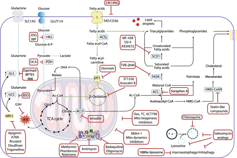
Cancer is now considered as a heterogeneous ecosystem in which tumor cells collaborate with each other and with host cells in their microenvironment. As circumstances change, the ecosystem evolves to ensure the survival and growth of the cancer cells. In this ecosystem, metabolism is not only a key player but also drives stemness. In this review, we first summarize our current understanding of how autophagy influences cancer stem cell phenotype. We emphasize metabolic pathways in cancer stem cells and discuss how autophagy-mediated regulation metabolism is involved in their maintenance and proliferation. We then provide an update on the role of metabolic reprogramming and plasticity in cancer stem cells. Finally, we discuss how metabolic pathways in cancer stem cells could be therapeutically targeted.

Keywords: Autophagy; Cancer stem cells; Lipid metabolism; Metabolic heterogeneity; Therapeutic target.


Conflict of interest statement

The authors declare that they have no competing interests.

Figures

Fig. 1
Fig. 1
The metabolic heterogeneity of cancer stem cells. Tumors are complex and dynamic structures encompassing populations of host cells (e.g., fibroblasts and immune cells) and cancer cells with different metabolic activities. These cells are affected in different ways by microenvironmental conditions and biological activities of other tumor cells. For example, cancer cells close to the vasculature show oxidative metabolism, whereas a shift toward a glycolytic metabolism is observed when glucose is present in cells residing in hypoxic areas. Despite metabolic heterogeneity, cancer cells cooperate to allow adaption to changes in conditions to ensure that metabolic requirements are met. Indeed, oxidative cancer cells, like proliferating cells, increase the consumption of glucose to produce ATP and generate biomass to support cell proliferation. The oxidative stress caused by rapidly proliferation of cancer cells induces glycolysis and autophagy/mitophagy in stromal cells and/or in glycolytic cancer cells leading to the release of high amounts of lactate, which fuels the metabolism of oxidative cancer cells. Key elements of lactate shuttles are the plasma membrane monocarboxylate transporters. MCT4 is involved in the export of lactate, and MCT1 and MCT2 are involved in the uptake of this catabolite. High levels of several factors including HIF-1α, NF-κB, TGF-β, and JNK/AP1 are associated with glycolytic phenotype. The metabolic status of a CSC depends on location. In actively growing regions of the tumor and in the presence of adequate levels of oxygen (normoxic conditions), CSCs rely on glycolytic and/or oxidative metabolism. Overexpression of HIF-1α in the hypoxic environment promotes upregulation of GLUT1, GLUT3, and glycolytic enzymes. In the metastatic niche, CSCs have increased utilization of extracellular catabolites. In nutrient-poor states, autophagy is activated to provide an alternative energy source. OXPHOS and the anabolic gluconeogenesis pathways control glucose homeostasis. Abbreviations: ATP, adenosine triphosphate; CSC, cancer stem cell; GLUT1/GLUT3, glucose transporter 1/3; HIF-1α, hypoxia-inducible factor 1α; HK2, hexokinase 2; JNK/AP1, c-Jun N-terminal kinases/activator protein 1; LDH, Lactate dehydrogenase; XMCT2/4, monocarboxylate transporter 2/4; NF-κB, nuclear factor-κB; OXPHOS, oxidative phosphorylation; PFKFB, phosphofructokinase/fructose bisphosphate; PKM2, pyruvate kinase isozyme M2; TGF-β, transforming growth factor β
Fig. 2
Fig. 2
Metabolic modulators with anti-CSC effects. Metabolic pathways such those involving glutamine, glycolysis, redox balance, lipids, and autophagy are potentially targetable in CSCs. Some of the metabolic enzymes that are currently being considered as therapeutic targets for CSC are indicated by blue rectangles in the figure. Transcription factor NRF2 plays a pivotal role in both intrinsic resistance and cellular adaptation to ROS and is shown in a yellow rectangle. The carnitine-dependent transporter, which inhibits the mitochondrial import of fatty acids is shown in a yellow ball. Inhibitors are indicated by red rectangles. Abbreviations: ACC, acetyl-CoA carboxylase; Ac-CoA, acetyl-coenzyme A; ACLY, ATP citrate lyase; ACSL, long-chain acyl-CoA synthetases; ATRA, all-trans retinoic acid; 3-BP, 3- bromopyruvate; BSO, L-buthionine-S,R-sulfoximine; CPT1, carnitine palmitoyltransferase; I/Q/II/III/IV/V, complexes of the electron transport chain; DCA, dichloroacetate; 2-DG, 2-deoxy-D-glucose; Doc, doxycycline; FASN, fatty acid synthetase; FAT/CD36, Fatty acid translocase; GCS, gamma glutamyl cysteine synthetase; GLS, glutaminase; GLUT1/4, glucose transporter 1/4; GSH, glutathione; HK2, hexokinase 2; HMG-CoAR, 3-hydroxy-3-methyl-glutaryl-coenzyme A reductase; 2M14NQ, 2-methylthio-1,4-naphthoquinone; Mito, mitochondrial; NRF2, nuclear factor erythroid 2-related factor 2; OAA, oxaloacetate; PDH, pyruvate dehydrogenase; ROS, reactive oxygen species; TC, tetracyclines; TCA, tricarboxylic acid cycle; SCD1, stearoyl-CoA desaturase-1; SLC1A5, solute carrier family 1 member 5;

References

    1. Martinez-Outschoorn UE, Peiris-Pages M, Pestell RG, Sotgia F, Lisanti MP. Cancer metabolism: a therapeutic perspective. Nat Rev Clin Oncol. 2017;14(2):113. doi: 10.1038/nrclinonc.2017.1. - DOI - PubMed
    1. Snyder V, Reed-Newman TC, Arnold L, Thomas SM, Anant S. Cancer stem cell metabolism and potential therapeutic targets. Front Oncol. 2018;8:203. doi: 10.3389/fonc.2018.00203. - DOI - PMC - PubMed
    1. Mowers EE, Sharifi MN, Macleod KF. Functions of autophagy in the tumor microenvironment and cancer metastasis. FEBS J. 2018;285(10):1751–1766. doi: 10.1111/febs.14388. - DOI - PMC - PubMed
    1. Poillet-Perez L, White E. Role of tumor and host autophagy in cancer metabolism. Genes Dev. 2019;33(11–12):610–619. doi: 10.1101/gad.325514.119. - DOI - PMC - PubMed
    1. Poillet-Perez L, Xie X, Zhan L, Yang Y, Sharp DW, Hu ZS, et al. Autophagy maintains tumour growth through circulating arginine. Nature. 2018;563(7732):569–573. doi: 10.1038/s41586-018-0697-7. - DOI - PMC - PubMed

Publication types