. 2020 Feb 6;13(1):50.
doi: 10.1186/s13071-020-3907-8.

Description, molecular characteristics and Wolbachia endosymbionts of Onchocerca borneensis Uni, Mat Udin & Takaoka n. sp. (Nematoda: Filarioidea) from the Bornean bearded pig Sus barbatus Müller (Cetartiodactyla: Suidae) of Sarawak, Malaysia

Affiliations

Description, molecular characteristics and Wolbachia endosymbionts of Onchocerca borneensis Uni, Mat Udin & Takaoka n. sp. (Nematoda: Filarioidea) from the Bornean bearded pig Sus barbatus Müller (Cetartiodactyla: Suidae) of Sarawak, Malaysia

Shigehiko Uni et al. Parasit Vectors. .

Abstract

Background: The genus Onchocerca Diesing, 1841 includes species of medical importance, such as O. volvulus (Leuckart, 1893), which causes river blindness in the tropics. Recently, zoonotic onchocercosis has been reported in humans worldwide. In Japan, O. dewittei japonica Uni, Bain & Takaoka, 2001 from wild boars is a causative agent for this zoonosis. Many filarioid nematodes are infected with Wolbachia endosymbionts which exhibit various evolutionary relationships with their hosts. While investigating the filarial fauna of Borneo, we discovered an undescribed Onchocerca species in the bearded pig Sus barbatus Müller (Cetartiodactyla: Suidae).

Methods: We isolated Onchocerca specimens from bearded pigs and examined their morphology. For comparative material, we collected fresh specimens of O. d. dewittei Bain, Ramachandran, Petter & Mak, 1977 from banded pigs (S. scrofa vittatus Boie) in Peninsular Malaysia. Partial sequences of three different genes (two mitochondrial genes, cox1 and 12S rRNA, and one nuclear ITS region) of these filarioids were analysed. By multi-locus sequence analyses based on six genes (16S rDNA, ftsZ, dnaA, coxA, fbpA and gatB) of Wolbachia, we determined the supergroups in the specimens from bearded pigs and those of O. d. dewittei.

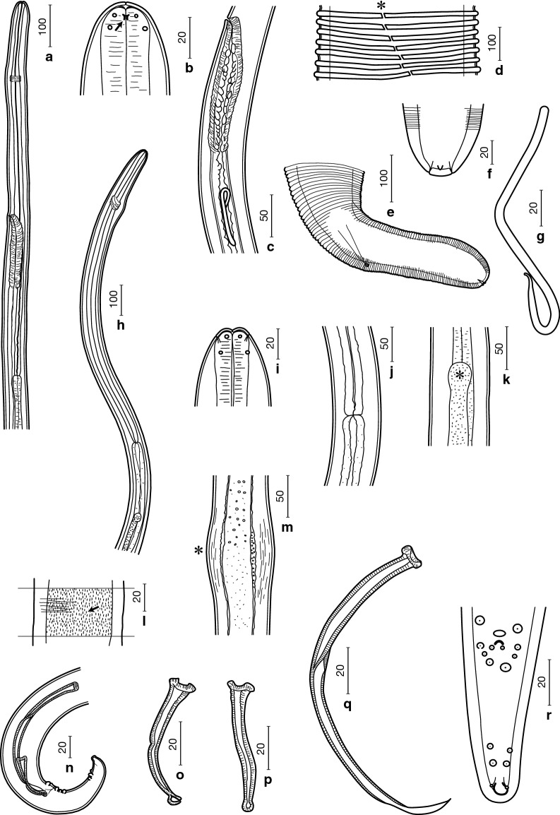
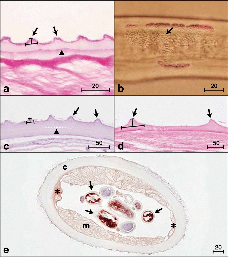
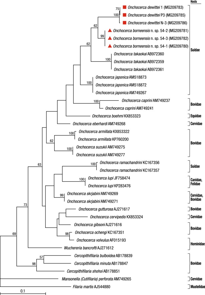
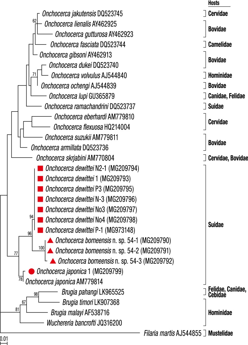
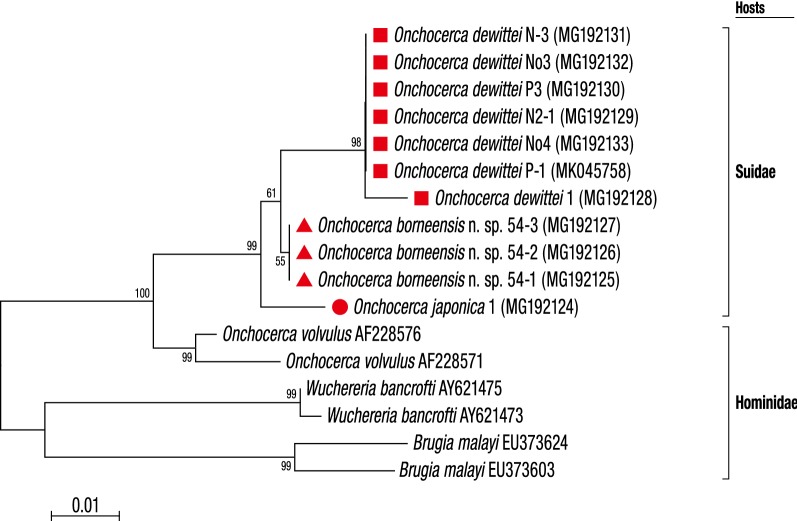
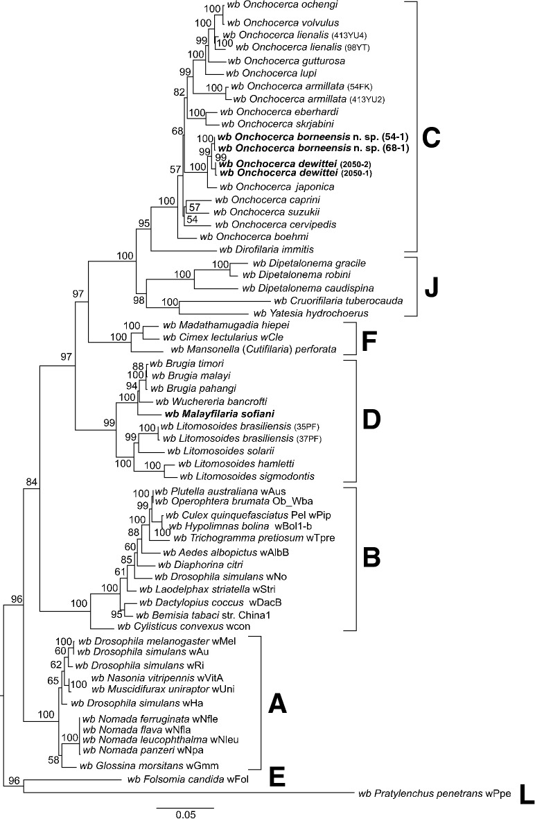
Results: Onchocerca borneensis Uni, Mat Udin & Takaoka n. sp. is described on the basis of morphological characteristics and its genetic divergence from congeners. Molecular characteristics of the new species revealed its close evolutionary relationship with O. d. dewittei. Calculated p-distance for the cox1 gene sequences between O. borneensis n. sp. and O. d. dewittei was 5.9%, while that between O. d. dewittei and O. d. japonica was 7.6%. No intraspecific genetic variation was found for the new species. Wolbachia strains identified in the new species and O. d. dewittei belonged to supergroup C and are closely related.

Conclusions: Our molecular analyses of filarioids from Asian suids indicate that the new species is sister to O. d. dewittei. On the basis of its morphological and molecular characteristics, we propose to elevate O. d. japonica to species level as O. japonica Uni, Bain & Takaoka, 2001. Coevolutionary relationships exist between the Wolbachia strains and their filarial hosts in Borneo and Peninsular Malaysia.

Keywords: Coevolution; Indomalayan realm; Malayfilaria sofiani; Onchocerca dewittei; Onchocerca japonica; Suidae.


Conflict of interest statement

The authors declare that they have no competing interests.

Figures

Fig. 1
Fig. 1
Line drawings of Onchocerca borneensis n. sp. Females (af), microfilaria (g) and males (hr). a Anterior end, left lateral view. b Anterior extremity, lateral view, showing amphid (arrow). c Vagina, left lateral view. d Transverse cuticular ridges at midbody region, showing lateral field (*). e Posterior end, left lateral view. f Posterior extremity, ventral view, showing internal terminal point and two subterminal phasmids. g Microfilaria without sheath. h Anterior end, lateral view. i Anterior extremity, dorsoventral view. j Oesophago-intestinal junction. k Apex of testis (*). l Short longitudinal cuticular crests (arrow) at midbody region. m Body swelling (*). n Posterior end, right lateral view. o Right spicule, lateral view. p Right spicule, dorsoventral view. q Left spicule, lateral view. r Posterior end, ventral view. Scale-bars are in micrometres
Fig. 2
Fig. 2
Light micrographs of Onchocerca borneensis n. sp. (a and b), O. dewittei dewittei (c and e) and O. d. japonica (d). a Longitudinal section of female at midbody: transverse cuticular ridges, triangular or trapezoid in shape (arrows; height and width, dark lines) and absence of internal striae at mid-line (arrowhead) (HE staining). b Short longitudinal cuticular crests (arrow) of male at midbody. c Longitudinal section of female at midbody: triangular transverse cuticular ridges (arrows; height and width, dark lines) and absence of internal striae (arrowhead) at mid-line (dotted line) (HE staining). d Longitudinal section of female at midbody: large triangular transverse cuticular ridges (arrows; height and width, dark lines), showing long distance between adjacent ridges (HE staining). e Transverse section of female with Wolbachia immunostaining. Dark red dots in ovaries and uterus (arrows) are Wolbachia. Lateral chords (*), muscles (m) and cuticle (c). Scale-bars are in micrometres
Fig. 3
Fig. 3
Taxonomic position of Onchocerca borneensis n. sp. and O. dewittei based on cox1 nucleotide sequences. The maximum-likelihood phylogenetic tree was generated under the Tamura-Nei model in MEGA7 with 500 bootstrap replicates. The scale-bar below the diagram indicates the number of inferred changes along each branch. Red triangles and squares indicate sequences generated in this study
Fig. 4
Fig. 4
Taxonomic position of Onchocerca borneensis n. sp., O. dewittei and O. japonica based on 12S rDNA nucleotide sequences. The maximum-likelihood phylogenetic tree was generated under the Tamura-Nei model in MEGA7 with 500 bootstrap replicates. The scale-bar below the diagram indicates the number of inferred changes along each branch. Red triangles, squares and circle indicate sequences generated in this study
Fig. 5
Fig. 5
Taxonomic position of Onchocerca borneensis n. sp., O. dewittei and O. japonica based on ITS nucleotide sequences. The maximum-likelihood phylogenetic tree was generated under the General Time Reversible model in MEGA7 with 500 bootstrap replicates. The scale-bar below the diagram indicates the number of inferred changes along each branch. Red triangles, squares and circle indicate sequences generated in this study
Fig. 6
Fig. 6
Phylogenetic tree of Wolbachia based on six markers using maximum-likelihood inference. Analysis based on concatenation of 16S rDNA, ftsZ, dnaA, coxA, fbpA and gatB (3086 bp in total length). The topology was generated using model TIM + F + I + G4 with IQ-TREE. The robustness of nodes was assessed with 1000 bootstrap replicates. The Wolbachia supergroups (A–F, J and L) were identified according to Lefoulon et al. [28] and Lo et al. [43]. The scale-bar indicates the distance in substitutions per nucleotide. Abbreviation: wb, Wolbachia

References

    1. WHO. Onchocerciasis. Geneva: World Health Organization; 2018. https://www.who.int/news-room/fact-sheets/detail/onchocerciasis. Accessed 30 Jan 2019.
    1. Otranto D, Sakru N, Testini G, Gürlü VP, Yakar K, Lia RP, et al. Case report: first evidence of human zoonotic infection by Onchocerca lupi (Spirurida, Onchocercidae) Am J Trop Med Hyg. 2011;84:55–58. doi: 10.4269/ajtmh.2011.10-0465. - DOI - PMC - PubMed
    1. Eberhard ML, Ostovar GA, Chundu K, Hobohm D, Feiz-Erfan I, Mathison BA, et al. Case report: zoonotic Onchocerca lupi infection in a 22-month-old child in Arizona: first report in the United States and a review of the literature. Am J Trop Med Hyg. 2013;88:601–605. doi: 10.4269/ajtmh.12-0733. - DOI - PMC - PubMed
    1. Mowlavi G, Farzbod F, Kheirkhah A, Mobedi I, Bowman DD, Naddaf SR. Human ocular onchocerciasis caused by Onchocerca lupi (Spirurida, Onchocercidae) in Iran. J Helminthol. 2014;88:250–255. doi: 10.1017/S0022149X13000060. - DOI - PubMed
    1. Takaoka H, Bain O, Uni S, Korenaga M, Kozek WJ, Shirasaka C, et al. Zoonotic onchocerciasis caused by a parasite from wild boar in Oita, Japan. A comprehensive analysis of morphological characteristics of the worms for its diagnosis. Parasite. 2004;11:285–292. doi: 10.1051/parasite/2004113285. - DOI - PubMed

MeSH terms

LinkOut - more resources