

Allo-antibody production after intraarticular administration of mesenchymal stem cells (MSCs) in an equine osteoarthritis model: effect of repeated administration, MSC inflammatory stimulation, and equine leukocyte antigen (ELA) compatibility
- PMID: 32028995
- PMCID: PMC7006079
- DOI: 10.1186/s13287-020-1571-8
Allo-antibody production after intraarticular administration of mesenchymal stem cells (MSCs) in an equine osteoarthritis model: effect of repeated administration, MSC inflammatory stimulation, and equine leukocyte antigen (ELA) compatibility
Abstract
Background: Antibody production after allogeneic administration of mesenchymal stem cells (MSCs) could impact their clinical application. Proinflammatory priming of MSCs can potentiate their regulatory ability in vivo but increased expression of major histocompatibility complex (MHC) might augment their immunogenicity, potentially leading to immune memory thus limiting repeated allogeneic administration. This study aimed at evaluating the production of cytotoxic allo-antibodies directed against donor's ELA (equine leukocyte antigen) in mismatched and halfmatched horses receiving repeated intraarticular administration of stimulated MSCs (MSC-primed) and unstimulated MSCs (MSC-naïve) in pathologic joints.
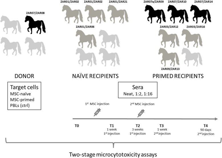
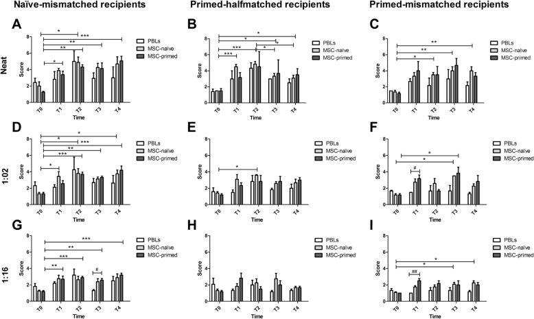
Methods: From available stored samples from a previous in vivo study, cells from one donor and serially collected sera (five time-points) from three groups of recipients were used based on their ELA haplotypes to perform microcytotoxicity assays: Group 1 recipients mismatched with the donor that received MSC-naïve (naïve-mismatched recipients); Group 2 recipients mismatched with the donor that received MSC-primed (primed-mismatched recipients); Group 3 recipients halfmatched with the donor (sharing 1/2 haplotypes) that received MSC-primed (primed-halfmatched recipients). Sera from recipients (neat, 1:2 and 1:16 dilution) were tested against target cells from the donor (cryopreserved and expanded MSC-naïve and MSC-primed) or from one animal presenting the same ELA haplotypes than the donor (fresh peripheral blood lymphocytes as control).
Results: One to three weeks after first MSC administration, all recipient groups produced allo-antibodies regardless of MSC received (naïve or primed) and matching degree with donor. However, secondary response after MSC re-exposure was less evident in halfmatched recipients (MSC-primed) than in mismatched ones (both MSC-naïve and MSC-primed). Recipients of MSC-primed (both mismatched and halfmatched) tended towards developing lower antibody response than MSC-naïve recipients in vivo, but MSC-primed were targeted to death in higher percentage in vitro in the microcytoxicity assay.
Conclusions: After first intraarticular allogeneic administration, the immunomodulatory profile of MSC-primed would have led to lower antibody production, but these antibodies would target more easily MSC-primed after second injection (re-exposure), likely because of their higher MHC expression.
Keywords: Allogeneic; Horse; Humoral response; Immunogenicity; Joint; MSC priming; Major histocompatibility complex (MHC).
Conflict of interest statement
The authors declare that they have no competing interests.
Figures
Similar articles
-
Equine allogeneic bone marrow-derived mesenchymal stromal cells elicit antibody responses in vivo.Stem Cell Res Ther. 2015 Apr 12;6(1):54. doi: 10.1186/s13287-015-0053-x. Stem Cell Res Ther. 2015. PMID: 25889095 Free PMC article.
-
Allogeneic major histocompatibility complex-mismatched equine bone marrow-derived mesenchymal stem cells are targeted for death by cytotoxic anti-major histocompatibility complex antibodies.Equine Vet J. 2017 Jul;49(4):539-544. doi: 10.1111/evj.12647. Epub 2016 Dec 13. Equine Vet J. 2017. PMID: 27862236 Free PMC article.
-
The systemic cellular immune response against allogeneic mesenchymal stem cells is influenced by inflammation, differentiation and MHC compatibility: in vivo study in the horse.Front Vet Sci. 2024 Jun 18;11:1391872. doi: 10.3389/fvets.2024.1391872. eCollection 2024. Front Vet Sci. 2024. PMID: 38957800 Free PMC article.
-
Immunoprivileged no more: measuring the immunogenicity of allogeneic adult mesenchymal stem cells.Stem Cell Res Ther. 2017 Dec 22;8(1):288. doi: 10.1186/s13287-017-0742-8. Stem Cell Res Ther. 2017. PMID: 29273086 Free PMC article. Review.
-
Anti-donor immune responses elicited by allogeneic mesenchymal stem cells: what have we learned so far?Immunol Cell Biol. 2013 Jan;91(1):40-51. doi: 10.1038/icb.2012.67. Epub 2012 Dec 4. Immunol Cell Biol. 2013. PMID: 23207278 Review.
Cited by
-
Comparing the immunomodulatory properties of equine BM-MSCs culture expanded in autologous platelet lysate, pooled platelet lysate, equine serum and fetal bovine serum supplemented culture media.Front Vet Sci. 2022 Aug 25;9:958724. doi: 10.3389/fvets.2022.958724. eCollection 2022. Front Vet Sci. 2022. PMID: 36090170 Free PMC article.
-
Translational Animal Models Provide Insight Into Mesenchymal Stromal Cell (MSC) Secretome Therapy.Front Cell Dev Biol. 2021 Mar 19;9:654885. doi: 10.3389/fcell.2021.654885. eCollection 2021. Front Cell Dev Biol. 2021. PMID: 33869217 Free PMC article. Review.
-
Cross-matching of allogeneic mesenchymal stromal cells eliminates recipient immune targeting.Stem Cells Transl Med. 2021 May;10(5):694-710. doi: 10.1002/sctm.20-0435. Epub 2020 Dec 25. Stem Cells Transl Med. 2021. PMID: 33369287 Free PMC article.
-
Equine Mesenchymal Stem Cells Influence the Proliferative Response of Lymphocytes: Effect of Inflammation, Differentiation and MHC-Compatibility.Animals (Basel). 2022 Apr 11;12(8):984. doi: 10.3390/ani12080984. Animals (Basel). 2022. PMID: 35454231 Free PMC article.
-
Case report: Equine metacarpophalangeal joint partial and full thickness defects treated with allogenic equine synovial membrane mesenchymal stem/stromal cell combined with umbilical cord mesenchymal stem/stromal cell conditioned medium.Front Vet Sci. 2024 May 22;11:1403174. doi: 10.3389/fvets.2024.1403174. eCollection 2024. Front Vet Sci. 2024. PMID: 38840629 Free PMC article.
References
-
- Colbath AC, Frisbie DD, Dow SW, Kisiday JD, McIlwraith CW, Goodrich LR. Equine models for the investigation of mesenchymal stem cell therapies in orthopedic disease. Oper Tech Sport Med. 2017;25(1):41–49. doi: 10.1053/j.otsm.2016.12.007. - DOI
-
- Cuerquis J, Romieu-Mourez R, Francois M, Routy JP, Young YK, Zhao J, et al. Human mesenchymal stromal cells transiently increase cytokine production by activated T cells before suppressing T-cell proliferation: effect of interferon-gamma and tumor necrosis factor-alpha stimulation. Cytotherapy. 2014;16(2):191–202. doi: 10.1016/j.jcyt.2013.11.008. - DOI - PubMed
Publication types
MeSH terms
Grants and funding
LinkOut - more resources
Full Text Sources
Research Materials
