Review
. 2020 Mar 1;34(5-6):263-284.
doi: 10.1101/gad.334631.119. Epub 2020 Feb 6.

(ADP-ribosyl)hydrolases: structure, function, and biology

Affiliations
Review

(ADP-ribosyl)hydrolases: structure, function, and biology

Johannes Gregor Matthias Rack et al. Genes Dev. .

Abstract

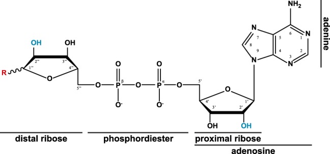
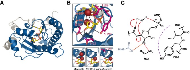
ADP-ribosylation is an intricate and versatile posttranslational modification involved in the regulation of a vast variety of cellular processes in all kingdoms of life. Its complexity derives from the varied range of different chemical linkages, including to several amino acid side chains as well as nucleic acids termini and bases, it can adopt. In this review, we provide an overview of the different families of (ADP-ribosyl)hydrolases. We discuss their molecular functions, physiological roles, and influence on human health and disease. Together, the accumulated data support the increasingly compelling view that (ADP-ribosyl)hydrolases are a vital element within ADP-ribosyl signaling pathways and they hold the potential for novel therapeutic approaches as well as a deeper understanding of ADP-ribosylation as a whole.

Keywords: ADP-ribose; ADP-ribosylation; ARH3; DNA damage; DraG; PARG; PARP; catalytic mechanism; genome stability; macrodomain; structural biology.


Figures

Figure 1.
Figure 1.
The chemical structure of ADP ribose, including atom and substructure labels, as used in this review. Throughout the ADP-ribosylation cycle different moieties (R, red) are attached to the anomeric carbon (C1′′); namely, the substrate β-NAD+ (nicotinamide is linked trans relative to the 2′′OH), the formed reaction products (linked cis [α] relative to the 2′′OH) (Table 1), and ADPr (1′′OH; anomeric mixture in aqueous solution). Linkage sites of consecutive ADP-ribose moieties within PAR are highlighted in blue ([2′] linear linkage; [2′′] branch point linkage).
Figure 2.
Figure 2.
Domain structure of macrodomains and (ADP-ribosyl)hydrolases. The hydrolytic domains are Macro (macrodomain), DUF2263, (microbial PARG), and Ribosyl_crysJ1 (ADP-ribosylation/Crystallin J1 fold), respectively. Subtype-specific sequence motifs are given above the first domain structure (red) of its type. Canonical PARGs contain an accessory domain (AD). In vertebrata, the AD contains a mitochondrial-targeting signal (MTS) and the N terminus is extended by a regulatory and targeting domain (RT domain), which holds the nuclear localization and export signal (NLS and NES, respectively) as well as a PCNA-interacting protein (PIP) box. Other domains: 3α, 3-α-helical bundle; SirTM, sirtuin of M class. Alternative splicing of the single PARG gene in humans is indicated above hPARG. Note that the PARG60 transcript involves splicing of exons 1 and 4 as well as exclusion of exon 5 leading to an altered N-terminal sequence, but including the MTS. The arrow indicates the position from which the primary sequence corresponds to the other splice variants. (†) PARG55 derives from the usage of an alternative start codon in the PARG60 transcript.
Figure 3.
Figure 3.
PAR degradation by PARG-like hydrolases. (A) Ribbon representation of the catalytic domains of canonical PARGs (depicted hPARG; PDB 4B1H) and microbial PARGs (depicted tcPARG; PDB 3SIG) in complex with ADPr. (B) Close up of the active site of hPARG. (Yellow) ADPr; (magenta) residues involved in ligand orientation and catalysis; (red) structural water (w2265); (dashed lines) selected polar interaction. (C) Potential reaction mechanism for PARG-like enzymes. Residue numbering is in accordance with human PARG111.
Figure 4.
Figure 4.
Comparison of adenine coordination across macrodomains and (ADP-ribosyl)hydrolases. Surface-liquorice representation of adenine coordination. The adenine base lies against the protein surface in most hydrolases with the exception of ARH3 in which it holds by π–π stacking perpendicular to the protein surface (view rotated [arrow] by ∼ 60° relative to the closeups). (Yellow) ADPr; (blue) coordinating residues; (red) waters; (dashed lines) selected polar contacts.
Figure 5.
Figure 5.
MacroD-type hydrolases. (A) Ribbon representation of hMacroD2 (PDB 4IQY) as typical representative of the MacroD-type class. (Blue) Macrodomain; (white) N-terminal extension; (yellow) ADPr. (B) The top panel shows closeup of the active site of hMacroD2. Color scheme as in A. (Magenta) Residues involved in ligand orientation and catalysis; (red) structural (w401) and catalytic water (w409); (dashed lines) selected polar interaction. The bottom panels show the replacement of the catalytic water from the active site in the hMacroD2:α-ADPr complex (PDB 4IQY), in the MERS-CoV macrodomain, due to cocrystallization with reaction product β-ADPr (PDB 5HOL), and in OiMacroD, due to p.G37V mutation (PDB 5LAU). (C) Potential reaction mechanism for MacroD-type enzymes. Residue numbering in accordance with hMacroD2. Note: Asp102 is part of the proposed His/Asp dyad and is not present in all MacroD-type hydrolases.
Figure 6.
Figure 6.
ALC1-like hydrolases. (A) Reaction mechanism for the nonenzymatic formation of a Schiff base and the Amadori rearrangement. (B) Closeup of the active site of hTARG1 in complex with the Amadori product of ADPr (yellow) and Lys84. (Magenta) Catalytic residues; (red) structural water (w310); (dashed lines) selected polar interaction. (C) Proposed reaction mechanism for TARG1 and related ALC1-like hydrolases. Residue numbering in accordance with hTARG1. (D) Electrostatic surface map of T. aquaticus DarG. (Red) Negative surface charge; (blue) positive surface charge; (white) neutral surface charge. Note that the prominent positively charged area, which runs perpendicular to the active site, was suggested as the DNA-binding surface. The cocrystallized ADPr is depicted in CPK coloring.
Figure 7.
Figure 7.
ARH structure and mechanism. (A) The left panel shows a ribbon representation of LchARH3 in complex with the ADPr analog ADP-HPD (CPK coloring; PDB 6HH3). The conserved 13 α-helical core motif is colored according to quasidomain classification. (Red) A; (green) B; (yellow) C; (blue) D. The right panels show a closeup of the metal coordination of LchARH3 in complex with Mg2+ (dark gray) and RruDraG in complex with Mn2+ (mauve). (B) Schematic representation of metal coordination defining the metal-to-metal distance. (C) Schematic representation of the dinuclear metal center. Both metals (dark gray) are octahedral coordinated. Ligands in the first coordination sphere are protein-derived monodentates (white), water (red), μ-aqua (purple), and synsyn-bridging carboxyl (yellow). Note that axial position 6 of MeII can be occupied by either water or glutamate, depending on the conformation of the Glu flap. (D) Potential reaction mechanisms for ARH3-type enzymes. Residue numbers according to hARH3.

References

    1. Aasland R, Abrams C, Ampe C, Ball LJ, Bedford MT, Cesareni G, Gimona M, Hurley JH, Jarchau T, Lehto VP, et al. 2002. Normalization of nomenclature for peptide motifs as ligands of modular protein domains. FEBS Lett 513: 141–144. 10.1016/S0014-5793(01)03295-1 - DOI - PubMed
    1. Abplanalp J, Leutert M, Frugier E, Nowak K, Feurer R, Kato J, Kistemaker HVA, Filippov DV, Moss J, Caflisch A, et al. 2017. Proteomic analyses identify ARH3 as a serine mono-ADP-ribosylhydrolase. Nat Commun 8: 2055 10.1038/s41467-017-02253-1 - DOI - PMC - PubMed
    1. Abraham R, Hauer D, McPherson RL, Utt A, Kirby IT, Cohen MS, Merits A, Leung AKL, Griffin DE. 2018. ADP-ribosyl-binding and hydrolase activities of the alphavirus nsP3 macrodomain are critical for initiation of virus replication. Proc Natl Acad Sci 115: E10457–E10466. 10.1073/pnas.1812130115 - DOI - PMC - PubMed
    1. Agnew T, Munnur D, Crawford K, Palazzo L, Mikoč A, Ahel I. 2018. MacroD1 is a promiscuous ADP-ribosyl hydrolase localized to mitochondria. Front Microbiol 9: 20 10.3389/fmicb.2018.00020 - DOI - PMC - PubMed
    1. Ahel D, Horejsi Z, Wiechens N, Polo SE, Garcia-Wilson E, Ahel I, Flynn H, Skehel M, West SC, Jackson SP, et al. 2009. Poly(ADP-ribose)-dependent regulation of DNA repair by the chromatin remodeling enzyme ALC1. Science 325: 1240–1243. 10.1126/science.1177321 - DOI - PMC - PubMed

Publication types

LinkOut - more resources