Review
. 2020 Mar 1;34(5-6):302-320.
doi: 10.1101/gad.334433.119. Epub 2020 Feb 6.

PARPs and ADP-ribosylation in RNA biology: from RNA expression and processing to protein translation and proteostasis

Affiliations
Review

PARPs and ADP-ribosylation in RNA biology: from RNA expression and processing to protein translation and proteostasis

Dae-Seok Kim et al. Genes Dev. .

Abstract

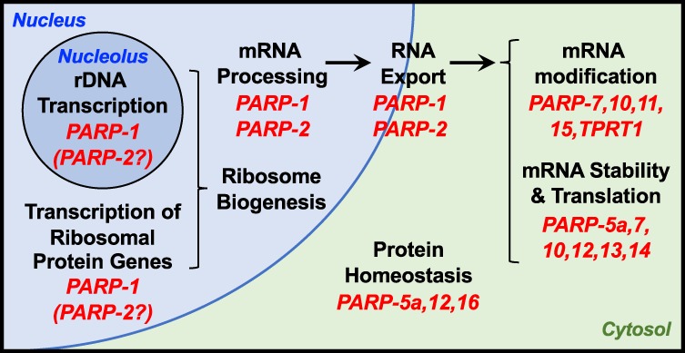
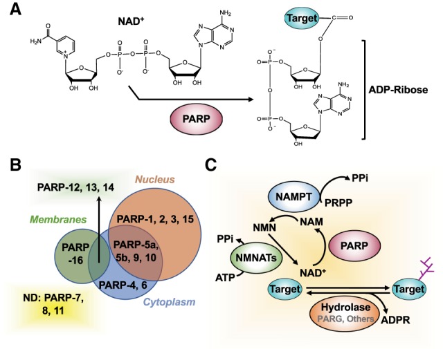
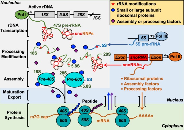
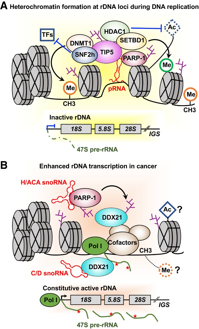
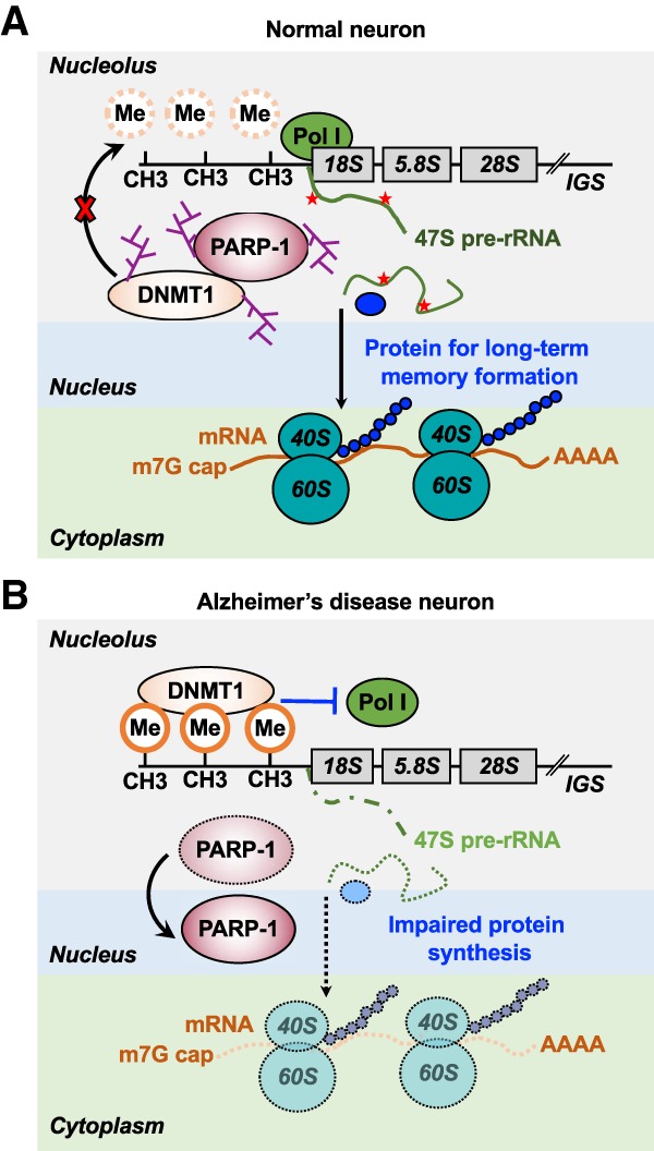
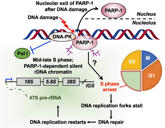
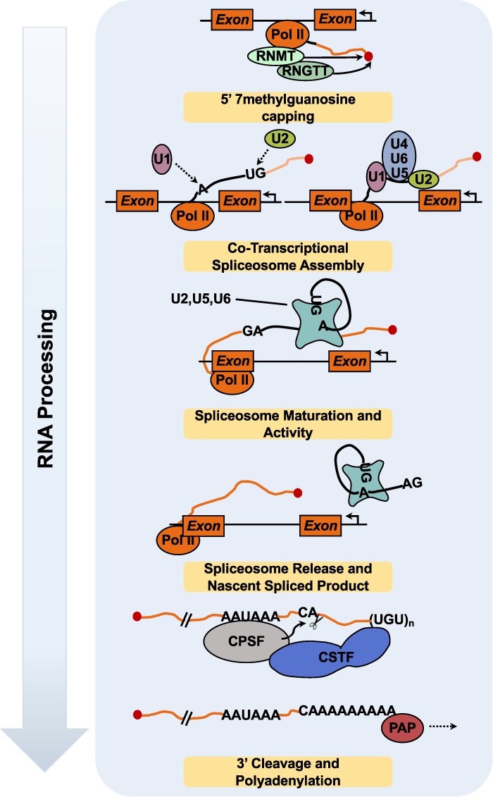
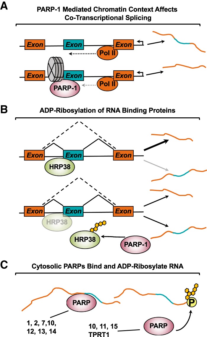
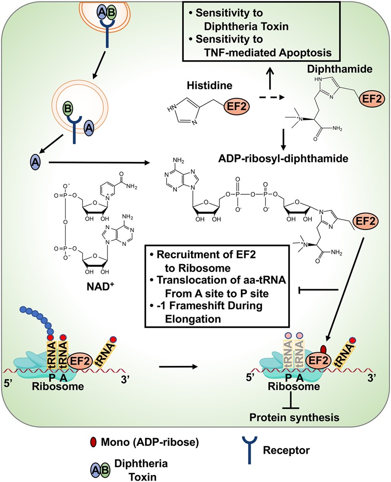
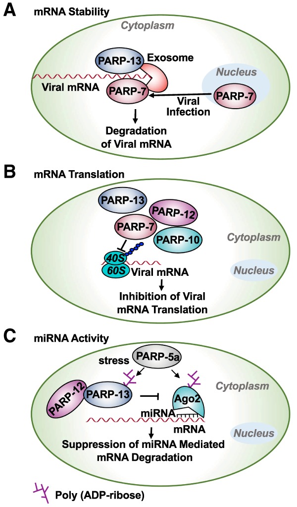
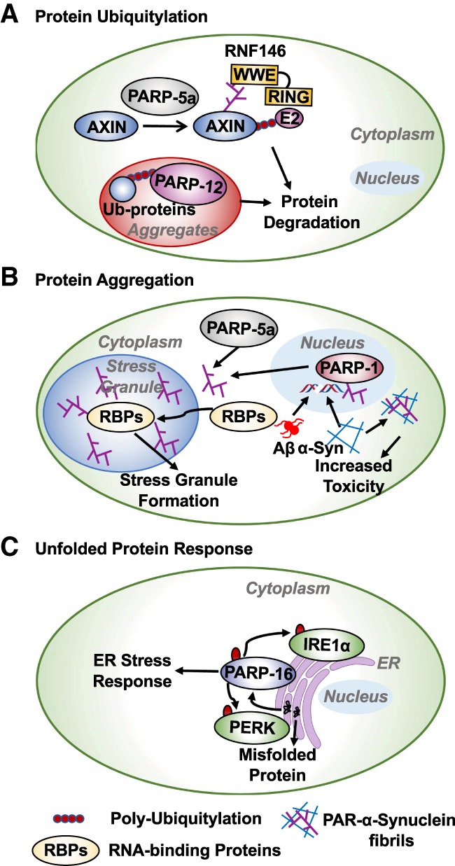
ADP-ribosylation (ADPRylation) is a posttranslational modification of proteins discovered nearly six decades ago, but many important questions remain regarding its molecular functions and biological roles, as well as the activity of the ADP-ribose (ADPR) transferase enzymes (PARP family members) that catalyze it. Growing evidence indicates that PARP-mediated ADPRylation events are key regulators of the protein biosynthetic pathway, leading from rDNA transcription and ribosome biogenesis to mRNA synthesis, processing, and translation. In this review we describe the role of PARP proteins and ADPRylation in all facets of this pathway. PARP-1 and its enzymatic activity are key regulators of rDNA transcription, which is a critical step in ribosome biogenesis. An emerging role of PARPs in alternative splicing of mRNAs, as well as direct ADPRylation of mRNAs, highlight the role of PARP members in RNA processing. Furthermore, PARP activity, stimulated by cellular stresses, such as viral infections and ER stress, leads to the regulation of mRNA stability and protein synthesis through posttranscriptional mechanisms. Dysregulation of PARP activity in these processes can promote disease states. Collectively, these results highlight the importance of PARP family members and ADPRylation in gene regulation, mRNA processing, and protein abundance. Future studies in these areas will yield new insights into the fundamental mechanisms and a broader utility for PARP-targeted therapeutic agents.

Keywords: ADP-ribosylation (ADPRylation); DNA damage; PARP inhibitors (PARPi); RNA stability; mRNA processing; mRNA splicing; mRNA translation; mono(ADP-ribose) (MAR); poly(ADP-ribose) (PAR); poly(ADP-ribose) polymerase (PARP); rRNA synthesis; ribosome biogenesis; stress responses.


Figures

Figure 1.
Figure 1.
Role of PARPs and ADP-ribosylation in RNA biology. PARPs and ADPRylation play broad roles in RNA-related processes, from rDNA transcription and ribosome biogenesis to mRNA processing, protein translation, and proteostasis. See the text for descriptions.
Figure 2.
Figure 2.
Overview of NAD+-dependent, PARP-mediated ADPRylation. (A) PARPs use NAD+ as a substrate to catalyze reversible ADPRylation of substrate proteins. (B) Subcellular localization of PARP family members. (C) NAD+ “feeds” PARP catalytic activity. This enzymatic activity generates nicotinamide (NAM) as a product, which is used by NAMPT in the NAD+ salvage pathway to regenerate NAD+. Several hydrolases that bind to specific structural features of MAR and PAR can hydrolyze the ADPR, returning the target proteins to their unmodified state.
Figure 3.
Figure 3.
The steps in ribosome biogenesis. rRNA molecules (28S, 18S, 5.8S, and 5S), small and large subunit ribosomal proteins, and assembly or processing factors are synthesized by RNA polymerases (Pol I–III), and are subsequently modified and processed. rRNAs assemble with ribosomal proteins to form preribosome particles, which are exported to the cytoplasm to form mature ribosomal subunits and active ribosomes.
Figure 4.
Figure 4.
Dual roles of PARP-1 in the regulation of rDNA transcription. (A) pRNA-mediates PARP-1 and NoRC complex (TIP5 and SNF2h) interactions, leading to PARP-1 automodification and subsequent TIP5 or histone ADPRylation. TIP5 recruits the histone deacetylase HDAC1, DNA methyltransferase DNMT1, and histone lysine methyltransferase SETBD1 to the establish a silent state on rRNA genes. (B) snoRNAs interact with PARP-1 to activate PARP-1 catalytic activity. Subsequently, snoRNA-activated PARP-1 PARylates DDX21 to promote DDX21 association with the rDNA locus and retention in the nucleolus, promoting enhanced rDNA transcription in cancer.
Figure 5.
Figure 5.
Role for PARP-1 in the regulation of rDNA transcription in neurons. (A) PARP-1-mediated DNMT1 ADPRylation prevents rDNA methylation by DNMT1, resulting in the production of rRNAs and subsequent ribosome biogenesis in normal neurons. (B) A substantial reduction in the nucleolar localization of PARP-1 leads to hypermethylation of rDNA by DNMT1, resulting in a reduction of rDNA transcription and ribosome biogenesis. Impaired ribosome biogenesis causes a disruption of long-term memory formation and the development of Alzheimer's disease.
Figure 6.
Figure 6.
DNA damage-induced formation of silent rDNA chromatin. DNA damage leads to the accumulation of cells in S phase, with DNA replication forks stalled at sites of DNA damage. DNA-PK acts upstream of PARP-1 to recruit it to chromatin. Subsequent PARP-1 activation plays an essential role in the formation of silent rDNA chromatin at the time of replication.
Figure 7.
Figure 7.
Overview of nuclear RNA processing events facilitating transformation from pre-mRNA to mRNA. Some RNA processing events occur cotranscriptionally, adding another level of regulation to the RNA processing. mRNAs are modified at their 5′ ends by the addition of a methylguanosine cap by RNA guanine-7 methyltransferase (RNMT) and RNA guanylyltransferase and 5′-phosphatase (RNGTT). RNMT recognizes and physically interacts with the CTD of RNA Pol II in close proximity to the 5′ end of the nascent RNA. RNMT (1) recruits RNGTT to add the guanosine to the 5′ end of the mRNA and (2) catalyzes the addition of a methyl group on the N7 position of the added guanosine. As RNA Pol II transcribes the length of the gene body, exon-intron boundaries are transcribed into the nascent pre-mRNA. The boundaries are identified via specific sequences that demarcate the exon–intron junctions. As the splice site motifs are transcribed, the spliceosome proteins snRNP U2 and U1 bind to their respective sequences, recruiting other spliceosome complex proteins (i.e., U5, U6, and U4). The spliceosome matures as U4 and U1 exit, followed by catalytic activation of the mature spliceosome. In a two-step process, the spliceosome forms a lariat structure with the exon and intron splice spites, cleaves the lariat, and ligates the exons together. Upon cleavage, the intron lariat spliceosome complex is released from the nascent spliced pre-mRNA. After RNA Pol II progresses into the 3′ UTR, the 3′ end is recognized by cleavage and polyadenylation specificity factor (CPSF) and cleavage stimulation factor (CSTF). These enzymes catalyze the cleavage of the pre-mRNA, leaving behind an exposed adenine, which is extended by polyadenylate polymerase (PAP).
Figure 8.
Figure 8.
Examples of RNA processing events modulated by PARP activity. (A) PARP-1 binding to chromatin is enriched at nucleosomes +1/+2 within active promoters and at intron/exon boundaries. PARP-1 bound nucleosomes decrease RNA Pol II processivity, resulting in increased cotranscriptional splicing. (B) RNA-binding protein, HRP38, typically functions to inhibit splicing events. PARP-1 binds to and ADPRylates HRP38 in its RNA-binding domain, thereby attenuating HRP38's ability to bind RNA. The outcome is increased splicing of target transcripts. (C) Both PARP-1 and PARP-2 bind to RNA, resulting in the stimulation of their catalytic activities. Other cytosolic PARP family members (e.g., PARP-7, PARP-10, PARP-11, PARP-12, PARP-13, PARP-14, PARP-15) are known to be or are predicted to bind RNA. Furthermore, PARP-10, PARP-11, PARP-15, and TRPT1 were recently found to ADPRylate single-stranded RNAs at phosphorylated ends.
Figure 9.
Figure 9.
Diphtheria toxin-mediated regulation of mRNA translation. Diphtheria toxin is internalized in cells by binding to cell surface receptors and is subsequently activated by cleavage of the regulatory B domain from the catalytic A domain. The activated A domain MARylates the diphthamide residue in EF2 protein using NAD+ as a substrate. Histidine is modified to diphthamide by a multi-step biosynthetic pathway. ADPRylation of EF2 attenuates protein synthesis by inhibiting (1) EF2 incorporation into ribosomes, (2) transfer of aminoacyl tRNA from the A site to P site, and (3) reverse translocation and −1 frameshifting of ribosomes during translational elongation. Loss of the members of the diphthamide biosynthesis pathway results in a loss of sensitivity to diphtheria toxin and enhanced sensitivity to TNFα-mediated apoptosis.
Figure 10.
Figure 10.
Role of PARPs in the regulation of mRNA functions. Several members of the PARP family control mRNA functions. These pathways play an important role in the regulation of response to viral infections and stress. (A) PARP-7 and PARP-13 bind to viral mRNAs and recruit the exosome complex, resulting in the degradation of the viral mRNAs, thus protecting against viral infections. (B) Interferon-responsive PARPs, such as PARP-7, PARP-10, PARP-12, and PARP-13, inhibit viral infections by inhibiting translation of the viral mRNAs. (C) In response to stress, PARP-13, PARP-12, and PARP-5a are recruited to stress granules, where PARP-5a PARylates PARP-13 and Ago2. These PARylation events inhibit miRNA silencing and increase expression of the genes required to elicit an antiviral response.
Figure 11.
Figure 11.
Role of PARPs in the regulation of protein homeostasis. The activity of both nuclear and cytosolic PARPs are required for protein homeostasis. (A) The WWE domain of RNF146 binds to PAR chains that are catalyzed by PARP-5a. RNF146 recruits an E2 ubiquitin-conjugating enzyme resulting in polyubiquitin-mediated degradation of PARP-5a substrates. PARP-12 binds to polyubiquitylated proteins in protein aggregates. (B) Toxic aggregates of β-amyloid and α-synuclein fibrils induce ROS-mediated DNA damage, causing the activation of PARP-1 and PAR synthesis. α-synuclein fibrils bound to PAR chains cause more cytotoxicity. The PAR chains generated by PARP-1 and PARP-5a recruit RNA-binding proteins (RBPs) to stress granules. PAR chains and modification of RBPs are required for stress granule assembly. (C) Unfolded proteins cause ER-stress and activate PARP-16. PARP-16 MARylates and activates PERK and IRE1α, which are required for ER stress response.

References

    1. Abeti R, Abramov AY, Duchen MR. 2011. β-Amyloid activates PARP causing astrocytic metabolic failure and neuronal death. Brain 134: 1658–1672. 10.1093/brain/awr104 - DOI - PubMed
    1. Abraham R, Hauer D, McPherson RL, Utt A, Kirby IT, Cohen MS, Merits A, Leung AKL, Griffin DE. 2018. ADP-ribosyl-binding and hydrolase activities of the alphavirus nsP3 macrodomain are critical for initiation of virus replication. Proc Natl Acad Sci 115: E10457–E10466. 10.1073/pnas.1812130115 - DOI - PMC - PubMed
    1. Aguilera-Gomez A, van Oorschot MM, Veenendaal T, Rabouille C. 2016. In vivo vizualisation of mono-ADP-ribosylation by dPARP16 upon amino-acid starvation. Elife 5: e21475 10.7554/eLife.21475 - DOI - PMC - PubMed
    1. Ahmed S, Bott D, Gomez A, Tamblyn L, Rasheed A, Cho T, MacPherson L, Sugamori KS, Yang Y, Grant DM, et al. 2015. Loss of the mono-ADP-ribosyltransferase, Tiparp, increases sensitivity to dioxin-induced steatohepatitis and lethality. J Biol Chem 290: 16824–16840. 10.1074/jbc.M115.660100 - DOI - PMC - PubMed
    1. Amé JC, Spenlehauer C, de Murcia G. 2004. The PARP superfamily. BioEssays 26: 882–893. 10.1002/bies.20085 - DOI - PubMed

Publication types

MeSH terms

LinkOut - more resources