Review
. 2020 Mar 1;34(5-6):341-359.
doi: 10.1101/gad.334425.119. Epub 2020 Feb 6.

The impact of PARPs and ADP-ribosylation on inflammation and host-pathogen interactions

Affiliations
Review

The impact of PARPs and ADP-ribosylation on inflammation and host-pathogen interactions

Anthony R Fehr et al. Genes Dev. .

Abstract

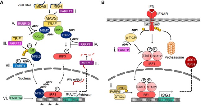
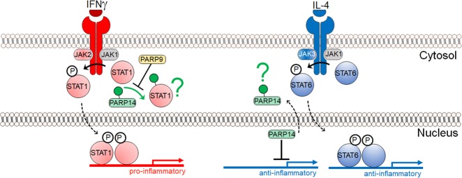
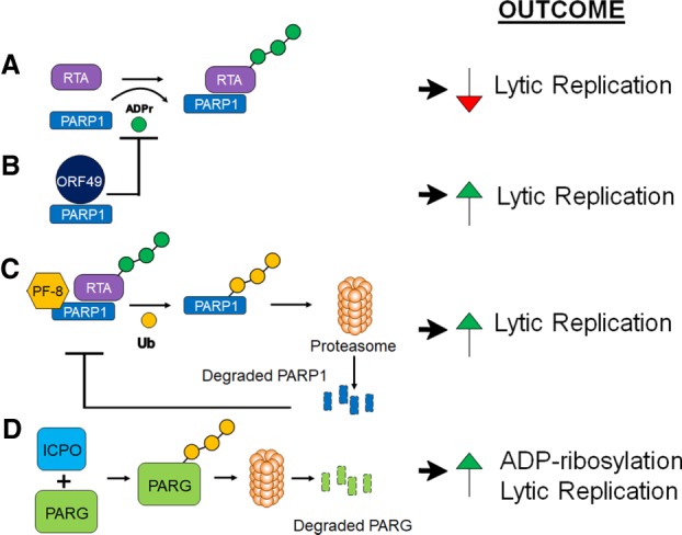
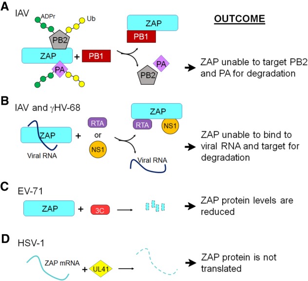
Poly-adenosine diphosphate-ribose polymerases (PARPs) promote ADP-ribosylation, a highly conserved, fundamental posttranslational modification (PTM). PARP catalytic domains transfer the ADP-ribose moiety from NAD+ to amino acid residues of target proteins, leading to mono- or poly-ADP-ribosylation (MARylation or PARylation). This PTM regulates various key biological and pathological processes. In this review, we focus on the roles of the PARP family members in inflammation and host-pathogen interactions. Here we give an overview the current understanding of the mechanisms by which PARPs promote or suppress proinflammatory activation of macrophages, and various roles PARPs play in virus infections. We also demonstrate how innovative technologies, such as proteomics and systems biology, help to advance this research field and describe unanswered questions.

Keywords: ADP-ribosylation; PARP; atherosclerosis; host–pathogen interactions; immunity; inflammation; macrophage; vascular disease.


Figures

Figure 1.
Figure 1.
PARPs regulate the innate immune response at many different points. (A) Mechanisms used by MARylating and nonenzymatic PARPs to modulate IFN and proinflammatory cytokine induction. (i) PARP13 can bind to RIG-I, which promotes its oligomerization and the initiation of the cascade. (ii) PARP13 can also bind to IFN mRNA and target it for degradation. (iii) PARP12 was shown to bind TRIF and enhance NFκB-dependent gene expression. (iv) PARP7 can ADP-ribosylate TBK-1, which inhibits it from phosphorylating IRF3. (v) PARP10 can interact with and ADP-ribosylate NEMO, which prevents the activation of IKKs. (vi) PARP14 promotes H3K27 acetylation and recruitment of Pol II to IFN promoters. (vii) Upon phosphorylation, PARP1 can poly-ADP-ribosylate NFκB and promote its activity. (B) Mechanisms used by MARylating and nonenzymatic PARPs to modulate IFN-I signaling. (i) PARP11 binds to and ADP-ribosylates the E3 ubiquitin ligase β-TrCP. This allows β-TrCP to interact with and ubiquitinate IFNAR, which targets it for proteasome-dependent degradation. (ii) PARP9 and DTX3L interact with and ubiquitinate histone protein H2BJ, which leads to chromatin remodeling that enhances expression of a subset of ISGs. (P) Phosphate group; (ADPr) ADP-ribose; (Ac) acetyl modification; (yellow ciricle) ubiquitin.
Figure 2.
Figure 2.
A partial model of PARP14 and PARP9 function in macrophage activation. In vivo and in vitro studies pertaining to IFNg signaling in primarily macrophages suggest that PARP14 mitigates proinflammatory phosphorylated STAT1 via ADP-ribosylation, and that PARP9 may act to inhibit PARP14's enzymatic activity (Iwata et al. 2016). In vitro studies pertaining to IL-4 signaling in the context of B-cell biology suggest that in nonstimulating conditions PARP14 is a suppressor of STAT6 target genes. In response to IL-4, PARP14 is thought to become enzymatically active and dissociate from the promoter(s), thereby allowing phosphorylated STAT6 to bind and activate target genes (Mehrotra et al. 2011). A green question mark indicates that the fate of ADP-ribosylated substrates is not known. The IFNγ and IL-4 mechanisms appear distinct, but they may be partial and complementary pictures of a complex biology.
Figure 3.
Figure 3.
Computational prediction of an association between the PARP9–PARP14 network and human inflammatory diseases. The network of PARP14 (blue)–PARP9 (purple) consists of proteins that directly interact with these PARPs (blue and orange nodes, respectively). P-values indicate the significance of closeness between the PARP14–PARP9 first neighbors in the interactome (the PARP9–PARP14 module) and gene modules of human diseases such as coronary artery disease compared with random expectation. Reproduced from Iwata et al. (2016).
Figure 4.
Figure 4.
Mechanisms used by herpesviruses to affect PARylation and their impact on replication. (A) PARP-1 can bind to and ADP-ribosylate the γHV RTA, which inhibits its ability to initiate lytic replication. (B) The γHV-68 protein ORF49 binds to PARP1 and prevents it from interacting with and ADP-ribosylating RTA, which allows RTA to initiate viral gene transcription. (C) The KSHV and γHV-68 PF-8 proteins bind to PARP1 and target it for ubiquitination and degradation. This again prevents ADP-ribosylation of RTA, which allows it to initiate lytic replication. (D) The HSV-1 ICP0 protein targets PARG for ubiquitination and degradation, resulting in enhanced PARylation during infection and increased replication. (ADPr) ADP-ribose; (Ub) ubiquitin.
Figure 5.
Figure 5.
Viral mechanisms of ZAP antagonism. (A) IAV protein PB1 binds to ZAP, which prevents its interaction with the PA and PB2 proteins that otherwise would lead to PARylation, ubiquitination, and degradation of these proteins. (B) IAV NS1 and γHV-68 RTA proteins interact with ZAP, preventing its association with viral RNA. (C) The EV-71 3C protease cleaves ZAP to prevent it from accumulating. (D) HSV-1 UL41 protein cleaves ZAP mRNA to prevent its translation. (ADPr) ADP-ribose; (Ub) ubiquitin.

References

    1. Abraham R, Hauer D, McPherson RL, Utt A, Kirby IT, Cohen MS, Merits A, Leung AKL, Griffin DE. 2018. ADP-ribosyl-binding and hydrolase activities of the alphavirus nsP3 macrodomain are critical for initiation of virus replication. Proc Natl Acad Sci 115: E10457–E10466. 10.1073/pnas.1812130115 - DOI - PMC - PubMed
    1. Aday AW, Ridker PM. 2019. Targeting residual inflammatory risk: a shifting paradigm for atherosclerotic disease. Front Cardiovasc Med 6: 16 10.3389/fcvm.2019.00016 - DOI - PMC - PubMed
    1. Aguiar RC, Yakushijin Y, Kharbanda S, Salgia R, Fletcher JA, Shipp MA. 2000. BAL is a novel risk-related gene in diffuse large B-cell lymphomas that enhances cellular migration. Blood 96: 4328–4334. 10.1182/blood.V96.13.4328 - DOI - PubMed
    1. Aguiar RC, Takeyama K, He C, Kreinbrink K, Shipp MA. 2005. B-aggressive lymphoma family proteins have unique domains that modulate transcription and exhibit poly(ADP-ribose) polymerase activity. J Biol Chem 280: 33756–33765. 10.1074/jbc.M505408200 - DOI - PubMed
    1. Aldinucci A, Gerlini G, Fossati S, Cipriani G, Ballerini C, Biagioli T, Pimpinelli N, Borgognoni L, Massacesi L, Moroni F, et al. 2007. A key role for poly(ADP-ribose) polymerase-1 activity during human dendritic cell maturation. J Immunol 179: 305–312. 10.4049/jimmunol.179.1.305 - DOI - PubMed

Publication types

Substances

LinkOut - more resources