Review
. 2020 Mar 1;34(5-6):360-394.
doi: 10.1101/gad.334516.119. Epub 2020 Feb 6.

PARP and PARG inhibitors in cancer treatment

Affiliations
Review

PARP and PARG inhibitors in cancer treatment

Dea Slade. Genes Dev. .

Abstract

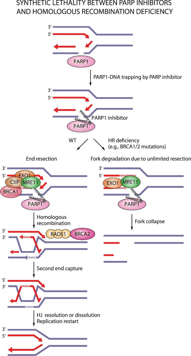
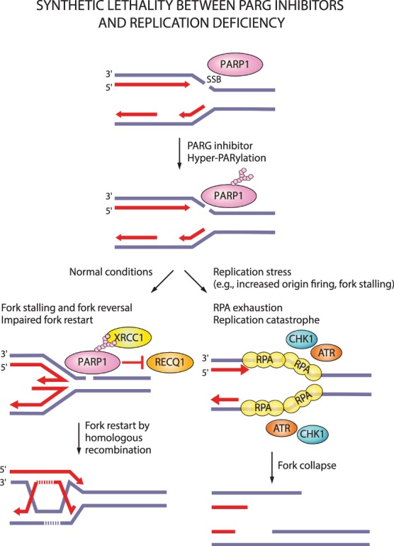
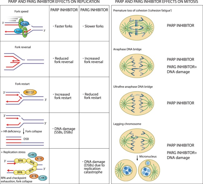
Oxidative and replication stress underlie genomic instability of cancer cells. Amplifying genomic instability through radiotherapy and chemotherapy has been a powerful but nonselective means of killing cancer cells. Precision medicine has revolutionized cancer therapy by putting forth the concept of selective targeting of cancer cells. Poly(ADP-ribose) polymerase (PARP) inhibitors represent a successful example of precision medicine as the first drugs targeting DNA damage response to have entered the clinic. PARP inhibitors act through synthetic lethality with mutations in DNA repair genes and were approved for the treatment of BRCA mutated ovarian and breast cancer. PARP inhibitors destabilize replication forks through PARP DNA entrapment and induce cell death through replication stress-induced mitotic catastrophe. Inhibitors of poly(ADP-ribose) glycohydrolase (PARG) exploit and exacerbate replication deficiencies of cancer cells and may complement PARP inhibitors in targeting a broad range of cancer types with different sources of genomic instability. Here I provide an overview of the molecular mechanisms and cellular consequences of PARP and PARG inhibition. I highlight clinical performance of four PARP inhibitors used in cancer therapy (olaparib, rucaparib, niraparib, and talazoparib) and discuss the predictive biomarkers of inhibitor sensitivity, mechanisms of resistance as well as the means of overcoming them through combination therapy.

Keywords: PARG inhibitor; PARP inhibitor; cancer therapy; poly(ADP-ribose) glycohydrolase; poly(ADP-ribose) polymerases.


Figures

Figure 1.
Figure 1.
Structures of PARP and PARG inhibitors. (A,C) Chemical structures. IC50 denotes half-maximal inhibitory concentration based on measurements of PARP/PARG activity in vitro. Cellular PAR synthesis EC50 denotes half-maximal effective concentration determined by measuring PAR levels in cellular extracts treated with inhibitors. Cytotoxicity CC50 denotes half-maximal cytotoxic concentration determined by measuring cell viability after PARP/PARG inhibitor treatment. (B,D) X-ray structures. (B) Veliparib bound to PARP1 active site (PDB: 2RD6). (D) JA2131 bound to PARG active site (PDB: 6OA3). Inhibitors are labeled in green, PARP1/PARG residues in the binding pocket are labeled in blue, water molecules are shown as red dots, and hydrogen bonds are represented by yellow dashes.
Figure 2.
Figure 2.
DNA lesions recognized by PARP1 as potential PARP-trapping sites. Unligated Okazaki fragments are DNA replication intermediates. Single-strand DNA breaks (SSBs) are a frequent form of endogenous DNA damage and are particularly hazardous for replication forks. Ribonucleotides incorporated into DNA need to be removed by RNase H2-mediated ribonucleotide excision repair. In RNase H2-deficient cells these ribonucleotides are removed by topoisomerase I (TOP1)-mediated excision. TOP1 cleavage results in nicks, covalent TOP1–DNA adducts, and single-strand DNA gaps that can engage PARP1.
Figure 3.
Figure 3.
Single-strand break (SSB) repair and replication fork protection by PARP1. PARP1 acts as a sensor of SSBs and recruits XRCC1. XRCC1 is a scaffold for the recruitment of proteins that process damaged termini, DNA polymerase β that fills the gap, and DNA ligase III that seals the nick. PARP1 rescues damaged replication forks through fork reversal or homologous recombination (HR). SSBs on the leading strand trigger fork reversal by fork remodeling proteins. PARP1 promotes fork reversal by inhibiting the RECQ1 helicase involved in fork restart. PARP1 stabilizes RAD51 filaments on reversed forks and together with BRCA1 and BRCA2 protects forks from degradation by the MRE11 nuclease. If forks collapse when encountering an SSB on the lagging strand, PARP1 promotes HR-mediated fork repair and restart by recruiting MRE11, EXO1, and BRCA1-CtIP for end resection, and BRCA2 for RAD51 filament formation.
Figure 4.
Figure 4.
Synthetic lethality between PARP inhibitors and homologous recombination deficiency. PARP entrapment on DNA lesions blocks replication machinery and loss of PARP activity prevents fork protection, fork reversal, and fork restart. This results in DSBs that need to be repaired by homologous recombination. In the case of homologous recombination deficiency due to, for example, mutations in BRCA1/2, PARP1-trapping lesions elicit excessive fork degradation by the MRE11 nuclease, the activity of which is unrestrained in the absence of BRCA1/2 and PARP1. This results in fork collapse.
Figure 5.
Figure 5.
Synthetic lethality between PARG inhibitors and replication stress. PARG inhibition increases PARylation levels and may prevent dissociation of PARP1 and PAR-binding repair proteins (e.g., XRCC1) from DNA damage sites. Loss of PARG activity causes fork stalling and impairs restart of reversed forks. Forks can presumably restart by homologous recombination. Under replication stress conditions, increased origin firing and prolonged fork stalling generates excessive ssDNA and causes RPA exhaustion. Such replication catastrophe results in fork collapse.
Figure 6.
Figure 6.
Cellular consequences of PARP and PARG inhibition on replication and mitosis. PARP inhibitors increase replication fork rate, reduce fork reversal, and cause premature fork restart. In HR-deficient cells, PARP inhibitors cause fork collapse and DSBs. PARG inhibitors reduce fork rate, increase fork reversal, and impair fork restart. In the presence of replication stress, PARG inhibitors cause replication catastrophe. Destabilization of replication forks causes mitotic defects and death by mitotic catastrophe. PARG inhibitors cause mitotic defects in combination with DNA-damaging agents.

References

    1. Abkevich V, Timms KM, Hennessy BT, Potter J, Carey MS, Meyer LA, Smith-McCune K, Broaddus R, Lu KH, Chen J, et al. 2012. Patterns of genomic loss of heterozygosity predict homologous recombination repair defects in epithelial ovarian cancer. Br J Cancer 107: 1776–1782. 10.1038/bjc.2012.451 - DOI - PMC - PubMed
    1. Ait Saada A, Lambert SAE, Carr AM. 2018. Preserving replication fork integrity and competence via the homologous recombination pathway. DNA Repair 71: 135–147. 10.1016/j.dnarep.2018.08.017 - DOI - PMC - PubMed
    1. Alexandrov LB, Nik-Zainal S, Wedge DC, Aparicio SA, Behjati S, Biankin AV, Bignell GR, Bolli N, Borg A, Børresen-Dale AL, et al. 2013. Signatures of mutational processes in human cancer. Nature 500: 415–421. 10.1038/nature12477 - DOI - PMC - PubMed
    1. Allison Stewart C, Tong P, Cardnell RJ, Sen T, Li L, Gay CM, Masrorpour F, Fan Y, Bara RO, Feng Y, et al. 2017. Dynamic variations in epithelial-to-mesenchymal transition (EMT), ATM, and SLFN11 govern response to PARP inhibitors and cisplatin in small cell lung cancer. Oncotarget 8: 28575–28587. - PMC - PubMed
    1. Alvarez-Gonzalez R, Althaus FR. 1989. Poly(ADP-ribose) catabolism in mammalian cells exposed to DNA-damaging agents. Mutat Res 218: 67–74. 10.1016/0921-8777(89)90012-8 - DOI - PubMed

Publication types

MeSH terms

Substances