. 2020 Mar 11;40(11):2357-2370.
doi: 10.1523/JNEUROSCI.2516-19.2020. Epub 2020 Feb 6.

Interferon-β Plays a Detrimental Role in Experimental Traumatic Brain Injury by Enhancing Neuroinflammation That Drives Chronic Neurodegeneration

Affiliations

Interferon-β Plays a Detrimental Role in Experimental Traumatic Brain Injury by Enhancing Neuroinflammation That Drives Chronic Neurodegeneration

James P Barrett et al. J Neurosci. .

Abstract

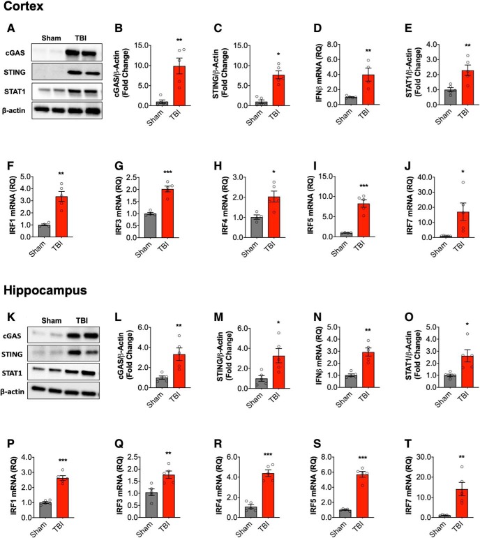
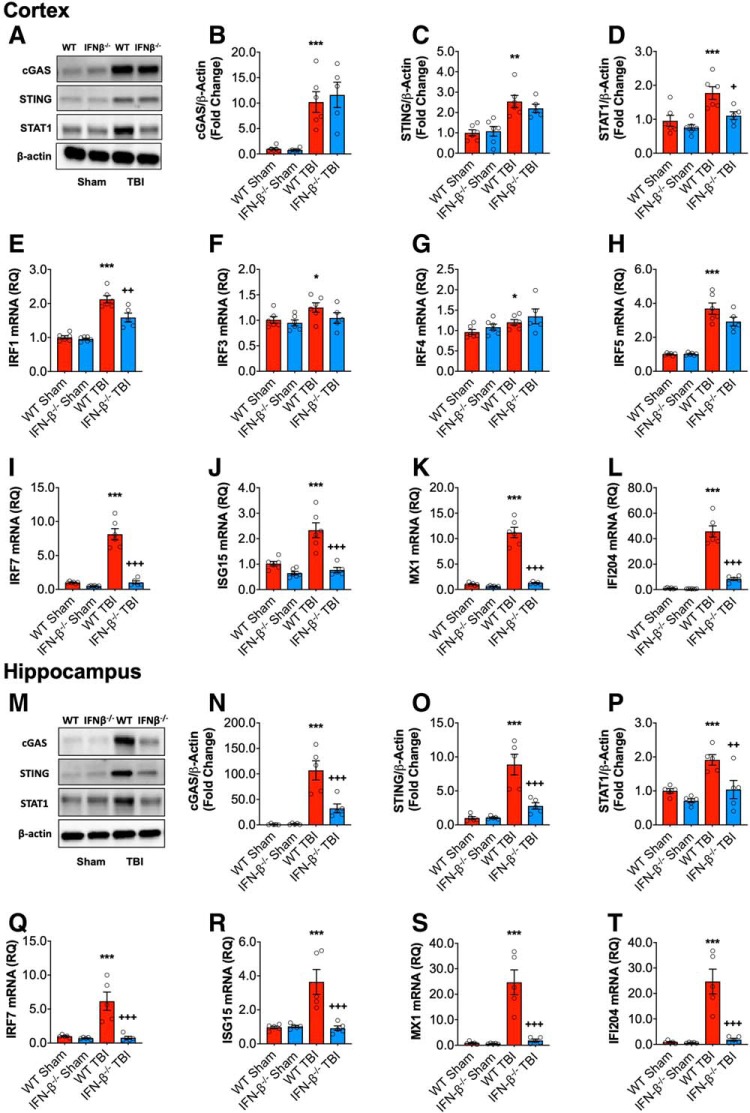
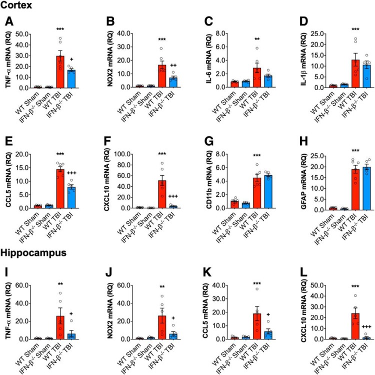
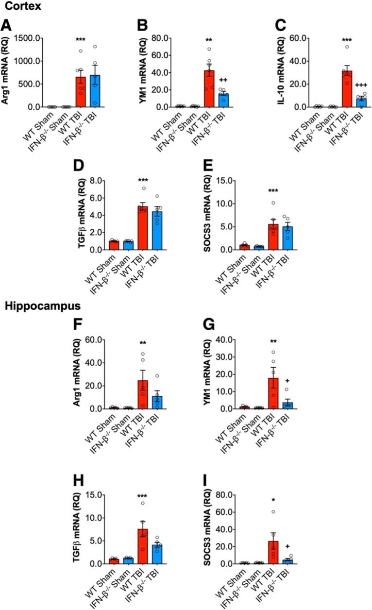
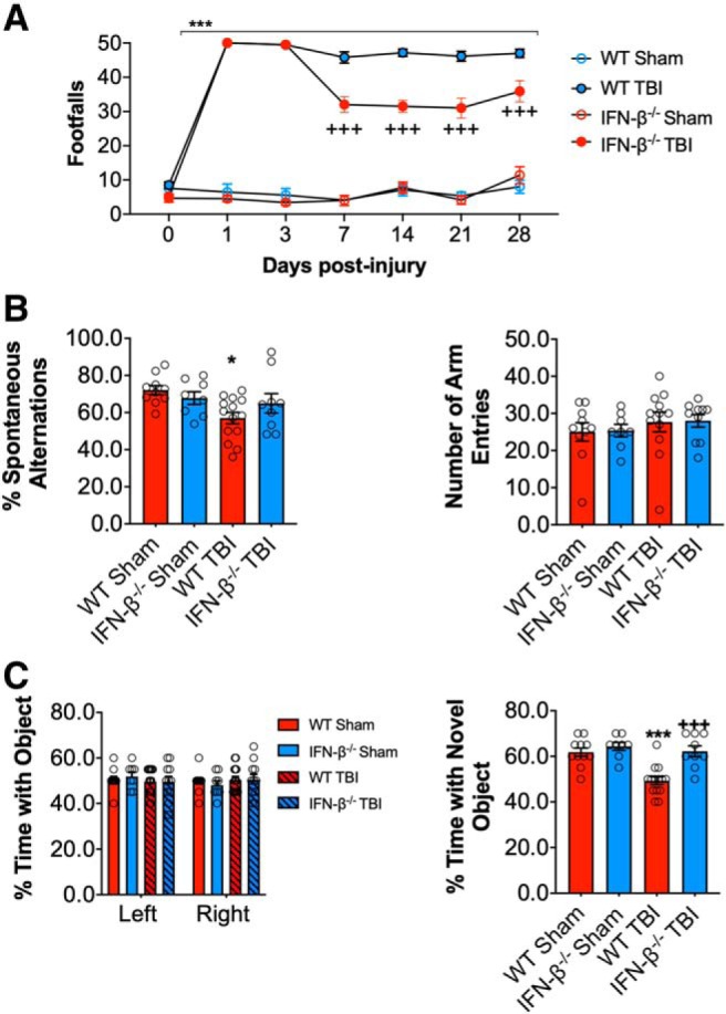
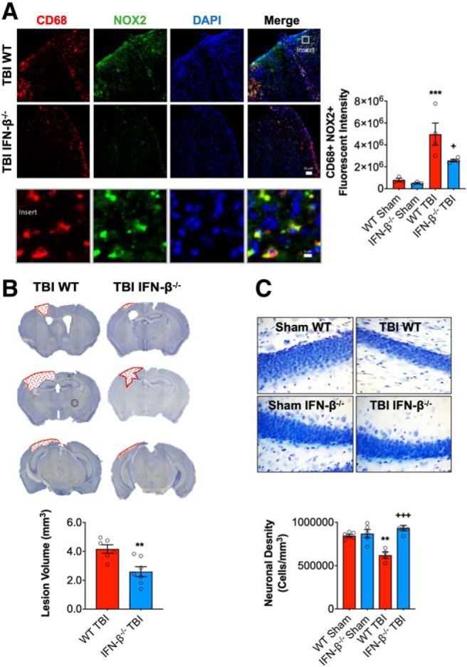
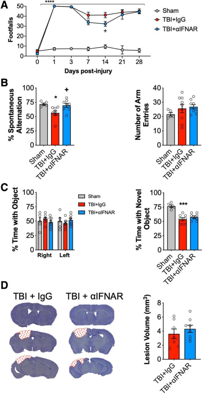
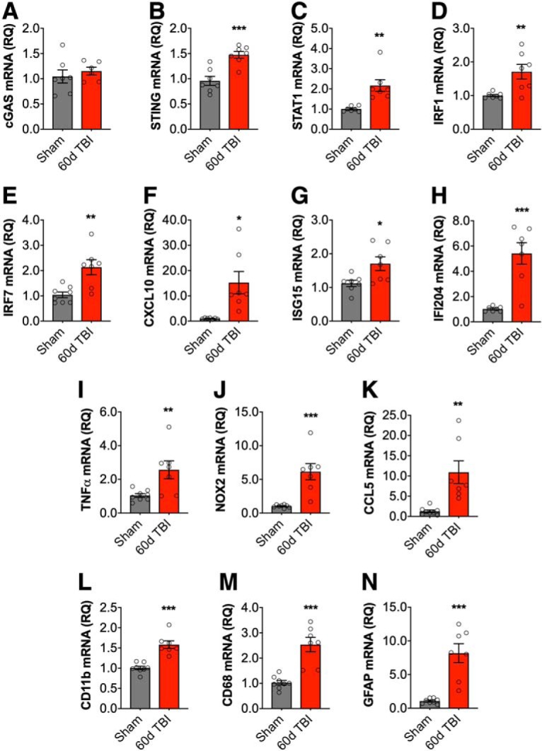
DNA damage and type I interferons (IFNs) contribute to inflammatory responses after traumatic brain injury (TBI). TBI-induced activation of microglia and peripherally-derived inflammatory macrophages may lead to tissue damage and neurological deficits. Here, we investigated the role of IFN-β in secondary injury after TBI using a controlled cortical impact model in adult male IFN-β-deficient (IFN-β-/-) mice and assessed post-traumatic neuroinflammatory responses, neuropathology, and long-term functional recovery. TBI increased expression of DNA sensors cyclic GMP-AMP synthase and stimulator of interferon genes in wild-type (WT) mice. IFN-β and other IFN-related and neuroinflammatory genes were also upregulated early and persistently after TBI. TBI increased expression of proinflammatory mediators in the cortex and hippocampus of WT mice, whereas levels were mitigated in IFN-β-/- mice. Moreover, long-term microglia activation, motor, and cognitive function impairments were decreased in IFN-β-/- TBI mice compared with their injured WT counterparts; improved neurological recovery was associated with reduced lesion volume and hippocampal neurodegeneration in IFN-β-/- mice. Continuous central administration of a neutralizing antibody to the IFN-α/β receptor (IFNAR) for 3 d, beginning 30 min post-injury, reversed early cognitive impairments in TBI mice and led to transient improvements in motor function. However, anti-IFNAR treatment did not improve long-term functional recovery or decrease TBI neuropathology at 28 d post-injury. In summary, TBI induces a robust neuroinflammatory response that is associated with increased expression of IFN-β and other IFN-related genes. Inhibition of IFN-β reduces post-traumatic neuroinflammation and neurodegeneration, resulting in improved neurological recovery. Thus, IFN-β may be a potential therapeutic target for TBI.SIGNIFICANCE STATEMENT TBI frequently causes long-term neurological and psychiatric changes in head injury patients. TBI-induced secondary injury processes including persistent neuroinflammation evolve over time and can contribute to chronic neurological impairments. The present study demonstrates that TBI is followed by robust activation of type I IFN pathways, which have been implicated in microglial-associated neuroinflammation and chronic neurodegeneration. We examined the effects of genetic or pharmacological inhibition of IFN-β, a key component of type I IFN mechanisms to address its role in TBI pathophysiology. Inhibition of IFN-β signaling resulted in reduced neuroinflammation, attenuated neurobehavioral deficits, and limited tissue loss long after TBI. These preclinical findings suggest that IFN-β may be a potential therapeutic target for TBI.

Keywords: interferon-β; neurodegeneration; neuroinflammation; neuroprotection; traumatic brain injury; type I interferons.


Figures

Figure 1.
Figure 1.
Type I IFN response in the injured brain following moderate-level controlled cortical impact. Cortical expression of cGAS and STING protein was assessed by Western immunoblotting (A, representative blot) in the ipsilateral cortex sham and TBI mice at 72 h post-injury. TBI increased cortical expression of cGAS (p = 0.0023; B) and STING (p = 0.023; C) protein compared with sham mice. TBI significantly increased the expression of IFN-β mRNA in the cortex of TBI mice (p = 0.006; D). The expression of STAT1 protein was significantly increased in the cortex of TBI mice compared with sham mice (p = 0.006; E; A, representative blot). mRNA expression of IRF family members was assessed, TBI significantly increased cortical IRF1 (p = 0.002; F), IRF3 (p = 0.0002; G), IRF4 (p = 0.016; H), IRF5 (p = 0.0002; I), and IRF7 (p = 0.046; J) mRNA expression. Expression of cGAS and STING protein was assessed by Western immunoblotting (K, representative blot) in the hippocampus of sham and TBI mice at 72 h post-injury. TBI increased expression of cGAS (t(8) = 3.645, p = 0.0065; L) and STING (t(8) = 2.956, p = 0.0182; M) protein compared with sham mice. IFN-β mRNA expression was significantly increased in the hippocampus of TBI mice (t(8) = 5.447, p = 0.0006; N). The expression of STAT1 protein was significantly increased in the hippocampus of TBI mice compared with sham mice (t(8) = 3.134, p = 0.014; O). mRNA expression of IRF family members was assessed, TBI significantly increased hippocampal IRF1 (t(8) = 9.671, p < 0.0001; P), IRF3 (t(8) = 3.523, p = 0.00078; Q), IRF4 (t(8) = 8.455, p < 0.0001; R), IRF5 (t(8) = 11.92, p < 0.0001; S), and IRF7 (t(8) = 3.921, p = 0.0044; T) mRNA expression. Data expressed as mean ± SEM (n = 5/group). *p < 0.05, **p < 0.01, ***p < 0.001, Student's t test.
Figure 2.
Figure 2.
Type I IFN response following TBI is reduced in IFN-β−/− mice. Cortical expression of cGAS and STING protein was assessed by Western immunoblotting (A, representative blot) in the ipsilateral cortex WT and IFN-β−/−sham and TBI mice at 72 h post-injury. TBI significantly increased cortical expression of cGAS (p < 0.0001; B) and STING (p < 0.0001; C) protein in WT and IFN-β−/− mice. The expression of STAT1 protein was significantly increased in the cortex of TBI mice compared with sham mice (p = 0.0004; D); this TBI effect was significantly reduced in IFN-β−/− mice (p = 0.015, WT TBI vs IFN-β−/− TBI). TBI significantly increased mRNA expression of IRF1 (p < 0.0001; E), IRF3 (p = 0.0478; F), IRF4 (p = 0.0208; G), IRF5 (p < 0.0001; H), and IRF7 (p < 0.0001; I) in WT and IFN-β−/− mice. The TBI-induced increase in IRF1 and IRF7 was significantly reduced in IFN-β−/− mice (IRF1: p = 0.0016, IRF7: p < 0.0001; WT TBI vs IFN-β−/− TBI). TBI significantly increased the expression of the viral response genes ISG15 (p = 0.0005; J), MX1 (p < 0.0001; K), and IFI204 (p < 0.0001; L) in WT and IFN-β−/− mice. This TBI effect was significantly reduced in IFN-β−/− mice, ISG15 (p < 0.0001, WT TBI vs IFN−/− TBI), MX1 (p < 0.0001, WT TBI vs I IFN-β−/− TBI), and IFI204 (p < 0.0001, WT TBI vs IFN-β−/− TBI). Expression of cGAS and STING protein was assessed by Western immunoblotting (M, representative blot) in ipsilateral hippocampal sham and TBI mice at 72 h post-injury. TBI increased expression of cGAS (p < 0.0001; N) and STING (p < 0.0001; O) protein compared with sham mice, this TBI effect was significantly reduced in IFN-β−/− mice (cGAS: p = 0.0007, WT TBI vs IFN-β−/− TBI; STING: p = 0.0004, WT TBI vs IFN-β−/− TBI). The expression of STAT1 protein was significantly increased in the hippocampus of TBI mice compared with sham mice (p = 0.0014; P), the expression of STAT1 was significantly attenuated in IFN-β−/− TBI mice compared with WT TBI mice (p = 0.0073). TBI significantly increased hippocampal IRF7 (p = 0.0015; Q), ISG15 (p = 0.0034; R), MX1 (p = 0.0001; S), and IFI204 (p = 0.0052; T) mRNA expression. The TBI effect on all of these genes was significantly reduced in IFN-β−/− mice, IRF7 (p = 0.0002, WT TBI vs IFN-β−/− TBI), ISG15 (p = 0.0006, WT TBI vs IFN-β−/− TBI), MX1 (p < 0.0001, WT TBI vs IFN-β−/− TBI), and IFI204 (p = 0.0078, WT TBI vs IFN-β−/− TBI). Data expressed as Mean ± SEM. *p < 0.05, **p < 0.01, ***p < 0.001 vs sham (effect of TBI) and +p < 0.05, ++p < 0.01, +++p < 0.001 WT TBI vs IFN-β−/− TBI. Two-way ANOVA (n = 6/group).
Figure 3.
Figure 3.
IFNβ deficiency reduces the proinflammatory response following TBI. Cortical expression of a number of proinflammatory genes was assessed in sham and TBI at 72 h post-injury. TBI significantly increased cortical mRNA expression of TNF-α (p < 0.0001; A), NOX2 (p < 0.0001; B), IL-6 (p = 0.0014; C), IL-1β (p < 0.0001; D), CCL5 (p < 0.0001; E), CXCL10 (p < 0.0001; F), CD11b (ITGAM) (p < 0.0001; G), and GFAP (p < 0.0001; H) in WT and IFN-β−/− mice. The TBI-induced increase in TNFα, NOX2, CCL5, and CXCL10 was significantly reduced in IFN-β−/− mice (TNF: p = 0.0131; NOX2: p = 0.0027; CCL5: p < 0.0001; CXCL10: p < 0.0001; WT TBI vs IFN-β−/− TBI). Hippocampal mRNA expression of TNF-α, NOX2, CCL5 and CXCL10 was measured in sham and TBI mice at 72 h post-injury. TBI significantly increased hippocampal mRNA expression of TNF-α (F(1,16) = 9.745, p = 0.066; I), NOX2 (F(1,16) = 11.33, p = 0.0039; J), CCL5 (F(1,19) = 15.08, p = 0.0013; K), and CXCL10 (F(1,19) = 22.82, p = 0.0002; L) in WT and IFN-β−/− mice. There was a genotype effect on these genes (NOX2: F(1,16) = 4.975, p = 0.0404; CCL5: F(1,16) = 5.200, p = 0.0366; CXCL10: F(1,16) = 20.79, p = 0.0003), and a significant interaction between TBI and genotype (NOX2: F(1,16) = 5.736, p = 0.0292; CCL5: F(1,16) = 5.839, p = 0.0280; CXCL10: F(1,16) = 18.94, p = 0.0005). The expression of all genes was significantly reduced in IFN-β−/− TBI mice compared with WT TBI mice (TNF-α: p = 0.0458; NOX2: p = 0.0224; CCL5: p = 0.0203; CXCL10: p < 0.0001; WT TBI vs IFN-β−/− TBI). Data expressed as mean ± SEM. **p < 0.01, ***p < 0.001 vs sham (effect of TBI) and +p < 0.05, ++p < 0.01, +++p < 0.001 WT TBI vs IFN-β−/− TBI. Two-way ANOVA (n = 6/group).
Figure 4.
Figure 4.
IFN-β deficiency alters the inflammatory response after TBI. Cortical mRNA expression of Arg1, YM1, IL-10, SOCS3, and TGFβ was assessed in WT and IFN-β−/− sham and TBI mice. TBI significantly increased cortical mRNA expression of Arg1 (p = 0.0002; A), YM1 (p < 0.0001; B), IL-10 (p < 0.0001; C), TGFβ (p < 0.0001; D), and SOCS3 (p < 0.0001; E) in WT and IFN-β−/− mice. The effect of TBI on YM1 (p = 0.0022, WT TBI vs IFN-β−/− TBI) and IL-10 (p < 0.0001, WT TBI vs IFN-β−/− TBI) was significantly reduced in IFN-β−/− TBI mice compared with WT TBI mice. Hippocampal mRNA expression of Arg1, YM1, SOCS3, and TGFβ was assessed in WT and IFN-β−/− Sham and TBI mice. TBI significantly increased hippocampal mRNA expression of Arg1 (F(1,16) = 11.70, p = 0.0035; F), YM1 (F(1,16) = 6.131, p = 0.0248; G), TGFβ (F(1,16) = 28.69, p < 0.0001; H), and SOCS3 (F(1,16) = 9.001, p = 0.0085; I) in WT and IFN-β−/− mice. There was a genotype effect on YM1 (F(1,16) = 4.737, p = 0.0449) and SOCS3 (F(1,16) = 4.907, p = 0.0416), as well as a significant interaction between TBI and genotype for YM1 (F(1,16) = 4.578, p = 0.0481) and SOCS3 (F(1,16) = 5.153, p = 0.0374). Post hoc analysis demonstrated decreased YM1 and SOCS3 expression in IFN-β−/− TBI mice (YM1: p = 0.0345; SOCS3: p = 0.0273; WT TBI vs IFN-β−/−TBI). Data expressed as mean ± SEM. *p < 0.05, **p < 0.01, ***p < 0.001 versus sham (effect of TBI) and +p < 0.05, ++p < 0.01, +++p < 0.001 WT TBI versus IFN-β−/− TBI. Two-way ANOVA (n = 6/group).
Figure 5.
Figure 5.
IFN-β deficiency improves motor and cognitive function recovery after TBI. Beam walk analysis of sham and TBI WT and IFN-β−/− mice. In WT mice TBI induced persistent deficits in fine motor coordination through 28 dpi (p < 0.0001, sham vs TBI; A). In contrast, IFN-β−/− TBI mice had significantly reduced fine motor coordination deficits at 14, 21, and 28 dpi (p < 0.0001, WT TBI vs IFN-β−/− TBI). TBI induces a significant decrease in the percentage spontaneous alternations in the Y-maze task in WT TBI mice compared with WT sham counterparts (p = 0.0134; B), at 8 dpi. In addition, there was no significant difference between groups in the number of entries in each arm of the Y-maze task. To assess nonspatial hippocampal-mediated memory, the NOR task was performed at 18 dpi. TBI mice exhibited a significant decrease in percentage time spent with the novel object compared with sham counterparts (p = 0.0008; C). However, the percentage time IFN-β−/− TBI mice spent with the novel object was comparable to sham animals and spent a significant more percentage of time with the novel object compared with WT TBI mice (p < 0.0001). Data expressed as mean ± SEM. *p < 0.05, ***p < 0.001 versus sham (effect of TBI) and +++p < 0.001 WT TBI versus IFN-β−/− TBI. A, Two-way repeated-measures ANOVA (n = 8–13/group) and (B, C) Two-way ANOVA (n = 8–13/group).
Figure 6.
Figure 6.
IFN-β deficiency reduces lesion volume and hippocampal neurodegeneration after TBI. Immunofluorescence analysis of NOX2 (green) and CD68 (red) demonstrates that injury-induced NOX2 expression in reactive microglia/macrophages was significantly decreased in IFN-β−/− TBI mice compared with WT TBI mice at 28 dpi (A). Scale bar, 50 μm. Representative images of cresyl violet stained coronal sections from WT and IFN-β−/− TBI mice at 28 dpi (B). Quantification of lesion volume in WT and IFN-β−/− TBI mice at 28 dpi. IFN-β−/− resulted in a significant reduction in TBI lesion volume (p = 0.003, B). Quantification of hippocampal neurodegeneration in WT and IFN-β−/− TBI mice at 28 dpi (C). In WT mice, TBI resulted in significant loss of hippocampal neurons compared with the WT sham group. In contrast, TBI in IFN-β−/− mice resulted in reduced neuronal loss, which was significantly different to the WT TBI group. Data expressed as mean ± SEM. ***p < 0.001 vs sham (effect of TBI) and +++p < 0.001 WT TBI vs IFN-β−/− **p = 0.0013 (Sham WT vs WT TBI), +p < 0.05 WT TBI vs IFN TBI. A, Two-way ANOVA (n = 4–5/group), B, Student's t test (n = 6–7/group), and C, Two-way ANOVA (n = 4–5/group).
Figure 7.
Figure 7.
Early post-injury inhibition of type I IFN signaling provides transient improvements in neurological function following TBI that are lost at later time points. Beam walk analysis of sham, IgG-, and αIFNAR-treated TBI mice at 28 dpi. TBI induced persistent deficits in fine motor coordination in IgG- and αIFNAR-treated TBI mice through 28 dpi (p < 0.0001, vs sham; A). TBI induces a significant decrease in the percentage spontaneous alternations in the Y-maze task in IgG-treated TBI mice compared with sham counterparts (p = 0.0281, vs sham; B), at 8 dpi. No injury effect was observed in αIFNAR-treated TBI mice; in addition to this, αIFNAR-treated TBI mice exhibited a significant improvement in performance compared with IgG-treated TBI mice (p = 0.0331 vs IgG-treated TBI mice). No significant differences were observed between groups in the number of entries in each arm of the Y-maze task. To assess nonspatial hippocampal-mediated memory, the NOR task was performed at 18 dpi. TBI induced a significant decrease in percentage time IgG- and αIFNAR-treated TBI mice spent with the novel object compared with sham counterparts (TBI+IgG: p < 0.0001; TBI+αIFNAR: p = 0.0002, vs sham; C). There was no effect of treatment. Representative images for lesion volume in IgG-treated- and αIFNAR-treated TBI mice at 28 dpi (D). Stereological analysis revealed lesion volume to be similar in IgG- and αIFNAR-treated mice. Data expressed as mean ± SEM. *p < 0.05, ***p < 0.001, ****p < 0.0001 versus sham (effect of TBI), and +p < 0.05 TBI + IgG versus TBI+IFNAR. Two-way ANOVA (A; n = 5–9/group), one-way ANOVA (B, C; n = 5–9/group), and Student's t test (D; n = 8/9group).
Figure 8.
Figure 8.
Type I IFN genes are chronically elevated in the injured cortex following TBI. Cortical mRNA expression of type I IFN-related genes was assessed at 60 dpi. Although there was no effect of TBI on cGAS gene expression (A), TBI significantly increased cortical STING (p = 0.0006; B), STAT1 (p = 0.0020; C), IRF1 (p = 0.0077; D), IRF7 (p = 0.046; E), CXCL10 (p = 0.0074; F), ISG15 (p = 0.0225; G), and IFI204 (p = 0.0002; H) mRNA expression. Chronic TBI also increased mRNA expression of TNF-α (p = 0.0101; I), NOX2 (p = 0.0005; J), CCL5 (p = 0.003; K), CD11b (p < 0.0001; L), CD68 (p = 0.0001; M), and GFAP (p = 0.0001; N). Data expressed as mean ± SEM (n = 7/group). *p < 0.05, **p < 0.01, ***p < 0.001, Student's t test.

Similar articles

Cited by

References

    1. Abdullah A, Zhang M, Frugier T, Bedoui S, Taylor JM, Crack PJ (2018) STING-mediated type-I interferons contribute to the neuroinflammatory process and detrimental effects following traumatic brain injury. J Neuroinflammation 15:323. 10.1186/s12974-018-1354-7 - DOI - PMC - PubMed
    1. Ahyi AN, Chang HC, Dent AL, Nutt SL, Kaplan MH (2009) IFN regulatory factor 4 regulates the expression of a subset of Th2 cytokines. J Immunol 183:1598–1606. 10.4049/jimmunol.0803302 - DOI - PMC - PubMed
    1. Akira S, Uematsu S, Takeuchi O (2006) Pathogen recognition and innate immunity. Cell 124:783–801. 10.1016/j.cell.2006.02.015 - DOI - PubMed
    1. Almine JF, O'Hare CA, Dunphy G, Haga IR, Naik RJ, Atrih A, Connolly DJ, Taylor J, Kelsall IR, Bowie AG, Beard PM, Unterholzner L (2017) IFI16 and cGAS cooperate in the activation of STING during DNA sensing in human keratinocytes. Nat Commun 8:14392. 10.1038/ncomms14392 - DOI - PMC - PubMed
    1. Aungst SL, Kabadi SV, Thompson SM, Stoica BA, Faden AI (2014) Repeated mild traumatic brain injury causes chronic neuroinflammation, changes in hippocampal synaptic plasticity, and associated cognitive deficits. J Cereb Blood Flow Metab 34:1223–1232. 10.1038/jcbfm.2014.75 - DOI - PMC - PubMed

Publication types

MeSH terms

Substances