. 2020 Apr 1;80(7):1461-1474.
doi: 10.1158/0008-5472.CAN-19-2578. Epub 2020 Feb 6.

Cancer Cell-Derived Matrisome Proteins Promote Metastasis in Pancreatic Ductal Adenocarcinoma

Affiliations

Cancer Cell-Derived Matrisome Proteins Promote Metastasis in Pancreatic Ductal Adenocarcinoma

Chenxi Tian et al. Cancer Res. .

Abstract

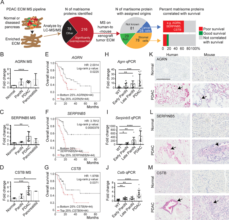
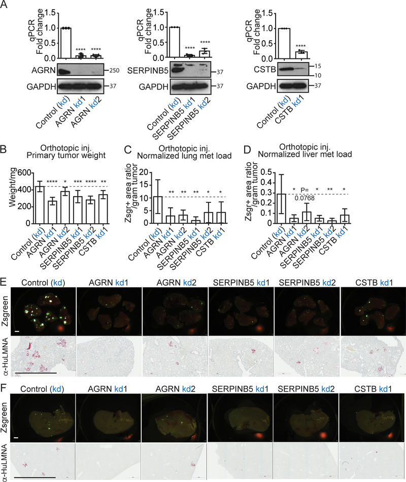
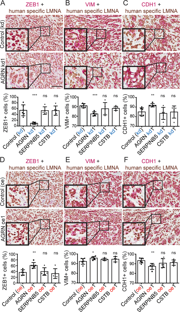
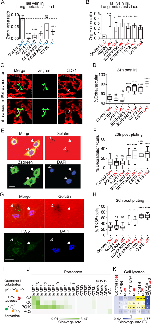
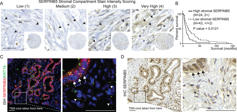
The prognosis for pancreatic ductal adenocarcinoma (PDAC) remains poor despite decades of effort. The abundant extracellular matrix (ECM) in PDAC comprises a major fraction of the tumor mass and plays various roles in promoting resistance to therapies. However, nonselective depletion of ECM has led to poor patient outcomes. Consistent with that observation, we previously showed that individual matrisome proteins derived from stromal cells correlate with either long or short patient survival. In marked contrast, those derived from cancer cells correlate strongly with poor survival. Here, we studied three cancer cell-derived matrisome proteins that are significantly overrepresented during PDAC progression, AGRN (agrin), SERPINB5 (serine protease inhibitor B5), and CSTB (cystatin B). Using both overexpression and knockdown experiments, we demonstrate that all three are promoters of PDAC metastasis. Furthermore, these proteins operate at different metastatic steps. AGRN promoted epithelial-to-mesenchymal transition in primary tumors, whereas SERPINB5 and CSTB enhanced late steps in the metastatic cascade by elevating invadopodia formation and in vivo extravasation. All three genes were associated with a poor prognosis in human patients and high levels of SERPINB5, secreted by cancer cells and deposited in the ECM, correlated with poor patient prognosis. This study provides strong evidence that cancer cell-derived matrisome proteins can be causal in promoting tumorigenesis and metastasis and lead to poor patient survival. Therefore, compared with the bulk matrix, mostly made by stromal cells, precise interventions targeting cancer cell-derived matrisome proteins, such as AGRN, SERPINB5, and CSTB, may represent preferred potential therapeutic targets. SIGNIFICANCE: This study provides insights into the biological roles of cancer cell-derived matrisome proteins in PDAC and supports the notion that these proteins are protumorigenic and better therapeutic targets.


Conflict of interest statement

The authors declare no potential conflicts of interest.

Figures

Figure 1.
Figure 1.. Expression of AGRN, SERPINB5 and CSTB in PDAC and correlation with patient survival.
A, a proteomic pipeline was used to identify overrepresented matrisome proteins in PDAC, and these proteins were then assigned as originating from human (cancer cell) or mouse (stroma) by MS analyses of human-to-mouse xenograft tumor ECM (13). Analyses of correlations with patient survival of individual matrisome proteins of different origins identified cancer-cell-derived matrisome proteins as being correlated with poor patient overall survival (e.g. AGRN, SERPINB5, and CSTB) whereas stromal-cell-derived proteins correlated either with good or poor survival and many showed no correlation with survival. B-D, quantitative TMT MS/MS reporter-ion ratios normalized to normal pancreas in human samples show increasing amounts of AGRN (B), SERPINB5 (C), and CSTB (D) proteins during PDAC progression. E-G, Kaplan-Meier analyses of TCGA RNA-seq data show that high expression levels of AGRN (E), SERPINB5 (F), and CSTB (G) correlate with poor overall patient survival. H-J, mRNA levels of all three genes, Agrn (H), Serpinb5 (I), and Cstb (J), are significantly elevated during PDAC progression in KC/KPC mouse samples, as shown by qRT-PCR. K-M, Immunohistochemistry confirmed protein overexpression in human and mouse PDAC compared to normal samples. Arrows point to epithelial regions. Scale bar is 200μm. *, p<0.05; **, p<0.01; ***, p<0.001; ****, p<0.0001. ns, not significant. All p-values come from two-tailed t-tests. All columns are represented by mean ± SD. This labeling scheme applies to all related figures.
Figure 2.
Figure 2.. Effects of knocking down expression of AGRN, SERPINB5, and CSTB on primary tumor growth and lung metastasis.
A, CRISPR-inactivation (kd) knocked down expression in the BxPC3 G1.1 cell line, of AGRN, SERPINB5 and CSTB, respectively, as shown by qPCR (top) and western blot (WB) (bottom). GAPDH is the loading control. B,C,D orthotopic injection of BxPC3 G1.1 cells knocked down for AGRN, SERPINB5, or CSTB showed that inhibition of each of them significantly reduced primary tumor weight (B), lung (C) and liver (D) metastasis load after normalization to primary tumor weight (C). N numbers are 12, 8, 9, 8, 10, 7 (left to right). E,F, lung (E) and liver (F) metastasis images from CRISPR-inactivation orthotopic xenograft mice are shown. Zsgreen (upper rows) and human-specific LMNA staining (bottoms rows) both highlight cancer cells; scale bar is 2mm.
Figure 3.
Figure 3.. Effects of overexpressing AGRN, SERPINB5, and CSTB on primary tumor growth and lung metastasis.
A, CRISPR-SAM-induced overexpression (oe) in the AsPC1 cell line, overexpressed AGRN, SERPINB5 and CSTB, respectively, as shown by qPCR (top) and western blot (WB) (bottom). GAPDH is the loading control. B, C, orthotopic injection of AsPC1 overexpressing AGRN, SERPINB5, or CSTB showed that overexpression of each of them failed to affect primary tumor weight (B). However, overexpression significantly increased lung metastasis load after normalization to primary tumor weight (C), except for CSTB oe2, for which P=0.0529. N numbers are 8, 6, 7, 6, 5, 5, 6 (left to right). D, lung images from CRISPR-SAM orthotopic xenograft mice are shown. Zsgreen (upper rows) and human-specific LMNA staining (bottom rows) both highlight cancer cells; scale bar is 2mm.
Figure 4.
Figure 4.. AGRN promote EMT in primary tumors.
A-F, double-color IHC with human-specific LMNA in brown and ZEB1 (A,D), VIM (B,E), or CDH1 (C,F) in red. The corresponding quantifications (see Materials and Methods) show that AGRN kd, but not SERPINB5 or CSTB kd, reduced ZEB1+ and VIM+ tumor-cell fractions and increased the CDH1+ tumor-cell fraction, while AGRN oe regulated EMT in the opposite direction.
Figure 5.
Figure 5.. SERPINB5 and CSTB promote formation of invadopodia and extravasation.
A, B, tail-vein injection showed that knockdown (kd, A) of each of the three genes restricted, while overexpression (oe, B) of each promoted experimental metastasis. N=5 for all. C, example images of cells that are inside or outside of vasculature 24h after tail-vein injection. Arrows point to cancer cells that are Zsgreen-positive. D, extravasation assay shows that overexpression of either SERPINB5 or CSTB (but not AGRN) caused a higher fraction of cancer cells to be extravasated at 24h post-injection. At least 40 cells were scored blindly per mouse. N=5 for all, except that N=10 for control group. E, example images of cells that degraded (arrows) or did not degrade (arrowhead) gelatin in the course of invadopodial assays. F, quantification of percentage of cells per field that have degraded gelatin at 20 hours post-plating for each cell line. G, colocalization of TKS5+ invadopodia and the degradation spots in AsPC1 cells that degraded (arrow) or did not degrade (arrowhead) gelatin 20 hours post-plating. H, quantification of percentage of cells per field that have TKS5+ invadopodia at 20 hours post-plating for each cell line. At least 20 fields were quantified per cell line for F and H (see Materials and Methods). I, protease activity assay uses peptides with quenched fluorescein that can be dequenched by protease cleavage. J,K, four peptides were cleaved by recombinant proteases (J) or cell lysates (K) in triplicate. Average cleavage rate is illustrated (J) and compared to the control cell lines (K).
Figure 6.
Figure 6.. Cancer-cell-derived SERPINB5 is a poor prognostic factor.
A, representative images for stromal SERPINB5 staining scoring system on a human patient tissue microarray. Arrows point to representative stromal regions that look like ECM, where the scoring was done. B, survival curves from patients categorized into two populations based on high and low stromal staining showed that high stromal signal correlates with significantly poorer patient survival. C, D, RNA in situ hybridization showed exclusively epithelial-cell origin of SERPINB5 RNA (C), whereas the adjacent section (D), which was stained for SERPINB5 by IHC showed protein staining over the extracellular ECM (D). Arrows point to the epithelial compartment, while arrowheads point to the IHC signal over the ECM. Scale bars are 50μm.

References

    1. Siegel RL, Miller KD, Jemal A. Cancer statistics, 2019. CA Cancer J Clin 2019;69:7–34 - PubMed
    1. Manji GA, Olive KP, Saenger YM, Oberstein P. Current and Emerging Therapies in Metastatic Pancreatic Cancer. Clinical cancer research : an official journal of the American Association for Cancer Research 2017;23:1670–8 - PubMed
    1. Cid-Arregui A, Juarez V. Perspectives in the treatment of pancreatic adenocarcinoma. World J Gastroenterol 2015;21:9297–316 - PMC - PubMed
    1. Rhim AD, Oberstein PE, Thomas DH, Mirek ET, Palermo CF, Sastra SA, et al. Stromal Elements Act to Restrain, Rather Than Support, Pancreatic Ductal Adenocarcinoma. Cancer cell 2014 - PMC - PubMed
    1. Ozdemir BC, Pentcheva-Hoang T, Carstens JL, Zheng X, Wu CC, Simpson TR, et al. Depletion of carcinoma-associated fibroblasts and fibrosis induces immunosuppression and accelerates pancreas cancer with reduced survival. Cancer cell 2014;25:719–34 - PMC - PubMed

Publication types

MeSH terms