. 2020 Feb 18;117(7):3704-3710.
doi: 10.1073/pnas.1917876117. Epub 2020 Feb 6.

Defective HIV-1 proviruses produce viral proteins

Affiliations

Defective HIV-1 proviruses produce viral proteins

Hiromi Imamichi et al. Proc Natl Acad Sci U S A. .

Abstract

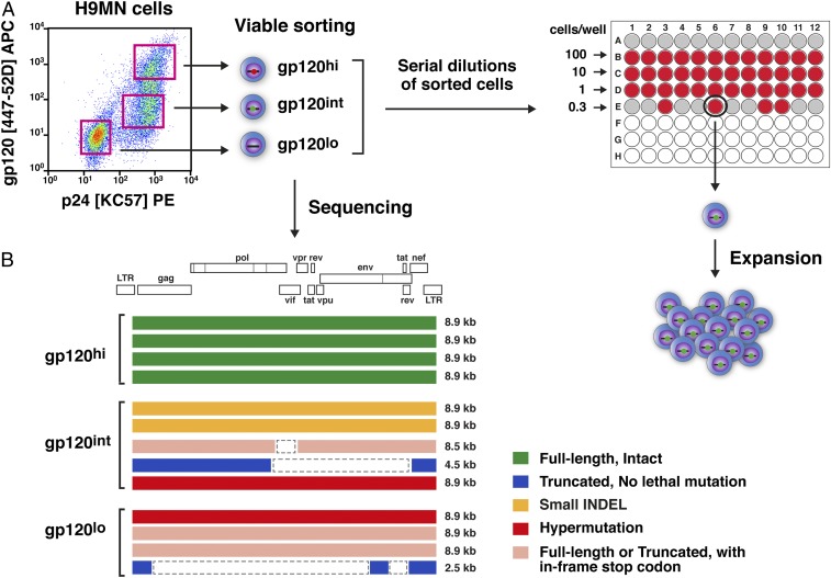
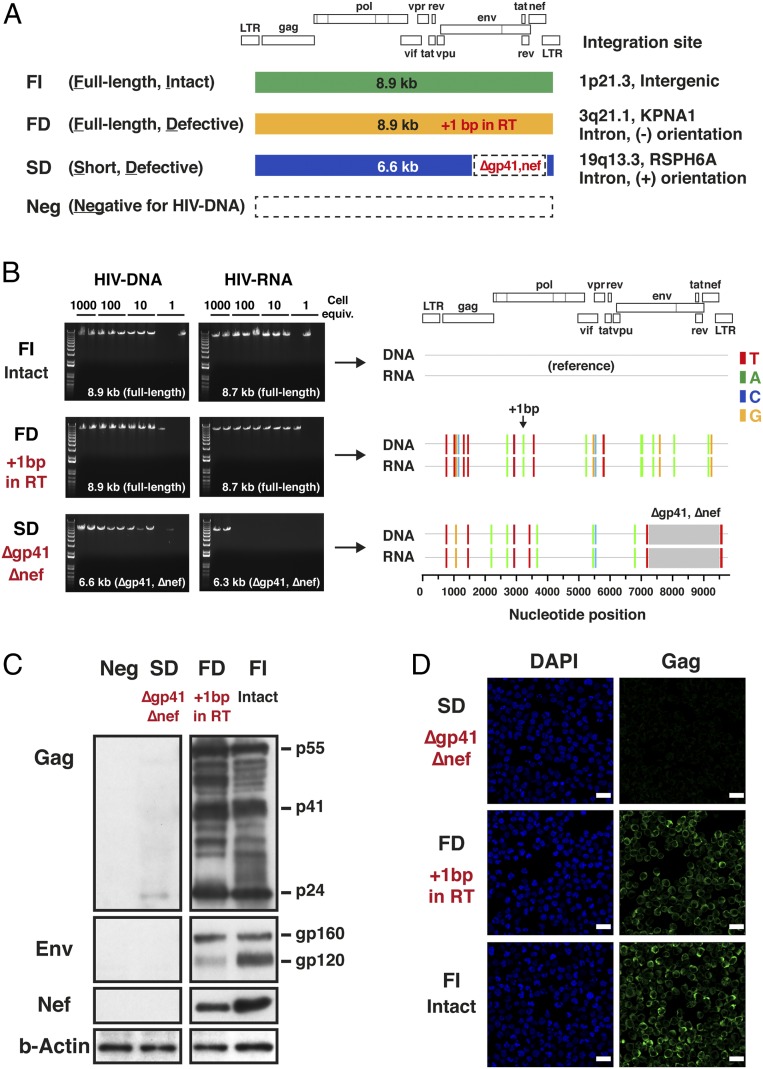
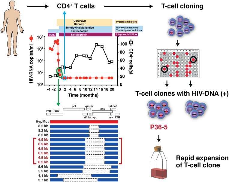
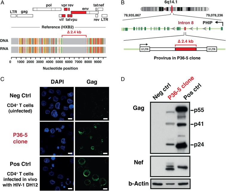
HIV-1 proviruses persist in the CD4+ T cells of HIV-infected individuals despite years of combination antiretroviral therapy (cART) with suppression of HIV-1 RNA levels <40 copies/mL. Greater than 95% of these proviruses detected in circulating peripheral blood mononuclear cells (PBMCs) are referred to as "defective" by virtue of having large internal deletions and lethal genetic mutations. As these defective proviruses are unable to encode intact and replication-competent viruses, they have long been thought of as biologically irrelevant "graveyard" of viruses with little significance to HIV-1 pathogenesis. Contrary to this notion, we have recently demonstrated that these defective proviruses are not silent, are capable of transcribing novel unspliced forms of HIV-RNA transcripts with competent open reading frames (ORFs), and can be found in the peripheral blood CD4+ T cells of patients at all stages of HIV-1 infection. In the present study, by an approach of combining serial dilutions of CD4+ T cells and T cell-cloning technologies, we are able to demonstrate that defective proviruses that persist in HIV-infected individuals during suppressive cART are translationally competent and produce the HIV-1 Gag and Nef proteins. The HIV-RNA transcripts expressed from these defective proviruses may trigger an element of innate immunity. Likewise, the viral proteins coded in the defective proviruses may form extracellular virus-like particles and may trigger immune responses. The persistent production of HIV-1 proteins in the absence of viral replication helps explain persistent immune activation despite HIV-1 levels below detection, and also presents new challenges to HIV-1 eradication.

Keywords: HIV; immune activation; provirus.


Conflict of interest statement

The authors declare no competing interest.

Figures

Fig. 1.
Fig. 1.
Isolation of single-cell clones from the H9MN cell line. (A) Schematic diagrams of the strategies used to isolate H9MN single-cell clones harboring defective proviruses. Flow cytometry plot of HIV-1 MN cells showing the expression of intracellular Gag p24 protein (clone: KC-57) and cell surface Env gp120 (clone: 447–52D) protein. The H9MN cells were flow-sorted into three populations based on cell surface expression of Env gp120 protein and CD3: gp120hi, gp120int, and gp120lo. The flow-sorted cells were serially diluted in 96-well plates and left to grow to a confluent status (2 weeks). Cells from the wells exhibiting growth (visually inspected by microscopy) were transferred to 48-well plates. A small aliquot of cells from the wells in the 48-well plates was taken to detect the presence of HIV-DNA by 5′LTR-to-3′LTR PCR. Cells from wells positive for HIV-DNA were further expanded in 24-well plates for an additional week. Upon completion of the expansion culture, one portion (1 × 10⁶) of the cells was used for simultaneous extraction of DNA and RNA (after cDNA synthesis), followed by 5′LTR-to-3′LTR single-genome amplification and direct amplicon sequencing; the other portion (2 × 10⁶) of the cells was used for Western blot and confocal microscopy (1 × 10⁶ cells) for detection of HIV-1 proteins. (B) Proviral sequences present in the three sorted fractions of H9MN cells.
Fig. 2.
Fig. 2.
Expression of HIV-DNA, HIV-RNA, and HIV-1 proteins in H9MN subclones harboring defective proviruses. (A) A total of four distinct single-cell clones were isolated: FI, a subclone containing an 8.9-kb full-length intact provirus; FD, a subclone containing an 8.9-kb full-length defective provirus with a 1-bp frameshift lethal mutation in the RT gene (HXB2 coordinate 3204); SD, a subclone containing a short defective provirus with a large (∼2.3-kb) internal deletion affecting the gp41 and nef coding regions; and Neg, a subclone negative for HIV-DNA. (B) Agarose gel pictures depicting sizes of HIV-DNA and HIV-RNA PCR fragments generated for FI (full-length intact provirus), FD (full-length defective provirus), and SD (short defective provirus) single-cell clones using 5′LTR-to-3′LTR PCR. The highlighter analysis was performed using the provirus sequence derived from the FI clone as a reference. The nucleotide positions that differ from the HIV-DNA sequence in the FI clone are indicated by color-coded bars. The gray areas indicate gaps. HIV-RNA sequences corresponded precisely to the HIV-DNA sequences for each clone. The integration sites of the proviruses present in the FI, FD, and SD clones were analyzed by restriction enzyme digestion of genomic DNA by BclI and inverse PCR. (C) Expression of HIV-1 proteins in three distinct H9MN single-cell clones by Western blot: SD, FD, and FI. A mouse monoclonal antibody for HIV-1 p24 (clone: 39/5.4A), a goat polyclonal antibody for HIV-1 Env, and a mouse monoclonal antibody for HIV-1 Nef (clone: EH1) were used. The SD clone expressed no Env or Nef and only a low level of p24 Gag. The FD and FI clones expressed Gag p55/p24 and Env gp160/gp120 proteins with molecular weights predicted by the HIV-RNA sequences. (D) Confocal microscopy analysis of the intracellular expression of Gag p24 in the three distinct single-cell clones. A mouse monoclonal antibody for HIV-1 p24 (clone: 39/5.4A) was used. Nuclei were visualized by DAPI (blue). Original magnification was ×63. (Scale bars, 20 µm.)
Fig. 3.
Fig. 3.
Isolation of CD4+ T cell clones harboring defective proviruses from an individual with advanced HIV-1 infection. Schematic diagram of the strategy used to isolate CD4+ T cell clones harboring defective proviruses from a patient with HIV-1 infection. CD4+ T cells were obtained from a time point 2 months after achieving a pVL <40 copies/mL (blue circle). Out of 6,000 cells initially plated, two T cell clones harboring the same defective provirus, P36-5, were isolated. This provirus, with a 2.4-kb internal deletion, was the predominant species present in the patient’s cells in vivo (green circle). By isolating T cell clones, the capacity to transcribe HIV-RNA and produce viral proteins at the single-cell level can be assessed.
Fig. 4.
Fig. 4.
Expression of HIV-DNA, HIV-RNA, and HIV-1 proteins in the CD4+ T cell clone harboring a defective provirus derived from an HIV-infected individual during cART. (A) HIV-RNA sequences corresponded precisely to the HIV-DNA sequences for the P36-5 clone. Analysis of the genome structure revealed a 2.4-kb internal deletion affecting the region encoding the HIV-1 accessory proteins (tat, rev, vpu, and others) and the gp120 portion of the Env protein (red). The Gag, Pol, and Nef regions remained intact in the provirus present in the P36-5 clone. (B) Whole-genome sequencing confirmed the presence of two intact LTRs and revealed the provirus to be integrated in intron 8 of the PHIP gene in the opposite orientation to the PHIP gene. (C) Confocal microscopy analysis of the intracellular expression of the Gag p24 protein. A mouse monoclonal antibody for HIV-1 p24 (clone: 39/5.4A) was used. Nuclei were visualized by DAPI (blue). Original magnification was ×63. (Scale bars, 5 µm.) (D) Expression of HIV-1 proteins by Western blot. Negative control (Neg Ctrl), uninfected CD4+ T cells; and positive control (Pos Ctrl), CD4+ T cells infected in vitro with the DH12 strain of HIV-1. A mouse monoclonal antibody for HIV-1 p24 (clone: 39/5.4A) and a mouse monoclonal antibody for HIV-1 Nef (clone: 3A2) were used. The predicted sizes of the Nef proteins, based on the provirus sequences for DH12 and P36-5, were 23.4 kDa and 22.6 kDa, respectively. The higher molecular weight band seen for Nef in P36-5 likely represents posttranslational modifications such as myristoylation.

References

    1. Imai H., et al. , A defective proviral DNA with a 2.6-kb deletion of human immunodeficiency virus type 1 (HIV-1) in a persistently HIV-1 infected cell clone. Virus Genes 5, 81–88 (1991). - PubMed
    1. Li Y., et al. , Molecular characterization of human immunodeficiency virus type 1 cloned directly from uncultured human brain tissue: Identification of replication-competent and -defective viral genomes. J. Virol. 65, 3973–3985 (1991). - PMC - PubMed
    1. Sanchez G., Xu X., Chermann J. C., Hirsch I., Accumulation of defective viral genomes in peripheral blood mononuclear cells of human immunodeficiency virus type 1-infected individuals. J. Virol. 71, 2233–2240 (1997). - PMC - PubMed
    1. Ho Y. C., et al. , Replication-competent noninduced proviruses in the latent reservoir increase barrier to HIV-1 cure. Cell 155, 540–551 (2013). - PMC - PubMed
    1. Imamichi H., et al. , Defective HIV-1 proviruses produce novel protein-coding RNA species in HIV-infected patients on combination antiretroviral therapy. Proc. Natl. Acad. Sci. U.S.A. 113, 8783–8788 (2016). - PMC - PubMed

Publication types