. 2020 Feb 7;367(6478):652-660.
doi: 10.1126/science.aay0542.

An AMPK-caspase-6 axis controls liver damage in nonalcoholic steatohepatitis

Affiliations

An AMPK-caspase-6 axis controls liver damage in nonalcoholic steatohepatitis

Peng Zhao et al. Science. .

Abstract

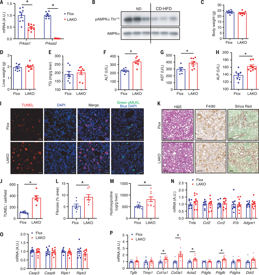
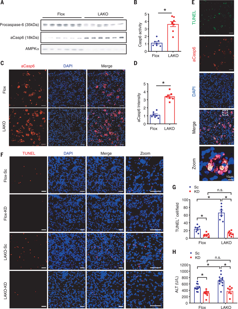
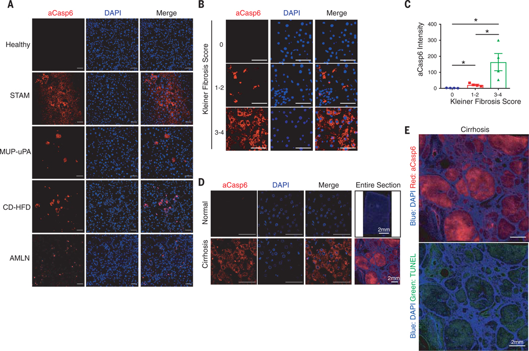
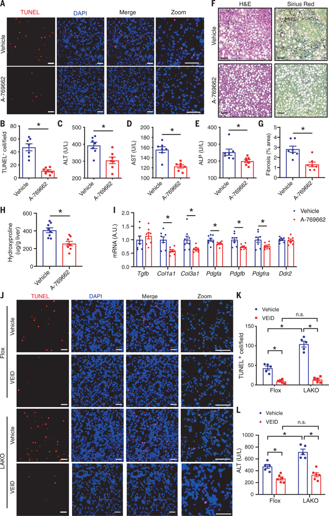
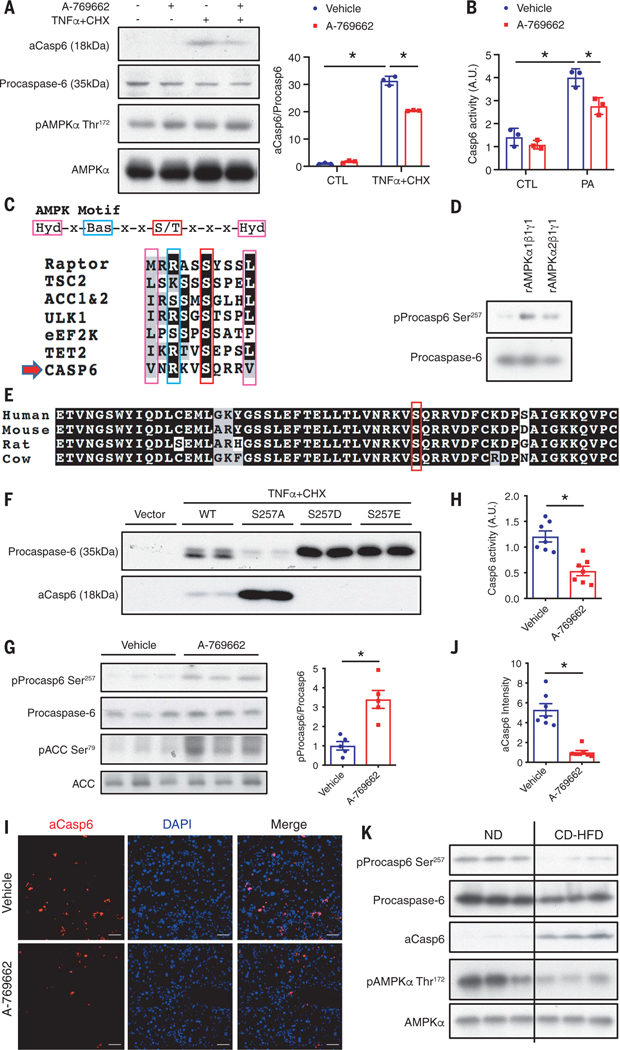
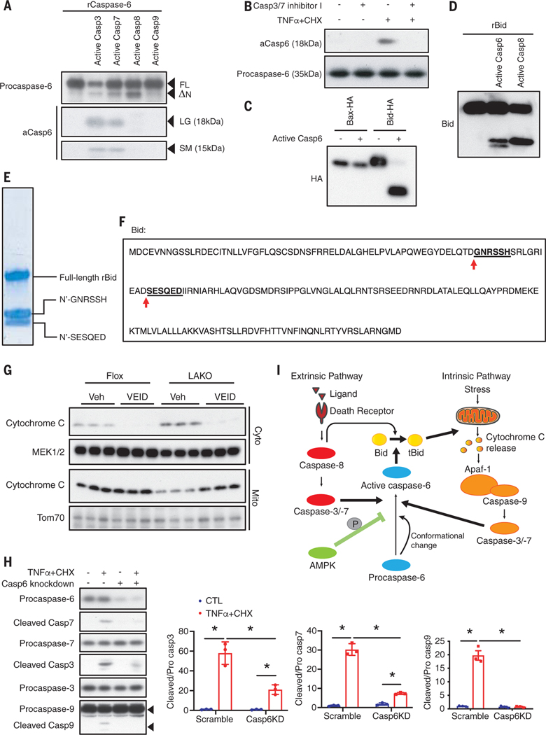
Liver cell death has an essential role in nonalcoholic steatohepatitis (NASH). The activity of the energy sensor adenosine monophosphate (AMP)-activated protein kinase (AMPK) is repressed in NASH. Liver-specific AMPK knockout aggravated liver damage in mouse NASH models. AMPK phosphorylated proapoptotic caspase-6 protein to inhibit its activation, keeping hepatocyte apoptosis in check. Suppression of AMPK activity relieved this inhibition, rendering caspase-6 activated in human and mouse NASH. AMPK activation or caspase-6 inhibition, even after the onset of NASH, improved liver damage and fibrosis. Once phosphorylation was decreased, caspase-6 was activated by caspase-3 or -7. Active caspase-6 cleaved Bid to induce cytochrome c release, generating a feedforward loop that leads to hepatocyte death. Thus, the AMPK-caspase-6 axis regulates liver damage in NASH, implicating AMPK and caspase-6 as therapeutic targets.


Conflict of interest statement

Competing interests: A.R.S. and P.Z. are named inventors of a patent application related to the use of AMPK and caspase-6 as therapeutic targets for NASH. J.L.W. is a founding member of Oxitope. M.K. and A.R.S. are founders of Elgia Therapeutics. Other authors declare no competing interests.

Figures

Fig. 1.
Fig. 1.. Liver-specific AMPK knockout exaggerates liver damage in NASH.
(A) Expression of Prkaa1 and Prkaa2 in liver; n = 8 to 9 mice. A.U., arbitrary units. (B) Immunoblot of liver lysate from ND or CD-HFD–fed mice; n = 3 mice. (C to P) Flox and LAKO mice fed CD-HFD for 11 weeks. (C) Body weight. (D) liver weight. (E) Liver TG. [(F) to (H)] Serum ALT (F), AST (G), ALP (H); n = 8 to 9 mice. (I) Liver sections stained TUNEL (red) and 4′,6-diamidino-2-phenylindole (DAPI) (blue) or pMLKL (Green). Scale bar, 50 μm. (J) Quantification of TUNEL-positive nuclei per field in (I); n = 7 mice. (K) H&E, F4/80, and Sirius red staining of liver sections. Scale bar, 100 μm; n = 7 mice. (L) Quantification of fibrosis area (percent of total area) shown in (K); n = 7 mice. (M) Liver hydroxyproline; n =8 to 9 mice. (N) Expression of TNFα, Ccl2, Ccr2, Il1b, and Adgre1 in liver; n = 8 to 9 mice. (O) Expression of Casp3, Casp8, Ripk1, and Ripk3 in liver; n =8 to 9 mice. (P) Expression of Tgfb, Timp1, Col1a1, Col3a1, Acta2, Pdgfa, Pdgfb, Pdgfra, and Ddr2 in liver; n = 8 to 9 mice. Mean ± SEM; *P < 0.05, Student’s unpaired t test.
Fig. 2.
Fig. 2.. AMPK deficiency increases caspase-6 cleavage to promote liver damage in NASH.
(A to E) Flox and LAKO mice fed CD-HFD for 11 weeks. (A) Immunoblot analysis of liver lysate; n = 6 mice. (B) Casp6 activity in liver lysate; n = 7 to 8 mice. (C) Liver sections stained active Caspase-6 (aCasp6, red) and DAPI (blue). Scale bar, 50 μm. (D) Quantification of aCasp6 staining in (C); n = 7 mice. (E) Liver sections stained TUNEL (green), aCasp6 (red), and DAPI (blue). Scale bar, 50 μm. *P < 0.05, Student’s unpaired t test. (F to H) Flox and LAKO mice fed with CD-HFD for 3 weeks, followed by intravenous injection of 1.5 mg/kg caspase-6 siRNA (KD) or scrambled RNA (Sc) twice per week for 3 weeks while fed continuous CD-HFD. (F) Liver sections stained TUNEL (red) and DAPI (blue). Scale bar, 50 μm. (G) Quantification of TUNEL-positive nuclei per field in (F); n =7 mice. (H) Serum ALT; n =7 to 10 mice. Mean ± SEM; *P < 0.05, two-way analysis of variance (ANOVA).
Fig. 3.
Fig. 3.. Caspase-6 is activated in mouse and human NASH.
(A) Healthy model: 24-weeks-old male C57BL/6J mice fed ND. STAM-NASH model: male C57BL/6J mice were subcutaneously injected 200 μg streptozotocin (STZ) within 48 hours after birth and fed HFD for 6 weeks starting at 4 weeks of age. MUP-uPA-NASH model: male MUP-uPA mice fed 60% HFD for 16 weeks. CD-HFD–NASH model: C57BL/6J mice were fed CD-HFD for 11 weeks. AMLN-NASH model: C57BL/6J mice fed AMLN diet for 30 weeks. Liver sections were stained with aCasp6 (red) and DAPI (blue). Scale bar, 50 μm. (B) Human liver sections were classified blindly by liver pathologist and stained with aCasp6 (Kleiner fibrosis score 0, 1 to 2, and 3 to 4). Scale bar, 50 μm. (C) Quantification of aCasp6 staining in (B), plotted against Kleiner fibrosis scores; n = 4 human subjects. (D) Human liver sections were stained aCasp6 to compare caspase-6 activation in healthy and cirrhotic donors. Scale bar, 50 μm. (E) Scanning of human liver section stained aCasp6 (red), TUNEL (green), and DAPI. Scale bar, 2 mm. Mean ± SEM; *P < 0.05, Student’s unpaired t test.
Fig. 4.
Fig. 4.. Both an AMPK agonist and a caspase-6 inhibitor therapeutically improve liver damage.
(A to I) C57BL/6J mice were fed CD-HFD for 6 weeks, followed by intraperitoneal injection of 25 mg/kg A-769662 or vehicle daily for 2 weeks while fed continuous CD-HFD. (A) Liver sections stained TUNEL (red) and DAPI (blue). Scale bar, 50 μm. (B) Quantification of TUNEL-positive nuclei per field in (A); n = 7 mice. [(C) to (E)] Serum ALT (C), AST (D), and ALP (E); n = 7 mice. (F) H&E and Sirius red staining of liver sections. Scale bar, 100 μm. (G) Quantification of liver fibrosis area (percent of total area) in (F); n = 7 mice. (H) Liver hydroxyproline; n = 8 mice. (I) Expression of Tgfb, Timp1, Col1a1, Col3a1, Pdgfa, Pdgfb, Pdgfra, and Ddr2 in livers; n = 8 mice. *P < 0.05, Student’s unpaired t test. (J to L) Flox and LAKO mice were fed CD-HFD for 6 weeks, followed by intraperitoneal injection of 5 mg/kg VEID or vehicle every other day for 2 weeks while continuously feeding. (J) Liver sections stained TUNEL (red) and DAPI (blue). Scale bar, 50 μm. (K) Quantification of TUNEL-positive nuclei per field in (J); n = 5 to 6 mice. Mean ± SEM; *P < 0.05, two-way ANOVA.
Fig. 5.
Fig. 5.. AMPK phosphorylates caspase-6 to inhibit its cleavage and activation.
(A) Primary hepatocytes were pretreated with 40 μM A-769662 for 1 hour then treated with 30 μg/ml CHX and 50 ng/ml TNFα for 2 hours. Shown is immunoblot analysis of cell lysates; n = 3 independent experiments. Mean ± SD; *P < 0.05, two-way ANOVA. (B) Primary hepatocytes were pretreated with 40 μM A-769662 for 1 hour then treated with 250 μM bovine serum albumin (BSA)–conjugated PA for 2 hours. Cell lysates were subject to caspase-6 activity assay. Mean ± SD; *P < 0.05. (C) Caspase-6 Ser257 locates within AMPK substrate motif. (D) In vitro kinase assay using recombinant caspase-6 and recombinant AMPKα1β1γ1 or AMPKα2β1γ1 active kinase. (E) Alignment of caspase-6 sequence. (F) HEK293T cells overexpressing caspase-6-myc wild type, S257A, S257D, or S257E mutant were treated with 10 μg/ml CHX and 25 ng/ml TNFα for 2 hours. Shown is immunoblot analysis of cell lysates. (Single-letter abbreviations for the amino acid residues are as follows: A, Ala; D, Asp; E, Glu; S, Ser. In the mutants, other amino acids were substituted at certain locations; for example, S257A indicates that serine at position 257 was replaced by alanine.) (G to J) C57BL/6J mice were fed CD-HFD for 6 weeks, followed by intraperitoneal injection of 25 mg/kg A-769662 or vehicle daily for 2 weeks while fed continuous CD-HFD. Mice were euthanized 6 hours after the last injection. (G) Immunoblot analysis of liver lysates; n = 5 mice. (H) Liver lysates were subject to caspase-6 activity assay; n = 7 mice. (I) Liver sections stained with aCasp6 (red) and DAPI (blue). Scale bar, 50 μm. (J) Quantification of aCasp6 staining per field in (I); n = 7 mice. Mean ± SEM; *P < 0.05, Student’s unpaired t test. (K) Immunoblot analysis of liver lysates from C57BL/6J mice fed ND or CD-HFD. AMPK and pAMPK blots are the same as in Fig. 1B.
Fig. 6.
Fig. 6.. Caspase-6 mediates a feedforward loop to sustain the caspase cascade.
(A) In vitro cleavage assay using recombinant procaspase-6 with active caspase-3, −7, −8, or −9. FL, full-length; ΔN, n terminus deleted form; LG, large; SM, small. (B) Primary hepatocytes were pretreated with 10 μM caspase-3/7 inhibitor I for 1 hour then treated with 30 μg/ml CHX and 50 ng/ml TNFα for 2 hours. Immunoblot analysis of cell lysates. (C) In vitro cleavage assay using purified Bid-HA or Bax-HA expressed in HEK293T cells, and active caspase-6. (D) In vitro cleavage assay using recombinant Bid with active caspase-6 or −8. (E) In vitro cleavage assay using recombinant Bid with active caspase-6. Bands for cleaved Bid were subject to Edman degradation. (F) Bid sequence and sites cleaved by active caspase-6. (G) Flox and LAKO mice were fed CD-HFD for 6 weeks, followed by intraperitoneal injection of 5 mg/kg VEID or vehicle every other day for 2 weeks while fed continuous CD-HFD. Livers were fractionated to separate cytosolic and mitochondrial extract for immunoblot analysis. (H) HepG2 cells transfected scrambled RNA or Caspase-6 siRNA were treated with vehicle or 30 μg/ml CHX and 50 ng/ml TNFα for 2 hours. Medium was changed to remove treatment for 5 hours. Cell lysates were subject to immunoblot analysis; n = 3 independent experiments. Mean ± SD; *P < 0.05, two-way ANOVA. (I) Proposed model for roles of AMPK–caspase-6 axis in apoptotic caspase cascade.

Comment in

References

    1. Hernandez-Gea V, Friedman SL, Annu. Rev. Pathol 6, 425–456 (2011). - PubMed
    1. Friedman SL, Neuschwander-Tetri BA, Rinella M, Sanyal AJ, Nat. Med 24, 908–922 (2018). - PMC - PubMed
    1. Michalopoulos GK, DeFrances M, Adv. Biochem. Eng. Biotechnol 93, 101–134 (2005). - PubMed
    1. Guicciardi ME, Gores GJ, Gut 54, 1024–1033 (2005). - PMC - PubMed
    1. Luedde T, Kaplowitz N, Schwabe RF, Gastroenterology 147, 765–783.e4 (2014). - PMC - PubMed

Publication types

MeSH terms